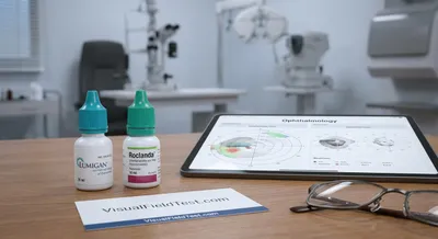
Lowering Eye Pressure: The Lumigan vs Roclanda Showdown
The two drops work differently. Lumigan (bimatoprost) is a prostaglandin analogue. Prostaglandin analogues act mainly by increasing the drainage of...
Deep research and expert guides on maintaining your visual health.
The two drops work differently. Lumigan (bimatoprost) is a prostaglandin analogue. Prostaglandin analogues act mainly by increasing the drainage of...
This article dives deep into what PDS is, how it can progress, and what it means for athletes and fitness enthusiasts. We’ll explain the eye’s...
Trabeculectomy (filtering surgery) creates a new drainage path for fluid (aqueous humor) to leave the eye under the eyelid. Surgeons remove a small...
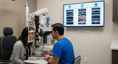
SLT is often recommended either before starting drops or after medications alone can’t reach the target pressure. Being “selective,” the laser...
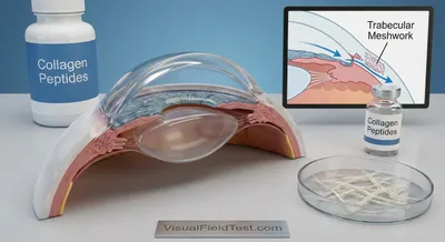
In a healthy eye, the TM and SC work together like a plumbing system. The TM is a spongy, porous tissue lined by endothelial cells, and it sits just...
Right now, no therapy has been proven to do this in patients. In large, decades-long studies only pressure lowering showed a clear benefit. In fact,...
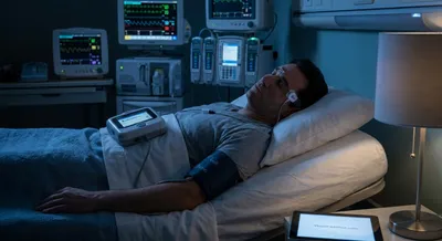
Importantly, body position and sleep also matter. Normally, when you lie down, intraocular pressure (IOP) tends to rise (by 10–20%) because eye fluid...

Research has shown that excessive night-time hypotension is associated with glaucomatous damage. In fact, Hayreh and colleagues found that night-time...
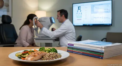
This article reviews the latest evidence on macronutrient patterns and glaucoma. We will survey epidemiologic studies of diet patterns...

Endothelin-1 (ET-1) is made by cells lining blood vessels throughout the body, and it helps regulate normal blood pressure and flow. In the eye, ET-1...

Indeed, multiple studies have found senescence markers in RGCs and optic nerve tissue in glaucoma models. Notably, removing those old RGCs has been...

The April 2026 trials can be grouped by their main intervention modality:

Earlier trial results. A recent Phase III study (NCX 470) and others confirmed that combined outflow enhancers can beat traditional drops. For...

Home readings can also serve as early safety signals. Protocols typically pre-specify pressure thresholds or warning rules. For instance, the...
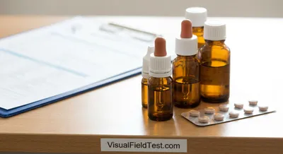
Beta-blockers (e.g. timolol): These are typically washed out by stopping the drop for 4 weeks. Research showed that a 2-week break is usually too...

In theory, brief exposure to high oxygen (like short HBOT sessions) could activate protective pathways inside eye cells. One key pathway involves the...
Playing soccer provides excellent cardiorespiratory exercise. The running, jogging, and game movement substantially raise heart rate and build...

However, hiking also brings challenges: long treks can lead to dehydration, strong sunlight, and difficult footing. Importantly for glaucoma...
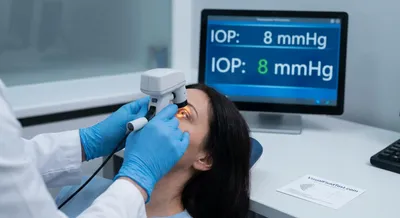
Intraocular pressure is the force exerted by the fluids inside the eye that keeps the eye firm and helps it maintain its shape. The eye constantly produces a clear fluid that nourishes tissues and then drains it away; intraocular pressure reflects the balance between production and drainage. Normal pressure varies between people but is usually within a range that is safe for the delicate tissues inside the eye. When fluid builds up because drainage is blocked or production is too high, pressure rises and can press on sensitive structures like the optic nerve. If pressure stays too high, it can damage nerve fibers and cause gradual vision loss without noticeable early symptoms. Measuring intraocular pressure is a routine part of eye exams and helps identify people at risk of certain eye conditions before vision is lost. Pressure can change over the course of a day and may be affected by body position, medications, and other health conditions. Treatments to manage elevated pressure include eye drops, laser procedures, and surgery that either reduce fluid production or improve drainage. Monitoring and controlling pressure is important because early detection and treatment can preserve vision and prevent irreversible damage.