Melatonin and the Eye: Nighttime IOP and Neuroprotection
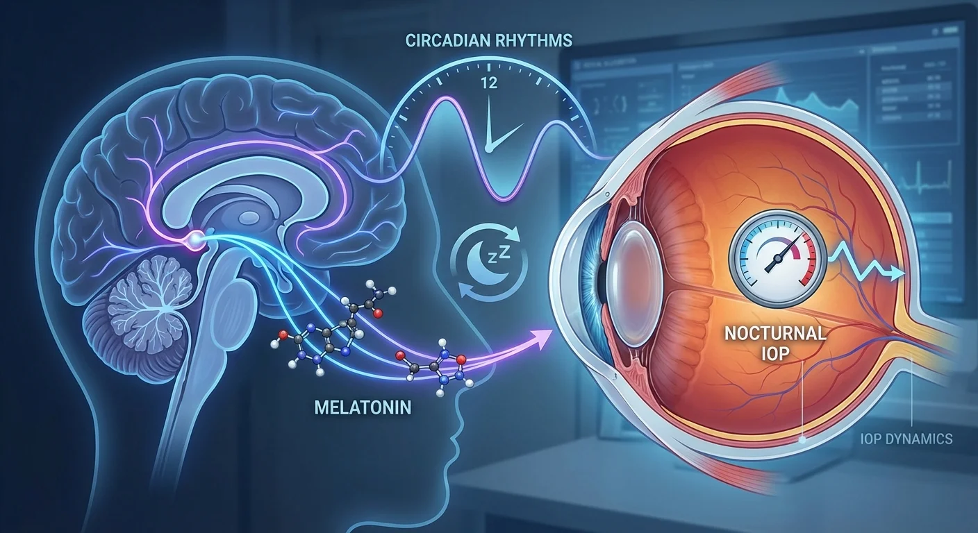
Melatonin is a neurohormone produced in a ~24-hour cycle (circadian rhythm) that plays key roles in sleep regulation and acts as a powerful antioxidant. In the eye, melatonin is synthesized locally (in the retina and ciliary body) and binds to MT1/MT2 melatonin receptors on ocular cells (pmc.ncbi.nlm.nih.gov). Its levels peak at night, coinciding with the normal drop in blood pressure and (in healthy individuals) the typical reduction in intraocular pressure (IOP) during sleep. These circadian patterns mean melatonin helps modulate aqueous humor (the watery fluid filling the front of the eye) dynamics. In turn, this affects nighttime IOP and retinal health, especially in aging. Recent studies suggest that impaired melatonin signaling may contribute to glaucoma risk, while melatonin analogs (drugs that mimic melatonin) show promise in lowering IOP and protecting retinal neurons (pmc.ncbi.nlm.nih.gov) (pmc.ncbi.nlm.nih.gov).
Ocular Melatonin and Circadian Control
Melatonin is not only made by the pineal gland but also produced in the eye itself. Photoreceptors in the retina generate melatonin at night, and the ciliary body (the gland that produces aqueous humor) also synthesizes melatonin and releases it into the aqueous (pmc.ncbi.nlm.nih.gov) (pmc.ncbi.nlm.nih.gov). This means melatonin levels in the aqueous humor rise in darkness, peaking around midnight to 2–4 AM (pmc.ncbi.nlm.nih.gov). By contrast, light exposure (especially blue light) suppresses melatonin via melanopsin-containing retinal ganglion cells. Thus, melatonin is a bridge between circadian signals (day–night) and intraocular physiology.
Receptors for melatonin (MT1, MT2 and possibly MT3) are found on cells of the eye, including the non-pigmented ciliary epithelial cells that secrete aqueous humor (pmc.ncbi.nlm.nih.gov). Activation of these receptors influences cellular pathways (via G-proteins) that control ion transport and fluid secretion. In simple terms, melatonin engagement tends to slow aqueous humor production, helping lower IOP. Conversely, loss of normal melatonin signaling (as may happen in glaucoma or with aging) can lead to higher nighttime IOP. For example, mice lacking the MT1 receptor have higher nocturnal IOP and suffer more retinal ganglion cell (RGC) loss (pmc.ncbi.nlm.nih.gov) (pmc.ncbi.nlm.nih.gov). Similarly, human glaucoma patients often secrete abnormally timed melatonin due to damage of light-sensitive retinal cells, suggesting a chicken-and-egg problem: glaucoma may disturb circadian rhythms, and disrupted melatonin may worsen glaucoma (pmc.ncbi.nlm.nih.gov) (pmc.ncbi.nlm.nih.gov).
Melatonin in Aqueous Humor Dynamics
The generation and drainage of aqueous humor set eye pressure. Melatonin influences both sides of this balance. As noted, melatonin slows aqueous production by ciliary epithelial cells via MT1/MT2 receptor signaling (which lowers cAMP inside the cells) (pmc.ncbi.nlm.nih.gov) (pmc.ncbi.nlm.nih.gov). Experiments in animals show melatonin analogs reduce IOP dramatically. For instance, the MT3 agonist 5-MCA-NAT produced a 43% IOP drop in rabbits (versus 24% by melatonin itself) (pmc.ncbi.nlm.nih.gov). In glaucoma-model monkeys, 5-MCA-NAT lowered IOP steadily over days, with effects lasting >18 hours (pmc.ncbi.nlm.nih.gov). Similarly, the MT2 agonist IIK7 and other analogs have shown significant pressure-lowering in animals. This suggests multiple melatonin receptors (especially MT3) mediate IOP control (pmc.ncbi.nlm.nih.gov) (pmc.ncbi.nlm.nih.gov).
In addition to reducing production, melatonin may help increase aqueous outflow. It modulates ion channels (e.g. chloride transport) and enzymes in the ciliary body. One study found melatonin boosted Cl⁻ transport in porcine ciliary cells, affecting fluid secretion (pmc.ncbi.nlm.nih.gov). Another showed a melatonin analog downregulated carbonic anhydrase enzymes (which normally drive aqueous creation), causing a 51% pressure drop lasting 4 days (pmc.ncbi.nlm.nih.gov). Melatonin also appears to interact with adrenergic (sympathetic) signals: melatonin analogs enhanced timolol’s IOP reduction by ~15% (pmc.ncbi.nlm.nih.gov) and brimonidine’s by ~30% (pmc.ncbi.nlm.nih.gov). In short, melatonin works synergistically with common glaucoma drugs to further lower IOP.
These findings help explain why normal nocturnal IOP often dips when melatonin is high. Healthy adults usually exhibit a small early-morning IOP trough concurrent with the dark-phase melatonin peak (pmc.ncbi.nlm.nih.gov). In glaucoma patients however, this dip may be blunted or shifted. Restoring melatonin (or using analogs) in the evening could reinforce the normal night-time pressure decrease.
Retinal Antioxidant and Neuroprotective Effects
Aside from IOP, melatonin is a potent retinal protector. It is a broad-spectrum antioxidant, scavenging reactive oxygen and nitrogen species far more effectively than many dietary antioxidants (pmc.ncbi.nlm.nih.gov). Melatonin’s metabolic breakdown products also remain antioxidant, creating a cascade of defense. Inside retinal cells and membranes, melatonin buffers oxidative stress from metabolism and light exposure. It upregulates antioxidant enzymes (glutathione peroxidase, superoxide dismutase, catalase) and boosts glutathione levels (pmc.ncbi.nlm.nih.gov). It stabilizes mitochondrial function, preserves membrane potential, and prevents harmful pore openings that would trigger cell death (pmc.ncbi.nlm.nih.gov). All told, melatonin curbs lipid, protein, and DNA damage in retinal neurons more effectively than vitamin C or E (pmc.ncbi.nlm.nih.gov) (pmc.ncbi.nlm.nih.gov).
Melatonin also modulates apoptosis and inflammation. It shifts Bcl-2 family proteins to favor cell survival, inhibits stress-activated protein kinases (JNK/p38), and activates SIRT1 pathways to mitigate cellular stress (pmc.ncbi.nlm.nih.gov). It dampens NF-κB signaling and reduces inflammatory cytokines (TNF-α, IL-6 etc.) in retinal tissue (pmc.ncbi.nlm.nih.gov). In models of glaucoma and optic nerve injury, melatonin treatment has reduced microglial activation, gliosis, and retinal ganglion cell death (pmc.ncbi.nlm.nih.gov). Notably, even when melatonin fails to lower eye pressure, it can still protect RGCs – for example, implanted melatonin prevented pressure-induced RGC loss in hypertensive glaucoma rats without changing IOP (pmc.ncbi.nlm.nih.gov). This indicates neuroprotection beyond hypotension.
By preserving RGCs and optic nerves, melatonin could help maintain visual function in glaucoma. Some animal studies found that melatonin analog eye drops preserved electroretinogram responses and retinal histology better than standard drops (pmc.ncbi.nlm.nih.gov). If translated to humans, this means melatonin-based therapy might slow visual field loss even when IOP is only partially reduced.
Human Studies: Melatonin Treatments and IOP
Clinical research on melatonin for eye health is emerging. Oral melatonin/analogs: A small pilot study gave 25 mg agomelatine (an MT1/MT2 agonist used for depression) daily to 10 glaucoma patients already on multiple drops (pubmed.ncbi.nlm.nih.gov). After 15–30 days, mean IOP dropped by about 30% above the baseline achieved with their existing therapy (pubmed.ncbi.nlm.nih.gov). All patients (with open-angle glaucoma) showed uniform reduction with agomelatine. This suggests melatonin agonists can add IOP lowering in patients who are otherwise well controlled.
Healthy volunteer studies are mixed. One trial found nightly oral melatonin (3–10 mg) lowered next-morning IOP by ~1–2 mmHg on average (pmc.ncbi.nlm.nih.gov). Another reported that 5 mg melatonin reduced IOP in human eyes unless bright light suppressed pineal output (pmc.ncbi.nlm.nih.gov). However, a placebo-controlled trial found no significant effect of oral melatonin on aqueous flow in healthy subjects (pmc.ncbi.nlm.nih.gov). These varied results may reflect differences in dose, timing, or light conditions.
Topical melatonin/analogs: No large human trials yet. In a clinical setting, melatonin is not yet approved as an eye drop. Preclinical studies are promising: rats treated with melatonin+agomelatine eye drops showed greater and longer IOP reduction than timolol drops (pmc.ncbi.nlm.nih.gov) (pmc.ncbi.nlm.nih.gov). The formulation reached the retina and inner eye tissues, reduced ganglion cell inflammation, and preserved retinal function better than controls (pmc.ncbi.nlm.nih.gov). These findings support further development, but human data are pending.
Other clinical uses: Melatonin is also explored for perioperative eye care. In cataract surgery, for example, a randomized trial found that 3 mg sublingual melatonin before surgery significantly lowered pain, anxiety, and intraoperative IOP compared to placebo (pubmed.ncbi.nlm.nih.gov). (Patients given melatonin had lower IOP at the end of the case, likely due to sedation and mild ocular hypotensive effect.) Such uses illustrate melatonin’s multiple benefits (anxiolysis, analgesia, IOP reduction) but also highlight dosing considerations.
Aging, Sleep, Glymphatic Flow, and Oxidative Stress
With age, endogenous melatonin production declines dramatically (pmc.ncbi.nlm.nih.gov). Older adults often have altered sleep–wake cycles (insomnia, phase shifts) and reduced nocturnal melatonin peaks. This can worsen glaucoma risk: poor sleep quality is itself linked to higher nocturnal IOP and poorer optic nerve perfusion. By synchronizing circadian rhythms, melatonin supplementation may improve sleep quality in seniors, indirectly benefiting ocular health. Better sleep enables optimal blood pressure dipping and may enhance clearance of metabolic waste from the retina and brain via the glymphatic system.
The glymphatic system – a paravascular CSF transport system in the brain – is most active during sleep. It clears toxic metabolites (e.g. amyloid-β, tau proteins, inflammatory molecules) that accumulate during wakefulness. Recent work shows melatonin can restore glymphatic function after injury (pmc.ncbi.nlm.nih.gov). In mice with brain hemorrhage, melatonin rescued glymphatic flow, reduced edema and blood–brain barrier damage, and improved cognitive outcomes (pmc.ncbi.nlm.nih.gov). These effects were tied to melatonin’s circadian regulation: it adjusted aquaporin-4 channels (water channels on astrocytes) which normally polarize during sleep to enable glymphatic clearance (pmc.ncbi.nlm.nih.gov).
By analogy, the retina’s waste clearance may also be enhanced during healthy sleep. (The eye lacks classic lymphatics, but arterio-venous pressure differences and Müller cell glial transport may serve a similar role.) Thus, circadian-aligned melatonin release (or supplementation) could help remove oxidative byproducts from the eye overnight. In aging eyes with disrupted rhythms, this “nightly brain/eye wash” may falter, accelerating damage. In this way, melatonin’s promotion of sleep quality and circadian alignment may complement its direct antioxidant and hypotensive effects. Optimized melatonin levels could reduce overall oxidative stress and neuroinflammation that contribute to glaucoma progression.
Dosing, Timing, and Interactions
For ocular benefit, timing melatonin correctly is important. Evening dosing (around bedtime) harnesses its natural role: a small oral dose 1–2 hours before sleep onset aligns with intrinsic melatonin rise. Oral melatonin has a short half-life (~1–2 hours) (www.ncbi.nlm.nih.gov), so immediate-release forms wear off by morning, minimizing “hangover” drowsiness. Extended-release or very high doses (eg, >10 mg) can cause residual sedation or dullness the next day (www.ncbi.nlm.nih.gov). Common side effects at high doses include dizziness, nausea, and daytime sleepiness (www.ncbi.nlm.nih.gov). Thus, start with low doses (1–3 mg) at night, titrating up if needed, and avoid morning dosing.
Melatonin analog drugs (like agomelatine, ramelteon, tasimelteon) also differ in half-life and receptor selectivity. Ramelteon (not typically used for IOP) has a brief action, whereas agomelatine’s metabolite might last longer. Any compound with long activity risks mild next-day sedation. Elderly patients may metabolize melatonin more slowly, so care is prudent.
Regarding drug interactions, no major contraindications exist between melatonin and glaucoma drops, but a few points deserve attention. Notably, melatonin analogs synergize with β-blockers: animal studies show melatonin receptor agonists modestly enhance timolol’s pressure-lowering effect (pmc.ncbi.nlm.nih.gov). No dangerous antagonism has been reported. Systemically, melatonin can interact with some antihypertensives: it slightly lowers nocturnal blood pressure in hypertensive patients (hellopharmacist.com), which might add to systemic beta-blocker effects. Conversely, beta-blockers (particularly oral propranolol) are known to blunt endogenous melatonin secretion, potentially worsening sleep. Topical timolol has minimal systemic absorption, but clinicians should be aware that concomitant systemic beta-blockade and melatonin use could affect blood pressure or sleep.
In summary, bedtime melatonin at modest doses appears safe for most patients, including those on ocular β-blockers. Just as importantly, preserving melatonin signaling might actually augment glaucoma therapy, improving both pressure control and retinal health.
Conclusion
Melatonin, through its circadian regulation, ocular receptors, and antioxidant actions, is emerging as an important modulator of IOP and retinal health. It helps slow aqueous humor production at night, augments standard glaucoma treatments, and defends retinal neurons from oxidative damage. Disrupted melatonin rhythms – due to aging, light pollution, or glaucoma-induced retinal damage – may contribute to harmful pressure spikes and neurodegeneration. Human data are still limited, but early trials suggest oral melatonin agonists (and future topical formulations) can lower IOP and protect vision (pubmed.ncbi.nlm.nih.gov) (pmc.ncbi.nlm.nih.gov). Clinically, optimizing melatonin (via supplements or analogs) should involve appropriate timing to align with the sleep cycle, monitoring for mild sedation, and considering interactions (particularly with systemic blood pressure). In the broader context of aging, improved sleep and glymphatic clearance from healthy melatonin rhythms may further shield the optic nerve from oxidative stress. As research continues, melatonin-based strategies could become valuable adjuncts in glaucoma care, bridging circadian biology and eye health.