Introduction
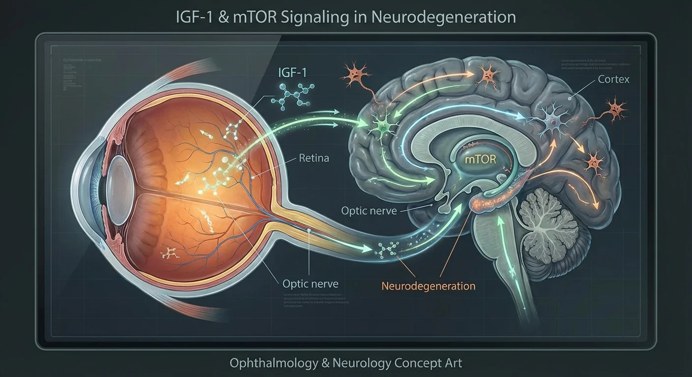
Glaucoma is now recognized not just as an eye pressure problem but as a neurodegenerative disease of the optic nerve. Retinal ganglion cells (RGCs) – the neurons that send visual signals from eye to brain – degenerate in glaucoma, much like neurons die in Alzheimer’s or Parkinson’s disease (pmc.ncbi.nlm.nih.gov) (pmc.ncbi.nlm.nih.gov). Researchers are uncovering how general health factors – hormones, metabolism, even stress levels – affect RGC survival. In particular, the IGF-1 (Insulin-like Growth Factor 1) and mTOR (mammalian Target Of Rapamycin) pathways, which normally promote cell growth and protein building, play important roles in eye health. Disturbances to these pathways (for example, from insulin resistance or poor nutrition) can converge on the axonal transport systems in neurons and stress RGCs. By comparing glaucoma to brain disorders, we can learn how these signals protect or harm nerves. This article reviews the evidence linking IGF-1, mTOR signaling, metabolic health, and nervous system balance to glaucoma risk, and highlights what blood or other tests might tell you about your eye–brain health.
IGF-1, Insulin, and the mTOR Pathway in Nerve Cells
IGF-1 is a small protein hormone closely related to insulin. It is made in the liver (and in some tissues) under the influence of growth hormone. In the body, IGF-1 encourages growth and survival of many cell types (pmc.ncbi.nlm.nih.gov). In the nervous system, IGF-1 is especially important for neuron growth and neuroprotection. For example, in lab studies IGF-1 significantly protected retinal ganglion cells (RGCs) from dying under stress (pmc.ncbi.nlm.nih.gov) (pmc.ncbi.nlm.nih.gov). When cultured RGCs were starved of oxygen (hypoxia), adding IGF-1 cut cell death by activating survival signaling pathways (the Akt/PI3K and Erk/MAPK routes) (pmc.ncbi.nlm.nih.gov). In other studies, boosting IGF-1 levels in injured optic nerves helped regenerate RGC axons (pmc.ncbi.nlm.nih.gov) (pmc.ncbi.nlm.nih.gov). In short, IGF-1 acts like a neurotrophic (nerve-growing) factor that helps keep nerve cells alive and even regrow.
The mTOR pathway is a central regulator of cell metabolism and growth. mTOR is a protein kinase (a “switch” enzyme) that senses nutrients, hormones, and energy. When nutrients and signals like insulin/IGF-1 are plentiful, mTOR becomes active (in two complexes, mTORC1 and mTORC2) and tells cells to grow and build protein (pmc.ncbi.nlm.nih.gov) (pmc.ncbi.nlm.nih.gov). Conversely, when nutrients are low, mTOR activity falls and the cell ramps up recycling (autophagy) to conserve resources. In neurons, mTOR helps maintain dendrites and synapses. For example, one study found that mTORC1 (through its target S6 kinase, S6K) and mTORC2 (via a subunit SIN1) controlled the branching and length of RGC dendrites (pmc.ncbi.nlm.nih.gov). This means normal insulin/IGF-1 signaling through mTOR supports the complex dendritic trees of RGCs.
In a powerful demonstration of this link, researchers showed that applying insulin directly to the eye in a mouse model of glaucoma stimulated RGC dendrite and synapse regeneration (pmc.ncbi.nlm.nih.gov). This treatment depended on the mTOR-S6K pathway: blocking S6K or its mTORC link (SIN1) prevented the regenerative effect (pmc.ncbi.nlm.nih.gov). In those experiments insulin rescued light responses and connectivity of RGCs, and improved the animals’ vision-like reflexes (pmc.ncbi.nlm.nih.gov). In sum, healthy IGF-1/insulin signaling through the mTOR pathway is crucial for RGC survival and function (pmc.ncbi.nlm.nih.gov) (pmc.ncbi.nlm.nih.gov).
Because the IGF/insulin and mTOR pathways are so intertwined, fitness and nutrition strongly influence nerve health. High anabolic (growth) signals tend to activate mTOR, whereas insulin resistance (as in metabolic syndrome or type 2 diabetes) weakens the pathway. In aging and obesity, IGF-1 and insulin signaling can become dysregulated. Intriguingly, human studies of Alzheimer’s and Parkinson’s disease also show links to these metabolic factors. In fact, age and conditions like obesity or diabetes are shared risk factors for “brain” neurodegenerative diseases (pmc.ncbi.nlm.nih.gov), suggesting a common metabolic mechanism – possibly via IGF-1/mTOR signaling – connecting systemic health to nerve cell vulnerability.
Glaucoma and Other Neurodegenerative Diseases: Shared Features
Glaucoma’s cell-level damage resembles that of Alzheimer’s, Parkinson’s, and other age-related brain diseases. In all cases, patients lose neurons (RGCs in glaucoma; cortical or basal ganglia neurons in AD/PD) over many years, often silently at first. These disorders share risk factors like age, obesity, and type 2 diabetes (pmc.ncbi.nlm.nih.gov). A 2024 review notes that obesity and diabetes raise the risk of both AD and PD, and that the insulin/IGF system may underlie this link (pmc.ncbi.nlm.nih.gov). Similarly, large-scale genetic and population studies find that diabetes increases glaucoma risk (pmc.ncbi.nlm.nih.gov) (pubmed.ncbi.nlm.nih.gov). In one Mendelian-Randomization analysis of over 20,000 glaucoma cases, a higher genetic predisposition to type 2 diabetes causally raised glaucoma odds by about 10–15% (pubmed.ncbi.nlm.nih.gov). Higher genetically predicted fasting glucose and HbA1c (markers of blood sugar control) also weakly predicted glaucoma (pubmed.ncbi.nlm.nih.gov). In practice, patients with diabetes often show worse glaucoma outcomes. (Indeed, retrospective data in one study showed diabetic patients on insulin had faster visual field loss than those on metformin (pmc.ncbi.nlm.nih.gov).) Overall this supports that high blood sugar and poor insulin action contribute to optic nerve damage, just as they do to brain disorders.
Inflammation and oxidative stress are other common threads. In glaucoma and Alzheimer’s alike, chronic oxidative stress builds up and overwhelms neurons. The mTOR pathway interacts with these processes: it both modulates oxidative stress and responds to it (pmc.ncbi.nlm.nih.gov). In retinal disease models (including glaucoma), inhibiting mTOR with rapamycin reduced oxidative damage and inflammation (pmc.ncbi.nlm.nih.gov) (pmc.ncbi.nlm.nih.gov). For example, rapamycin eye drops in rats reduced microglial activation (immune cells in the retina) and preserved RGCs under high-eye-pressure stress (pmc.ncbi.nlm.nih.gov). Likewise, rapamycin has been found to protect neurons in AD/PD models under oxidative conditions (pmc.ncbi.nlm.nih.gov). These parallels suggest that strategies which bolster IGF/mTOR signaling (in balance) or otherwise combat metabolic stress could benefit both brain and eye health.
Insulin Resistance, Metabolic Health, and Glaucoma Risk
Because IGF-1 and insulin are so similar in structure and signaling, insulin health is closely tied to RGC survival. Insulin and IGF-1 bind to related receptors and activate the same downstream cascades (via IRS→PI3K→Akt→mTOR) (pmc.ncbi.nlm.nih.gov) (pmc.ncbi.nlm.nih.gov). In the retina, insulin receptors are present on RGCs (pmc.ncbi.nlm.nih.gov), and insulin signaling affects retinal metabolism. When the body develops insulin resistance (as in prediabetes or type 2 diabetes), brain and retinal neurons receive less effective growth signaling. Experimental disruption of insulin signaling in rodents can raise eye pressure and kill RGCs (pmc.ncbi.nlm.nih.gov). Conversely, improving insulin sensitivity seems neuroprotective: it is speculated that good diabetes control might reduce glaucoma risk.
Epidemiological data back this up. People with type 2 diabetes have a significantly higher glaucoma risk (pmc.ncbi.nlm.nih.gov). In one large review, diabetes (and longer duration of it) was linked to more glaucoma even after adjusting for age (pmc.ncbi.nlm.nih.gov). As noted, a recent genetic study also supports diabetes as an independent causal risk factor (pubmed.ncbi.nlm.nih.gov). This could be due to many mechanisms: high blood sugars damage microvasculature (reducing blood flow to optic nerve), advanced glycation products accumulate, and insulin resistance deprives RGCs of supportive signaling.
Testing for insulin resistance. For practical patient screening, certain blood tests can assess metabolic risk. The most direct are fasting glucose and HbA1c, which measure blood sugar levels, and fasting insulin. From insulin and glucose one can calculate HOMA-IR (a rough insulin-resistance index). A high HOMA-IR suggests metabolic syndrome. Typical labs can include:
- Fasting glucose and HbA1c: High values (>100 mg/dL or HbA1c >5.7% up to diabetic levels) imply poor sugar control, which is a glaucoma risk factor (pubmed.ncbi.nlm.nih.gov).
- Fasting insulin: Normal is around 2–20 µU/mL (varies by lab). Elevated fasting insulin indicates insulin resistance. Durable high insulin together with glucose implies cells aren’t responding well.
- HOMA-IR: Calculated as (fasting insulin × fasting glucose)/405. Values above ~2 suggest insulin resistance. If these markers are abnormal, lifestyle changes or medication may reduce eye risk (and cardiac risk).
Autonomic Nervous System Balance and Ocular Blood Flow
Glaucoma patients often have signs of autonomic imbalance, especially sympathetically driven stress. A key measure is heart rate variability (HRV), which quantifies fluctuations between heartbeats. High HRV is a healthy sign of strong parasympathetic (calming) tone and adaptability; low HRV implies sympathetic (stress) dominance. Studies find that glaucoma patients – including those with normal eye pressure (“normal-tension glaucoma”) – often have reduced HRV and signs of vascular dysregulation. For example, in one study NTG patients had a “predominance of sympathetic activity” on a stress test compared to healthy controls (pmc.ncbi.nlm.nih.gov). These patients also showed reduced blood flow (lower diastolic velocity) in the central retinal and ciliary arteries (pmc.ncbi.nlm.nih.gov). In other words, stressed subjects had more constricted retinal blood vessels.
Even more striking, a retrospective clinical study divided glaucoma patients by HRV. Those with low HRV (high stress) had much faster nerve-fiber loss and worse visual field decline than patients with high HRV (pubmed.ncbi.nlm.nih.gov). The low-HRV group had on average a 1.44 µm/year retinal nerve fiber thinning versus 0.29 µm/year in the high-HRV group (nearly five times faster) (pubmed.ncbi.nlm.nih.gov). They also had more IOP fluctuations and lower overall eye perfusion pressure. This suggests that autonomic dysfunction – measurable by heart rate tests – accelerates glaucoma damage, likely by impairing ocular blood flow and increasing pressure variability (pubmed.ncbi.nlm.nih.gov) (pmc.ncbi.nlm.nih.gov).
Measuring and improving HRV. While not a standard lab test, HRV can be measured with consumer devices (chest straps or smartwatches) that track beat-to-beat intervals. Patients interested in comprehensive risk profiling could measure their resting HRV (often reported as “SDNN” or “RMSSD”) using guided protocols. Higher HRV (more variability) is better; lower HRV signals chronic stress. Improving HRV through regular exercise, stress reduction, and sleep hygiene could help balance the autonomic system.
In summary, stress and autonomic imbalance are plausible contributors to glaucoma, converging on RGC health by worsening blood flow and metabolic stress. This ties back to insulin/IGF-1: stress hormones and insulin signals cross-talk (stress tends to raise blood sugar and insulin resistance). Thus a multi-pronged view – metabolic health, autonomic balance, and anabolic signaling – is needed for RGC protection.
Axonal Transport and Retinal Ganglion Cell Survival
RGCs have very long axons (the optic nerve), relying on continuous transport of nutrients and proteins from the cell body to the far synapses in the brain. Healthy IGF-1/insulin/mTOR signaling supports the axonal transport machinery. For instance, IGF-1 activates the PI3K/Akt pathway which in turn stabilizes microtubules (the “rails” for axon transport) and promotes production of tubulin, a key structural protein (pmc.ncbi.nlm.nih.gov) (pmc.ncbi.nlm.nih.gov). In experiments with optic nerve injury, activating IGF-1/mTOR signaling boosted RGC axon regrowth (pmc.ncbi.nlm.nih.gov) (pmc.ncbi.nlm.nih.gov). Conversely, insulin deficiency or resistance can impair this support. In prediabetes or diabetes, neurons may lose sensitivity to insulin, analogous to insulin-resistant tissues. One review notes that inability of cells to respond to insulin (as in type 2 diabetes) can increase RGC vulnerability (pmc.ncbi.nlm.nih.gov). In practice, that could mean slowed axonal transport and buildup of toxic waste.
Tau protein and axons: Another connection is tau, a microtubule-associated protein that helps maintain axon structure. Glaucoma patients have been found to have abnormal, hyperphosphorylated tau both in their eyes and cerebrospinal fluid (pmc.ncbi.nlm.nih.gov). This is the same kind of tau pathology seen in Alzheimer’s. Under high eye pressure, animals showed tau mislocalization in RGCs. Experimentally knocking down tau improved RGC survival (pmc.ncbi.nlm.nih.gov), highlighting how metabolic stress on axons (like from disrupted insulin signaling) can involve tau-related transport failures.
In all, anabolic signals like IGF-1 preserve axonal transport and synapses. When these signals drop (insulin resistance, nutrient stress) or when tau is dysregulated, RGCs lose their “connection” and degenerate. This underscores why systemic conditions impact eye nerves.
Caloric Restriction, Fasting, and “Mimetic” Therapies
Caloric restriction (CR) and its mimetics can broadly influence the IGF/mTOR axis by lowering nutrient signals. Many animal studies point to benefits of CR or fasting on retinal aging. For example, one mouse study used an every-other-day fasting regimen (a form of CR) in a glaucoma-like model. The fasted mice had much less RGC death and retinal degeneration than normal-fed mice, even though eye pressure was unchanged (pmc.ncbi.nlm.nih.gov). Their vision-related function was better preserved too. Mechanistically, fasting boosted blood levels of β-hydroxybutyrate (a ketone body) and increased markers of autophagy and stress resistance in the retina (pmc.ncbi.nlm.nih.gov). In short, periods of low calorie intake “reprogrammed” the retinal neurons to survive stress, by enhancing antioxidant defenses and growth-factor expression. Reviews conclude that CR activates protective processes like autophagy and reduced oxidative stress that are known to slow neural aging (pmc.ncbi.nlm.nih.gov) (pmc.ncbi.nlm.nih.gov).
Because long-term fasting is hard for most people, researchers are also studying caloric restriction mimetics – drugs or compounds that trigger similar pathways. Two prominent examples are rapamycin and metformin.
-
Rapamycin is a drug that directly inhibits mTORC1. In eye research, rapamycin has shown powerful neuroprotective effects. In glaucoma models, rapamycin reduced RGC death and inflammation (pmc.ncbi.nlm.nih.gov). Topical rapamycin eye drops even lowered IOP slightly by relaxing eye drainage tissue (pmc.ncbi.nlm.nih.gov). Notably, rapamycin’s benefit in retina is linked to enhancing autophagy (the cell’s recycling process) and quelling oxidative damage (pmc.ncbi.nlm.nih.gov) (pmc.ncbi.nlm.nih.gov). However, experiments suggest autophagy’s role can differ: one report found that in a glaucoma model, rapamycin-induced autophagy actually correlated with increased RGC loss (pmc.ncbi.nlm.nih.gov). The overall takeaway is still that moderate mTOR inhibition (as with rapamycin) often protects stressed neurons in animal studies (pmc.ncbi.nlm.nih.gov) (pmc.ncbi.nlm.nih.gov). (Rapamycin is being tested in eye diseases clinically, but it is an immune-suppressant drug and not currently a standard therapy for glaucoma.)
-
Metformin is a widely used diabetes drug that acts partly by activating AMPK, a cellular energy sensor, thereby mimicking some effects of CR. A 2025 study showed that giving metformin to mice protected their RGCs in an ischemic eye injury model (pmc.ncbi.nlm.nih.gov) (pmc.ncbi.nlm.nih.gov). Metformin greatly preserved RGC number and retinal structure after injury, likely by activating AMPK and boosting autophagy/mitophagy (cleaning up damaged cell parts) in the retina (pmc.ncbi.nlm.nih.gov). In the same paper, a small patient study found that diabetic glaucoma patients on metformin had stable visual fields over 6 months, whereas those on insulin (but not metformin) showed worsening fields (pmc.ncbi.nlm.nih.gov). This real-world hint suggests metformin may slow glaucoma progression. Importantly, metformin is fairly safe and accessible, so it is an attractive candidate for eye protection in metabolic patients (though formal trials are still needed).
-
Other compounds: Natural substances like resveratrol (found in red grapes) have been studied. In rodent models, resveratrol reduced oxidative stress and preserved RGCs under pressure or ischemia (pmc.ncbi.nlm.nih.gov). It works partly by activating SIRT1 (a “longevity” enzyme) and the PI3K/Akt survival pathway (pmc.ncbi.nlm.nih.gov). While resveratrol is less potent than a drug like metformin, it exemplifies the general idea: antioxidant and nutrient-sensing treatments that come from diet can protect retinal neurons.
In sum, interventions that modestly dampen the IGF/mTOR growth signal – such as fasting, drugs like rapamycin or metformin, or even nutritional compounds – tend to activate cellular clean-up pathways and bolster neuron resilience. These have shown neuroprotective effects in the retina. They are still experimental for glaucoma, but they validate the principle that metabolic state and nutrition can directly influence eye health (pmc.ncbi.nlm.nih.gov) (pmc.ncbi.nlm.nih.gov).
Candidate Biomarkers and Practical Testing
Given these insights, what can patients measure in blood or via simple tests to get a read on their IGF/mTOR axis and metabolic risk? Here are some candidate biomarkers and how to interpret them:
-
IGF-1 (Blood test): A standardized blood test for IGF-1 exists (often done when evaluating growth issues). Levels are age-dependent (peak in youth, decline with age). Typical adult values range roughly 80–350 ng/mL (varies by lab). A low IGF-1 for age might indicate poor growth hormone signaling or under-nutrition; a high IGF-1 might occur in acromegaly or high-protein diets. In theory, extremely low IGF-1 could mean less neurotrophic support, whereas very high IGF-1 chronically might increase growth-related risks (like certain cancers). In practice, one study did not find a difference in blood IGF-1 between glaucoma patients and controls (pubmed.ncbi.nlm.nih.gov). That suggests circulating IGF-1 alone doesn’t diagnose glaucoma risk. However, an IGF-1 test could still be part of an overall endocrine panel. If your IGF-1 comes back low on a screening, it could be worth checking related hormones (growth hormone, nutrition status).
-
Insulin and HOMA-IR: As noted, high fasting insulin indicates insulin resistance. If you have a fasting glucose and insulin, even a patient without diabetes can calculate HOMA-IR. For example, insulin (µU/mL) × fasting glucose (mg/dL) / 405. Values above ~2 suggest reduced insulin sensitivity. Patients can often get these through annual check-ups or direct-to-consumer labs. High HOMA-IR or elevated insulin + glucose signals metabolic strain, which correlates with glaucoma risk (pubmed.ncbi.nlm.nih.gov) and general vascular risk.
-
Hemoglobin A1c (HbA1c): This is a routine test for average blood sugar over 3 months. Values above 5.7% indicate prediabetes; above 6.5% means diabetes. The MR study (pubmed.ncbi.nlm.nih.gov) suggests even moderate rises in blood sugar (fasting glucose or HbA1c) were linked to higher glaucoma odds. Keeping HbA1c in the normal range (<5.7%) is a goal not just for diabetes prevention but possibly for eye health.
-
Beta-Hydroxybutyrate (Ketone levels): This can be measured in blood (via a lab or home meter) or urine (ketone sticks). Higher levels of the ketone β-hydroxybutyrate (e.g. >0.5 mM fasting) indicate a shift to fat metabolism, as occurs in fasting or ketogenic diets. In the mouse study above, higher β-hydroxybutyrate was a marker of the beneficial starvation response (pmc.ncbi.nlm.nih.gov). It also has direct neuroprotective signaling roles. Thus, a moderate elevation of ketones (during fasting or ketogenic diet) is generally considered positive (“metabolic flexibility”). Persistently high ketone levels outside of dietary context could signal unmanaged diabetes (ketoacidosis), so always interpret with context.
-
Adiponectin, Leptin and Lipid Panel: These are broader metabolic biomarkers. Adiponectin (a protein from fat tissue) usually falls with insulin resistance; higher adiponectin is protective for blood vessels. Leptin levels rise with obesity. While not used clinically for glaucoma, abnormal patterns (high leptin, low adiponectin) would imply metabolic syndrome, which is bad for eye health. Checking cholesterol and blood pressure is also wise, as the MR study (pubmed.ncbi.nlm.nih.gov) hinted high blood pressure has some glaucoma risk.
-
Inflammatory Markers (CRP, IL-6): Chronic low-level inflammation may link to neurodegeneration. A simple C-reactive protein (CRP) test (part of many annual labs) can reveal systemic inflammation. Elevated CRP is not specific, but patients might notice if systemic stress/inflammation is present.
-
HRV Measurement: As discussed, HRV is not a blood test but an accessible test using wearable technology. Devices like smartwatches or chest-straps (Polar, Garmin, Apple Watch, etc.) can record HRV under resting conditions. Patients should follow standardized measurement (e.g. morning supine, average over 5+ minutes). A notably low HRV reading (especially over time) suggests sympathetic dominance. Any consistent pattern of low HRV might prompt a conversation with your doctor about stress management or cardiovascular check-up.
-
Eye-specific tests: While not blood tests, keep in mind that retinal imaging (OCT scans) and visual field tests are direct ways to profile glaucoma risk already in use. For example, loss of the retinal nerve fiber layer on OCT or changes in visual field perimetry are direct biomarkers of neurodegeneration in the eye (pubmed.ncbi.nlm.nih.gov). These would fall under “multi-target profiling” too.
In practice, a multi-target approach would combine systemic and local data. For example, a patient with high fasting glucose, low IGF-1, and low HRV (along with some optic nerve thinning on OCT) might be flagged as high risk for glaucoma progression. Conversely, someone with well-controlled blood sugar, normal IGF-1, and healthy HRV may have a better prognosis.
Interpreting Results:
- Normal ranges vary by lab. Always compare IGF-1 to age-adjusted norm; consult a healthcare provider to interpret high or low values.
- Glucose/insulin tests: use clinical cutoffs (glucose >100 mg/dL, insulin >15–20 µU/mL often warrant follow-up).
- HRV: healthy individuals typically have SDNN (a global HRV measure) above 50 ms. Values below 20 ms are quite low (seen in severe stress or disease) (pubmed.ncbi.nlm.nih.gov). There is no single “normal” HRV, but trends (improving or worsening) are informative.
Obtaining these tests is often possible through routine health care or direct-to-consumer labs. For example, many commercial labs offer an IGF-1 test and insulin/glucose panel. Always do these tests fasting in the morning. If you plan to use wearable HRV, pick a reliable app or device and measure regularly to get a baseline.
Conclusion
Taken together, the IGF-1/insulin/mTOR signaling system is a central link between metabolism and nerve health across eye and brain. Strong evidence shows that healthy anabolic signaling (good insulin action and moderate IGF-1 levels) helps maintain retinal ganglion cell function, whereas insulin resistance and metabolic stress undermine it. At the same time, autonomic balance (as tracked by HRV) influences ocular blood flow and disease progression. Interventions that improve metabolic health – from diet and exercise to drugs like metformin or approaches that mimic fasting – show neuroprotective effects in glaucoma models.
Patients and clinicians can use these insights by combining traditional eye exams (eye pressure, OCT, visual field) with systemic biomarkers. Checking blood sugar control, lipid levels, and even IGF-1 can give clues to optic nerve vulnerability. Monitoring heart rate variability offers a window into bodywide stress. While no single test will predict glaucoma, a multi-target profile incorporating metabolic, hormonal, and neural data could help identify high-risk individuals early, potentially guiding more aggressive neuroprotective strategies.
Future research will refine which biomarkers best flag impending glaucoma (beyond IOP) and test if metabolic or CR-mimetic therapies can slow disease. For now, patients can focus on known factors: keep blood sugar, blood pressure, and weight in check, reduce chronic stress, and consider discussing with their doctor whether drugs like metformin (if diabetic) or lifestyle changes could have the added benefit of protecting vision (pmc.ncbi.nlm.nih.gov) (pmc.ncbi.nlm.nih.gov). In this way, eye care is becoming holistic: it’s not just about the eyeball, but about the whole body’s growth and energy balance.