Aging, Senescence, and Glaucoma
Glaucoma is a leading cause of blindness and its risk rises with age. In aged eyes, cells can enter a senescent state – they stop dividing but stay alive – and release harmful signals called the senescence-associated secretory phenotype (SASP). Senescent cells in the eye can worsen disease. For example, aged trabecular meshwork cells (the filter in front of the eye) become stiff and clogged, raising eye pressure (pmc.ncbi.nlm.nih.gov). In the retina and optic nerve, senescent cells release cytokines (like IL-6, IL-8, IL-1β) and enzymes (MMPs) that cause inflammation, tissue remodeling, and nerve cell death (pmc.ncbi.nlm.nih.gov) (pmc.ncbi.nlm.nih.gov). These SASP factors have been found in human glaucomatous eyes and animal models of eye pressure, where they drive retinal ganglion cell (RGC) damage (pmc.ncbi.nlm.nih.gov) (pmc.ncbi.nlm.nih.gov). Targeting these cells is a new idea: removing or quieting them may help protect the optic nerve.
Senescence in the Eye
Senescent cells build up in key eye tissues. In the trabecular meshwork (TM), senescence stiffens the meshwork and increases resistance to fluid outflow (pmc.ncbi.nlm.nih.gov). This raises intraocular pressure, a main risk factor for glaucoma. In humans with glaucoma, more senescent TM cells (marked by enzymes like SA-β-gal, or proteins p16^INK4a and p21^CIP1) have been measured compared to normal eyes (pmc.ncbi.nlm.nih.gov). High p16 and p21 in TM cells correlate with glaucoma and fewer TM cells survive into old age (pmc.ncbi.nlm.nih.gov).
In the optic nerve head and retina, aging and stress cause RGCs and supporting cells (astrocytes, microglia) to become senescent. These cells then secrete SASP factors – pro-inflammatory cytokines (IL-6, IL-1β, IL-8), chemokines (CCL2, CXCL5), and matrix metalloproteinases – which poison nearby neurons and propagate senescence to neighbors (pmc.ncbi.nlm.nih.gov) (pmc.ncbi.nlm.nih.gov). In mouse models of high eye pressure and in human glaucoma tissue, elevated levels of IL-6, IL-1β, IL-8 and other SASP markers have been found, linked to chronic inflammation and RGC death (pmc.ncbi.nlm.nih.gov). Thus, senescence and SASP contribute to TM dysfunction and optic nerve damage in glaucoma.
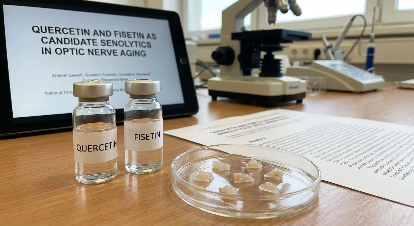
Quercetin and Fisetin as Senolytics
Senolytic drugs are agents that kill senescent cells, while senomorphics suppress their harmful secretions. Quercetin and fisetin are natural flavonoids with senolytic or senomorphic activity. Quercetin is found in many fruits and vegetables and is known as an antioxidant. Research shows quercetin can selectively eliminate senescent cells and tone down the SASP (pmc.ncbi.nlm.nih.gov). It has anti-inflammatory and anti-oxidant properties in the eye as well. In models of retinal stress, quercetin increases protective enzymes (like Nrf2 and HO-1) and reduces cell death (pmc.ncbi.nlm.nih.gov). As a senolytic, quercetin suppresses NF-κB (a key SASP regulator) and lowers secretion of IL-6 and other cytokines from senescent cells (pmc.ncbi.nlm.nih.gov).
Fisetin, a sibling of quercetin, is emerging as a powerful senolytic. In one report, fisetin outperformed quercetin in killing senescent cells in culture and mice (pmc.ncbi.nlm.nih.gov). Fisetin treatment in old mice reduced p16^INK4a and other senescence markers in tissues, improved health span, and extended lifespan (pmc.ncbi.nlm.nih.gov) (pmc.ncbi.nlm.nih.gov). Fisetin is already used as a dietary supplement and appears safe. Its strong senolytic effect suggests it might clear senescent eye cells too. Both quercetin and fisetin have favorable safety profiles in humans, so they are attractive for trials (pmc.ncbi.nlm.nih.gov) (pmc.ncbi.nlm.nih.gov).
Preclinical Evidence in Glaucoma
In glaucoma models, removing senescent cells has shown clear benefit. In a mouse model of acute ocular hypertension, many RGCs became senescent and activated SASP genes after eye pressure was raised (pmc.ncbi.nlm.nih.gov). In that study, researchers used a genetic trick and the senolytic drug dasatinib to ablate (kill) the p16^INK4a-positive senescent RGCs. Remarkably, early clearance of those cells rescued the remaining RGCs: treated mice preserved their visual responses and RGC counts much better than controls (pmc.ncbi.nlm.nih.gov). In other words, senolysis protected the optic nerve from ongoing damage.
While dasatinib was the drug used there, it shows lent credence to the idea: if senescent RGCs and glia are culprits, then targeting them should help. Quercetin or fisetin might serve similarly. There is some evidence that dietary fisetin improves eye function in a genetic glaucoma mouse (DBA/2J strain) by reducing retinal inflammation and saving neurons (though this finding needs more confirmation).
Another study even looked at human patients incidentally exposed to senolytics. In a retrospective review of glaucoma patients who happened to be taking senolytic agents (for other conditions), no harm was seen: patients on senolytics did not have worse vision, higher pressures, or faster visual field loss than matched controls (pmc.ncbi.nlm.nih.gov). In fact, this suggests senolytics did not injure the eye, and supports further study of their protective effects.
Benefits and Risks of Senolysis in the Eye
Potential benefits: Clearing senescent cells in the TM and optic nerve could reduce inflammation and tissue dysfunction. In the TM, it might restore healthier outflow channels and lower eye pressure. In the retina/nerve head, it may break the cycle of SASP-driven damage, preserving RGCs that would otherwise die. Senolysis could complement existing glaucoma treatments (pressure-lowering) by targeting the aging component of the disease.
Potential risks: The optic nerve is delicate neural tissue. In theory, killing cells – even if they are “zombie” senescent cells – might have unintended effects. For instance, some supporting cells might become partly senescent to limit damage, and their sudden removal could trigger inflammation. Also, systemic senolytic drugs sometimes affect other tissues. A known senolytic navitoclax can cause low platelets, so drug choice and dose must be careful. Quercetin and fisetin are generally well-tolerated, but high doses or long-term effects in the eye are untested. Any trial must watch for retinal or optic nerve inflammation, bleeding, or loss of function. So far, limited data (e.g. the human report above) show no obvious ocular toxicity (pmc.ncbi.nlm.nih.gov), which is encouraging.
Trial Design, Biomarkers, and Monitoring
To test senolytics in glaucoma or ocular aging, a carefully designed trial is needed. Possible design: a randomized, placebo-controlled trial in patients with early glaucoma or ocular hypertension. The senolytic (e.g. intermittent high-dose fisetin or quercetin) would be given orally or by eye drop tablets (if topical formulations become available).
Endpoints: Key outcomes would include standard glaucoma measures – intraocular pressure (IOP), retinal nerve fiber layer (RNFL) thickness on OCT imaging, visual field tests, and pattern electroretinogram (PERG) or visual evoked potentials (VEPs) to assess nerve function. Improvement or slower decline in these would be primary signals of neuroprotection.
Biomarkers: On the lab side, tracking senescence biomarkers would help show target engagement. A leading marker is p16^INK4a. This cell-cycle inhibitor is upregulated in senescent cells. In a trial, one could measure p16^INK4a RNA or protein levels in surrogate samples. For example, blood T-cells or skin cells often reflect organismal senescence and might decrease p16 after therapy (pmc.ncbi.nlm.nih.gov) (pmc.ncbi.nlm.nih.gov). In the eye specifically, researchers could analyze excised TM cells (if any are removed in routine glaucoma surgery) for p16 or SA-β-gal. Tear fluid or the aqueous humor could be assayed for SASP factors like IL-6, IL-8 and MMPs (pmc.ncbi.nlm.nih.gov). A drop in these cytokines after treatment would suggest reduced SASP. Serial optical coherence tomography (OCT) angiography might also show changes in blood flow or extracellular matrix in the TM region.
Safety monitoring: Participants would receive regular eye exams with dilated fundoscopy to check for inflammation, vascular changes, or pigmentary alterations. Circulating inflammatory markers (CRP, IL-6) and blood counts should be monitored (in case of off-target effects like those seen with other senolytics). If an ocular formulation is used, measures like corneal thickness and endothelial cell counts could be added. The retrospective study [Source 7] reassured that vision and IOP stayed stable with senolytic exposure (pmc.ncbi.nlm.nih.gov), but a trial would need more intense monitoring.
Interim analyses should watch for any drop in visual acuity or new ocular symptoms. Because neural tissue turnover is slow, trials might need many months to see structural change, so the design could be an initial 6–12 month pilot phase. Positive results could then lead to larger, longer trials.
Conclusion
As we seek new therapies for glaucoma and optic nerve aging, senolytics like quercetin and fisetin offer a novel angle. By targeting the harmful senescent cells and their SASP factors in the trabecular meshwork and optic nerve head, these compounds could reduce age-related damage. Preclinical studies show that clearing senescent cells preserves retinal ganglion cells and vision (pmc.ncbi.nlm.nih.gov), and early human data suggest senolytics do not harm the eye (pmc.ncbi.nlm.nih.gov). Careful clinical trials, with IOP and nerve imaging endpoints, senescence biomarkers (p16^INK4a and SASP cytokines), and vigilant safety checks will be needed to test this idea. If successful, senolysis could become a powerful adjunct to our arsenal against glaucoma and other optic neuropathies of aging.