Introduction
Glaucoma is an eye disease in which retinal ganglion cells (RGCs) – the nerve cells that carry visual signals from the eye to the brain – slowly die. This causes gradual, irreversible vision loss. Doctors usually focus on lowering eye pressure to slow glaucoma, but research now shows that oxidative stress (a kind of chemical stress in the body) and imbalances in the autonomic nervous system (the “automatic” nervous system that controls things like heart rate) also play a role. In glaucoma patients, blood levels of certain redox markers – substances that indicate oxidative damage – tend to be higher than normal. At the same time, many glaucoma patients have depressed heart rate variability (HRV), a sign of autonomic imbalance. Together, raised oxidative stress and poor autonomic regulation may worsen RGC damage.
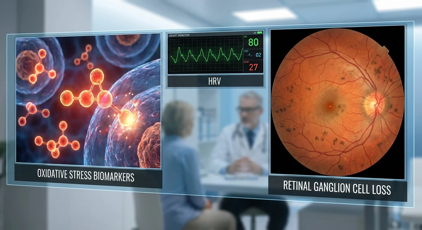
In this article, we explain what oxidative stress markers like F2-isoprostanes, malondialdehyde (MDA), and 8-hydroxy-2’-deoxyguanosine (8-OHdG) are, and how they are found in glaucoma. We define HRV (heart rate variability) and review how it is altered in glaucoma. We describe possible biological pathways linking oxidative stress and autonomic imbalance to faster RGC death. We then summarize what studies of antioxidants (nutrients that fight oxidative stress) have shown on glaucoma outcomes. Finally, we suggest future “multi-omics” studies that combine blood or urine redox markers, HRV measurements, and advanced retinal imaging for new insights.
Throughout, we focus on information that patients can understand and act on. We also explain which oxidative stress tests can be ordered (via blood or urine) and what high or low readings might mean for someone concerned about glaucoma.
Oxidative Stress Markers in Glaucoma
Oxidative stress means there are too many “free radicals” (reactive oxygen molecules) in the body, causing damage to cells. We cannot directly measure free radicals easily, so doctors and researchers use biomarkers in blood or urine that indicate oxidative damage. Three important markers in glaucoma are F2-isoprostanes, malondialdehyde (MDA), and 8-hydroxy-2’-deoxyguanosine (8-OHdG). All three rise when oxidative stress increases.
-
F2-Isoprostanes (8-iso-PGF2α) – these are stable molecules formed when fats (polyunsaturated fats in cell membranes) oxidize. F2-isoprostanes are considered a “gold standard” for measuring lipid (fat) oxidation (pmc.ncbi.nlm.nih.gov). Higher blood or urine levels of these suggest that cells are under oxidative attack. Although not all glaucoma studies measure them, high F2-isoprostane levels have been found in many diseases and are thought to reflect strong oxidative stress (pmc.ncbi.nlm.nih.gov). (In practice, labs can measure F2-isoprostanes in urine or plasma using specialized equipment, but this is mostly done in research settings.)
-
Malondialdehyde (MDA) – this chemical is produced when reactive oxygen species break down fats in the body. Like F2-isoprostanes, it signals fat damage from oxidation. Multiple glaucoma studies have found that glaucoma patients have higher MDA in their blood than healthy people (pmc.ncbi.nlm.nih.gov) (pmc.ncbi.nlm.nih.gov). In fact, a large review found that MDA was the most consistently elevated oxidative stress marker in glaucoma patients’ blood (pmc.ncbi.nlm.nih.gov). In one study of angle-closure glaucoma, patients had significantly higher MDA than control subjects (pmc.ncbi.nlm.nih.gov). Notably, that study showed patients with very high MDA levels had faster vision loss: those with MDA above about 12 units had much more rapid visual field decline (pmc.ncbi.nlm.nih.gov).
-
8-Hydroxy-2’-deoxyguanosine (8-OHdG) – this marker indicates oxidative damage to DNA (the genetic material in cells). When oxidative stress cuts or changes DNA, 8-OHdG levels rise and can be measured in blood or urine. Studies of glaucoma patients (in normal-tension and pseudoexfoliation glaucoma) have found significantly higher plasma 8-OHdG levels than in control subjects (pmc.ncbi.nlm.nih.gov) (pmc.ncbi.nlm.nih.gov). For example, one study found average blood 8-OHdG around 17 ng/mL in healthy people and ~23 ng/mL in glaucoma patients (pmc.ncbi.nlm.nih.gov). Another report showed glaucoma risk was over 4 times higher in people whose 8-OHdG was above a certain cutoff (pmc.ncbi.nlm.nih.gov) (pmc.ncbi.nlm.nih.gov). In short, high 8-OHdG means more DNA damage from reactive oxygen, and this is seen in glaucoma patients.
Other markers sometimes measured include total antioxidant levels (like “total antioxidant status” or enzymes such as superoxide dismutase (SOD) and glutathione peroxidase). In many glaucoma studies, these antioxidants are low (because they’ve been used up fighting free radicals) while markers like MDA, 8-OHdG, or H₂O₂ are high (pmc.ncbi.nlm.nih.gov). (For brevity we focus on F2-isoprostanes, MDA, and 8-OHdG here, but many studies report lower antioxidant enzymes and vitamins in glaucoma.)
Summary: In glaucoma patients, studies consistently see higher oxidative damage in the body. Markers like MDA and 8-OHdG tend to be above the normal range seen in healthy people (pmc.ncbi.nlm.nih.gov) (pmc.ncbi.nlm.nih.gov). Researchers believe this extra oxidative stress contributes to glaucoma’s effects on the optic nerve.
Measuring Oxidative Stress: Available Tests
While these markers are important in research, they are not yet routine clinical tests. However, some specialized labs and health clinics offer oxidative stress panels. Here is what patients should know:
-
8-OHdG test: Can be measured in blood plasma or in urine. Commercial kits (ELISA tests) exist to measure urine 8-OHdG (for example, Genox “8-OHdG Check” kit (www.genox.com)). A healthcare provider can arrange this through specialized labs. There is no universal “normal” level, but studies give some idea. For example, one glaucoma study found control patients averaged ~17 ng/mL while glaucoma patients averaged ~23 ng/mL (pmc.ncbi.nlm.nih.gov). If your 8-OHdG comes back much higher than typical healthy values, it suggests elevated DNA damage from oxidative stress.
-
F2-isoprostanes test: Typically measured in a 24-hour urine sample or sometimes blood. It is considered very reliable but requires lab equipment (mass spectrometry). Normal values depend on age and method, but again a much higher result suggests increased lipid peroxidation. This test is mainly done in research or specialized centers.
-
MDA test: Malondialdehyde can be measured in blood plasma (often by the “thiobarbituric acid reactive substances” or TBARS method, or by chromatography). Normal lab ranges vary, but one glaucoma study used a cutoff of ~12 µmol/L to flag higher risk (pmc.ncbi.nlm.nih.gov). If your lab report shows MDA elevated above typical values (ask the lab’s reference range), that may indicate excess oxidative stress on fats.
-
Total Antioxidant or Enzyme tests: Some labs measure “total antioxidant capacity” or levels of SOD or glutathione peroxidase. Lower-than-normal results again point to oxidative stress, since antioxidants are being consumed.
In practice, these tests are not standard like cholesterol or blood sugar. If you want them checked, you may need to request a specialty lab or consult a doctor who can order them. Be aware that results must be interpreted in context by a professional. Factors like diet, time of day, or exercise can affect these markers.
As one review points out, oxidative stress is not routinely assessed in daily practice (pmc.ncbi.nlm.nih.gov) because the reactive oxygen species themselves are short-lived. Instead doctors look at indirect markers (like above) or focus on reducing stress through lifestyle. If you get test results, compare them to any provided “normal range,” and discuss with your doctor. Generally, higher-than-normal 8-OHdG, MDA, or isoprostanes points toward increased oxidative stress, whereas levels in the normal range are reassuring.
Autonomic Function and Heart Rate Variability in Glaucoma
The autonomic nervous system (ANS) controls involuntary functions like heart rate, blood vessel tone, and digestion. It has two branches – sympathetic (often called “fight or flight”) and parasympathetic (rest/digest). A healthy balance between them causes a constantly varying heart rate. Heart rate variability (HRV) is a measure of how much the time between heartbeats fluctuates. In simple terms, higher HRV means the heart is responding flexibly (often a sign of good health), while very low HRV suggests autonomic imbalance, usually too much sympathetic activity or reduced parasympathetic tone.
Recent studies show that glaucoma patients often have reduced HRV compared to people without glaucoma (pmc.ncbi.nlm.nih.gov) (pmc.ncbi.nlm.nih.gov). For example, in one large study, patients with exfoliation glaucoma (a form of open-angle glaucoma) had notably lower HRV metrics than healthy controls (pmc.ncbi.nlm.nih.gov). Another analysis found that glaucoma patients with the lowest HRV (strongest sympathetic dominance) had much faster thinning of the optic nerve layer in the retina than those with higher HRV (pmc.ncbi.nlm.nih.gov). In that study, the low-HRV group lost about 1.4 μm of nerve fiber thickness per year (vs only 0.3 μm/yr in the high-HRV group) (pmc.ncbi.nlm.nih.gov). They also had more fluctuations in eye pressure and lower eye perfusion pressure, indicating that autonomic imbalance affects eye blood flow.
In summary, glaucoma—especially certain types like exfoliation glaucoma—tends to be accompanied by ANS dysfunction. Studies consistently observe that glaucoma patients have smaller variations in heart rate than healthy people (pmc.ncbi.nlm.nih.gov) (pmc.ncbi.nlm.nih.gov). Lower HRV is a signal of chronic stress or overactive sympathetic nerves. Importantly, low HRV in glaucoma has been linked to worse outcomes: patients with depressed HRV had faster retinal nerve fiber loss and more central vision defects (pmc.ncbi.nlm.nih.gov).
Measuring HRV: A person can measure HRV with devices like heart monitors or even some smartwatches and smartphones that use pulse sensors. Clinics sometimes use a short ECG or a handheld HRV analyzer (like a fingertip sensor). If your HRV is lower than average for your age and sex, it suggests autonomic stress. For example, the study [26] used SDNN (a standard HRV measure) to split patients into “low” vs “high” HRV groups. While there are no simple cutoffs publicized, an SDNN under roughly 50 ms is often considered low in adults. However, consult your doctor with raw HRV data; they may use it together with other health info rather than alone.
Connection to Oxidative Stress: A low HRV means the body is in a higher stress state. In many conditions (like chronic kidney disease or heart disease), researchers have found that higher oxidative stress biomarkers go hand-in-hand with lower HRV (pmc.ncbi.nlm.nih.gov). In one study of kidney disease patients, those with high plasma F2-isoprostane levels (a measure of oxidative stress) had significantly reduced HRV (pmc.ncbi.nlm.nih.gov). While this exact link hasn’t been extensively studied in glaucoma, it suggests a cycle: oxidative stress can affect blood vessels and nerves, leading to autonomic imbalance, which in turn can worsen blood flow and stress on the eyes.
How Oxidative Stress and Autonomic Imbalance Can Accelerate RGC Loss
To understand how oxidative stress and ANS imbalance might cause retinal ganglion cells (RGCs) to die faster, consider these intertwined pathways:
-
Direct oxidative damage to RGCs: RGCs are neurons with very high energy demands (especially their long unmyelinated axons inside the retina) (pmc.ncbi.nlm.nih.gov) (pmc.ncbi.nlm.nih.gov). They rely heavily on mitochondria (the cell’s power plants) to produce ATP. Mitochondria naturally leak reactive oxygen species (ROS) during energy production. If ROS production is too high or the cell’s antioxidant defenses are weak, excess ROS accumulate. In RGCs, this means oxidative damage to DNA, proteins, and lipids. For example, 8-OHdG is formed when ROS damage DNA in RGCs. Once DNA and mitochondrial membranes are damaged, key cell processes fail. Chronically high ROS trigger the cell’s built-in death program (apoptosis) by releasing factors like cytochrome c from mitochondria (pmc.ncbi.nlm.nih.gov). In plain terms, too much oxidative stress poisons RGCs and makes them self-destruct. This has been seen in many eye studies: excess ROS was found in retinal cells after injury, and adding antioxidants can block the damage in animal models (pmc.ncbi.nlm.nih.gov) (pmc.ncbi.nlm.nih.gov).
-
Vascular (blood flow) effects: Autonomic imbalance (sympathetic overactivity) can narrow blood vessels and reduce blood flow to the eye. In glaucoma, adequate blood supply is critical for RGC survival. For example, the study [26] showed that patients with low HRV had lower ocular perfusion pressure (the effective blood pressure supplying the optic nerve) and more year-to-year fluctuations in eye pressure. Low blood pressure or spiking eye pressure can starve RGCs of oxygen intermittently. Ischemia (shortage of oxygen) itself causes oxidative stress – when oxygen supply returns, it generates ROS (ischemia-reperfusion injury). Thus, ANS-driven vasoconstriction and blood flow instability create a cycle of hypoxia and oxidative damage to RGCs (pmc.ncbi.nlm.nih.gov).
-
Inflammation and cell stress: Oxidative stress can inflame supporting cells in the retina (glial cells). These cells then release inflammatory molecules that further stress RGCs. Meanwhile, autonomic dysfunction is often linked to systemic low-grade inflammation. Together, excessive ROS and a sympathized state might enhance harmful inflammation around the optic nerve head, accelerating RGC death.
-
Mechanical stress interactions: High eye pressure (IOP) itself deforms the optic nerve head, stretching RGC axons. Stressed axons become energy-starved and produce more ROS. If antioxidants are low (as seen in glaucoma patients), the extra ROS tip the balance toward cell death. ANS imbalance can worsen IOP swings and reduce the eye’s ability to regulate IOP and blood flow, magnifying this effect (pmc.ncbi.nlm.nih.gov) (pmc.ncbi.nlm.nih.gov).
In sum, oxidative stress damages RGCs at a cellular level, while autonomic/autonomic vascular issues impair RGC blood supply and healing. A major glaucoma review put it succinctly: RGC apoptosis in glaucoma is driven by raised IOP, poor blood flow (“vascular insufficiency”), and oxidative stress (pmc.ncbi.nlm.nih.gov). These factors work together: oxidative stress injures RGC mitochondria and DNA (pmc.ncbi.nlm.nih.gov) (pmc.ncbi.nlm.nih.gov), while autonomic stress causes retinal ischemia and nutrient shortage, leading to faster RGC apoptosis. In patients, this shows up as faster loss of optic nerve fibers and vision when HRV is low (pmc.ncbi.nlm.nih.gov) or oxidative markers are high (pmc.ncbi.nlm.nih.gov).
Antioxidant Interventions and Glaucoma Outcomes
Because glaucoma involves oxidative damage, many studies have asked whether antioxidant supplements can help protect the eye. Antioxidants include vitamins (C, E), nutrients like coenzyme Q10, flavonoids (in fruits/tea), omega-3 fatty acids, and plant extracts (like Gingko biloba). These substances can neutralize free radicals, at least in theory.
Laboratory and animal findings: In animal models of glaucoma or eye injury, giving antioxidants often reduced RGC loss. For example, in rats with glaucoma or retinal ischemia, supplements such as vitamin A, Ginkgo, alpha-lipoic acid, coenzyme Q10, omega-3 fatty acids, and resveratrol all showed some protection of retinal cells (pmc.ncbi.nlm.nih.gov) (pmc.ncbi.nlm.nih.gov). One review’s table lists many experiments: e.g. Ginkgo biloba extract reduced RGC death in high-pressure rat eyes (pmc.ncbi.nlm.nih.gov); coenzyme Q10 and vitamin E protected cultured retinal cells from oxidative toxins (pmc.ncbi.nlm.nih.gov); and dietary antioxidants (like anthocyanins from fruits) helped preserve retinal structure in animal glaucoma models (pmc.ncbi.nlm.nih.gov). These studies suggest that antioxidants can help retinal cells survive stress.
Human clinical trials: Trials in glaucoma patients have been smaller and mixed, but some are encouraging. A recent systematic review of 15 randomized trials found that antioxidant supplements significantly improved glaucoma-related outcomes (pmc.ncbi.nlm.nih.gov). On average, patients taking antioxidants (vitamins, coenzyme Q10, lutein, etc.) had lower eye pressure, slower visual field loss, and better ocular blood flow than those on placebo (pmc.ncbi.nlm.nih.gov). Importantly, there were no more side effects (like blood pressure changes) in the antioxidant group than placebo, so they appeared safe (pmc.ncbi.nlm.nih.gov).
Some specific human findings: in a 2003 trial, glaucoma patients taking Ginkgo biloba extract had modest improvements in visual field indices (pmc.ncbi.nlm.nih.gov). A later study confirmed that NVG (normal-tension glaucoma) patients on Gingko had better blood flow around the optic nerve (pmc.ncbi.nlm.nih.gov). Other small trials found benefits for green tea extract (epigallocatechin gallate) on retinal function, or blackcurrant anthocyanins boosting ocular circulation (though IOP or vision did not change much) (pmc.ncbi.nlm.nih.gov) (pmc.ncbi.nlm.nih.gov). A combination of botanical extracts (forskolin+rutin) even reduced IOP by about 10% beyond usual drops (pmc.ncbi.nlm.nih.gov).
However, it’s fair to say results are variable. Some antioxidant trials show modest gains or none. Differences in dose, patient type, and study size matter. Overall, most evidence suggests that adding antioxidants is promising and safe, but not yet a standalone cure. Major reviews conclude they may help slow glaucoma damage (pmc.ncbi.nlm.nih.gov) (pmc.ncbi.nlm.nih.gov), but larger definitive studies are still needed.
Practical takeaway: At minimum, a healthy diet rich in antioxidants (fruits, leafy greens, omega-3s) seems wise for eye health. Some ophthalmologists already recommend supplements like vitamins C/E, lutein, or omega-3 to glaucoma patients as an extra measure. Check with your doctor before starting any pills, especially in high doses. The research so far implies such supplements won’t hurt and might help aspects of glaucoma like blood flow or nerve health (pmc.ncbi.nlm.nih.gov).
Future Directions: Integrating Redox, HRV, and Retinal Imaging
Researchers now envision more integrative studies – so-called multi-omics – to outsmart glaucoma. This means collecting many types of data together: blood (or urine) markers of redox balance, continuous HRV recordings, detailed retinal images, and even genetic or metabolic profiles. By putting all pieces together, one could find patterns not visible in isolation.
For example, modern metabolomics (measuring dozens of small molecules in blood) has already revealed unique signatures in glaucoma. A review of human metabolomic studies found altered levels of amino acids, lipids, and related pathways in glaucoma patients (pmc.ncbi.nlm.nih.gov). These metabolic changes hint at underlying processes that could be targeted. Combining that with HRV data (gut-brain-ANS interactions) and high-resolution OCT imaging of the optic nerve and retinal layers, researchers could classify patients into subgroups. Perhaps some patients have a “high-oxidative-stress” profile (very elevated 8-OHdG, low HRV, and thinner retinal nerve fiber layer on OCT), while others do not.
A parallel can be seen in research on diabetic retinopathy: one recent study in mice used a multi-omics approach, combining retinal tissue transcriptomics, blood serum metabolomics, and genetic data (GWAS), to link blood metabolite changes with early retina inflammation (pmc.ncbi.nlm.nih.gov). Applying a similar strategy to glaucoma – e.g. linking peripheral metabolism to retinal nerve fiber loss – could uncover new drug targets or screening markers. For instance, if certain blood metabolites consistently rise before any vision loss, they could become early warning biomarkers.
What this means for patients: In the future, a patient visit might involve a panel of simple blood (or urine) tests for several oxidative stress markers, an HRV measurement (like a five-minute ECG or a wearable monitor at home), and advanced eye imaging. Analyzing all results together could predict who is at highest risk of progression. Moreover, if a specific biomarker (say, very high F2-isoprostanes) is found to drive damage, one might tailor treatment to reduce that stress or use targeted antioxidants.
For now, we are not there yet, but multi-omics glaucoma research is a promising direction. The hope is to move beyond focusing solely on eye pressure, and build a fuller picture of each patient’s disease.
Conclusion
Glaucoma is more than just high eye pressure – it is linked to widespread oxidative stress in the body and dysfunction of the autonomic nervous system. Glaucoma patients tend to have higher levels of blood markers like MDA and 8-OHdG, indicating cell and DNA damage (pmc.ncbi.nlm.nih.gov) (pmc.ncbi.nlm.nih.gov). At the same time, they often show depressed heart rate variability, reflecting sympathetic overdrive (pmc.ncbi.nlm.nih.gov) (pmc.ncbi.nlm.nih.gov). These two factors likely act together to kill retinal ganglion cells faster. Oxidative stress damages RGC mitochondria and DNA (pmc.ncbi.nlm.nih.gov) (pmc.ncbi.nlm.nih.gov), while autonomic imbalance leads to poor eye blood flow and pressure swings (pmc.ncbi.nlm.nih.gov) (pmc.ncbi.nlm.nih.gov).
There is good evidence that antioxidants can help – animal studies consistently show RGC protection (pmc.ncbi.nlm.nih.gov) (pmc.ncbi.nlm.nih.gov), and human trials indicate supplements can improve visual field and eye blood flow (pmc.ncbi.nlm.nih.gov) (pmc.ncbi.nlm.nih.gov). Patients can discuss antioxidant vitamins and nutrients with their ophthalmologist as part of a healthy-lifestyle approach.
Importantly, some oxidative stress tests (blood or urine 8-OHdG, MDA, isoprostanes) are accessible, though not routine. If these are measured, elevated values (like 8-OHdG well above ~20 ng/mL or MDA above known lab ranges) should prompt attention to diet, lifestyle, and possibly antioxidant support. Similarly, measuring HRV (with a simple home monitor or clinic ECG) can indicate autonomic health; low HRV may mean extra stress on the eyes.
In the future, combining these measures with advanced retinal imaging and genetic data in integrative studies may offer a new era of personalized glaucoma care. For now, staying informed about oxidative stress and heart health in glaucoma is wise. Good nutrition, stress reduction, and regular check-ups remain key to protecting your vision.