
Therapeutic Mechanisms Debuting in April 2026 Glaucoma Trials
Earlier trial results. A recent Phase III study (NCX 470) and others confirmed that combined outflow enhancers can beat traditional drops. For...
Deep research and expert guides on maintaining your visual health.
Earlier trial results. A recent Phase III study (NCX 470) and others confirmed that combined outflow enhancers can beat traditional drops. For...
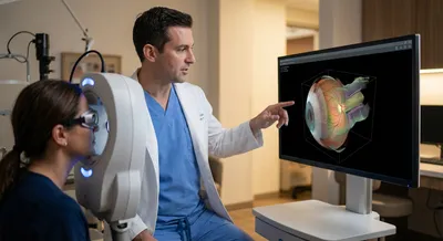
To check the RNFL, doctors commonly use optical coherence tomography (OCT), a non-invasive imaging test that takes cross-sectional “slice” pictures...
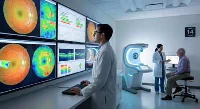
Doctors don’t judge an OCT scan in isolation. Instead, the scan machine compares your eye measurements to a built-in reference database of healthy...
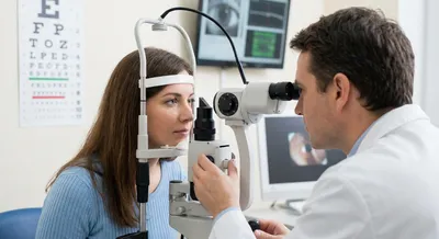
Goldmann Applanation Tonometry (GAT) is the gold standard for IOP measurement. () In this test, a tiny probe gently flattens (“applanates”) the...

This article reviews all human studies on magnesium in glaucoma. Specifically, we look at trials and reports of magnesium levels or supplementation...
Patient History and Questionnaires: Start by asking targeted questions every visit. Simple questions like “Do you have trouble reading or driving at...
Our visual field test is inspired by the perimetry methods eye care professionals use. Check for blind spots and track changes over time.
Test Your VisionOptical coherence tomography is a medical imaging technique that uses light to create detailed cross-sectional pictures of the eye. It works by sending low‑power light into the eye and measuring reflections from different layers to build a high-resolution image. The resulting views show thin slices of structures such as the retina, optic nerve, and cornea, allowing clinicians to see layer-by-layer detail. Because it captures microscopic structure, it reveals subtle changes that are not visible with routine exams or photos. The test is quick, noninvasive, and painless, usually performed in a clinic without special preparation. Optical coherence tomography matters because it helps doctors diagnose and monitor eye problems like glaucoma, macular degeneration, and diabetic eye disease earlier and more accurately. It provides precise measurements—such as the thickness of retinal layers—that make it easier to track small changes over time and to judge whether treatments are working. The images guide treatment decisions, for example when to start or adjust medications, perform laser treatment, or consider surgery. There are some limits: dense cataracts or poor fixation can reduce image quality, and the scans still need expert interpretation. Overall, it has transformed eye care by improving detection, follow-up, and personalized treatment plans.