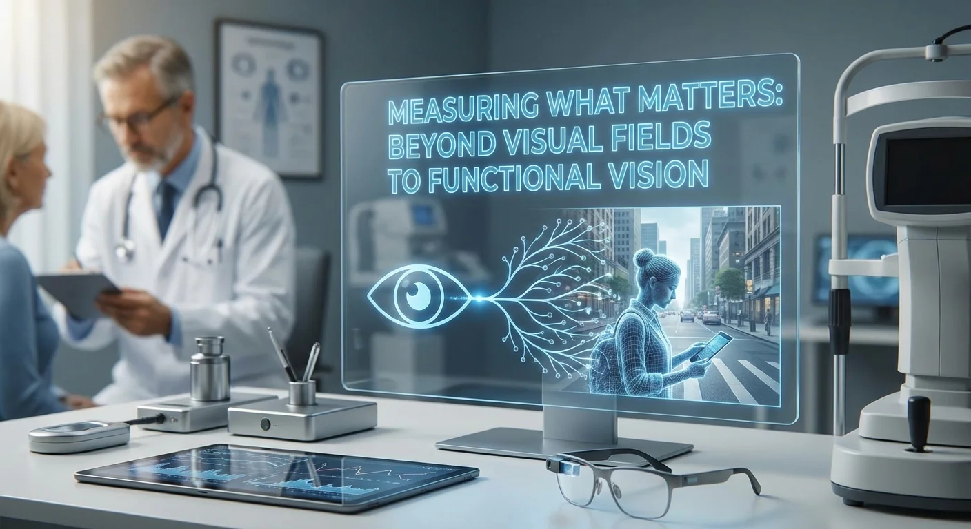
Introduction
Eyesight is more than letters on a chart. While visual field testing is the gold standard for glaucoma and other eye diseases, patients often notice vision problems in day-to-day life before standard tests change. For example, people may struggle to read a book in low light or feel unsafe driving at night, even if their Snellen acuity or visual field is “normal.” In this article we explore functional vision assessments beyond routine perimetry – tests of contrast sensitivity, reading speed, and even driving ability – and show how they reveal real-world vision loss earlier. We also review new home-based visual field platforms and explain how to track their results over time alongside OCT scans. Understanding these tools helps catch vision decline sooner and shows how impairments affect quality of life. Importantly, we offer practical tips so doctors can blend these extra tests into appointments without overwhelming patients or staff.
Limits of Standard Visual Field Testing
Standard perimetry (visual field testing) maps out how well you see at many points in each eye. While crucial, perimetry has limits. It is done in a quiet exam room on a lit screen, which might not catch all problems people face. For instance, a glaucoma patient may pass a field test but still have trouble driving in bright sun or reading faded print. Research shows routine tests like visual acuity charts or basic perimetry don’t always predict how safely someone will drive (pmc.ncbi.nlm.nih.gov). In fact, one study notes that quick, high-contrast charts and fields focus on ideal conditions, whereas real-world tasks (driving at night or in traffic) involve motion, low contrast, and divided attention (pmc.ncbi.nlm.nih.gov). Because everyday vision involves more than stationary charts, we need complementary tests of functional vision – measures tied to daily activities.
Key Functional Vision Assessments
Contrast Sensitivity Testing
Contrast sensitivity (CS) measures how well you can distinguish shades of light and dark, not just black letters on white. This reflects everyday tasks like seeing a curb in dim light or spotting a gray car on a foggy road. It is often tested with charts (e.g. Pelli-Robson) that use letters of decreasing contrast. Contrast sensitivity tends to drop early in diseases like glaucoma or macular degeneration, sometimes before standard tests change (pmc.ncbi.nlm.nih.gov). A 2024 study explains that decline in CS can explain many vision complaints that standard acuity misses (pmc.ncbi.nlm.nih.gov). CS is vital for safe mobility and driving: as one review notes, it “refers to the ability to visually discern objects from their background and is a critical component of mobility, stability, and safe driving” (pmc.ncbi.nlm.nih.gov). In practice, a quick CS test can be done in a minute or two. Poor CS scores can alert doctors to early functional loss – for example, a classic study found that an oscilloscope-measured CS test detected disability in early glaucoma before visual fields changed (pmc.ncbi.nlm.nih.gov).
Reading Speed Assessments
Reading speed is another practical measure of vision. Tests like the MNREAD or IReST charts quantify how many words per minute a person can read comfortably under a fixed font. Reading uses fine central vision and eye movements, so it may slow even if the canopy field is largely intact. In an aging eye study, glaucoma patients with more advanced bilateral disease read about 30–35 words per minute slower than those without glaucoma (pmc.ncbi.nlm.nih.gov). That study defined “impaired” reading as less than 90 words/min; many glaucoma patients fell below this despite mild field loss. Other research shows a mismatch between what patients feel about their reading ability and measured speed (pmc.ncbi.nlm.nih.gov). In one trial, some patients read slowly in tests yet reported only slight difficulty, or vice versa, highlighting why both objective reading speed tests and patient questionnaires matter (pmc.ncbi.nlm.nih.gov). Clinically, timing a paragraph out loud is quick and can flag central vision problems or visual processing delays. If results slow over time, it suggests vision is worsening even if acuity and fields seem stable.
Driving Simulation and On-road Metrics
Driving is a complex visual task involving peripheral vision, glare sensitivity, and rapid decision-making. Simulated driving tests offer a way to measure how vision loss translates to real-world driving performance. Research using driving simulators finds that these tests “can be used as a performance-based test for evaluation of functional impairment” in glaucoma and other eye diseases (pmc.ncbi.nlm.nih.gov). In other words, by recreating streets and hazards on a computer, doctors can directly see whether a person reacts more slowly or misses stop signs and pedestrians. For example, studies in glaucoma patients have shown they respond more slowly to hazards and make more lane deviations than healthy drivers, even if visual acuity was similar. Importantly, standard in-clinic visual tests (Snellen acuity, simple contrast charts, or field tests) often fail to predict true driving safety (pmc.ncbi.nlm.nih.gov). As one review points out, those quick tests “may not be strong predictors of driving safety,” so simulators fill this gap. Availability of driving sims varies, and many are still research tools, but even asking targeted questions about driving (night glare, difficulty noticing cars) is valuable. Some clinics also use tests like the Useful Field of View (UFOV) test on a computer to assess visual processing speed, which links strongly to accident risk. In summary, driving-related metrics (whether questionnaires or simulated tests) can uncover vision limitations that matter for independence well before a field defect is severe (pmc.ncbi.nlm.nih.gov) (pmc.ncbi.nlm.nih.gov).
Emerging Home-Based Visual Field Monitoring Devices
New technology lets patients measure vision at home between visits. These smartphone or tablet-based tools make frequent monitoring possible. For glaucoma, platforms like Melbourne Rapid Fields (MRF), Eyecatcher, and VF-Home allow patients to check parts of their visual field using a tablet or virtual-reality headset. Several studies report that these home tests give results very similar to the in-clinic Humphrey field test (pmc.ncbi.nlm.nih.gov). In practice, a patient can take a field test at home on their computer or phone screen, and the data is sent to their doctor. Researchers have found that patients are willing to do this regularly: for example, monthly home testing with the Eyecatcher program was well accepted, with performance very close to clinic exams (pmc.ncbi.nlm.nih.gov). An important bonus is frequency: with at-home testing, a patient can take more tests per year than the usual one or two clinic fields. Modeling shows this catch more rapid changes sooner. Indeed, one systematic review notes that using home tests significantly improved early detection of fast field loss, simply by allowing more tests per year (pmc.ncbi.nlm.nih.gov).
Telemedicine studies also report high adherence and satisfaction. Home devices were “feasible and reliable for home use with good patient adherence” and did not suffer much from distractions or lighting changes (pmc.ncbi.nlm.nih.gov) (pmc.ncbi.nlm.nih.gov). Patients felt in control and clinics saw fewer missed visits. Of course, clinics should still schedule occasional in-person field exams for confirmation, but home monitoring can reduce travel and waiting times while keeping closer watch on vision change (pmc.ncbi.nlm.nih.gov) (pmc.ncbi.nlm.nih.gov). In short, home-based field testing is an emerging tool that helps bridge the gaps between appointments.
Interpreting Home- and Clinic-based Data with OCT
Home vision tests and traditional clinical tests can be charted together over time. Doctors often keep track of trends: for example, serial OCT scans of the optic nerve or macula are graphed to see if nerve fiber layers are thinning. Functional measurements can be added to these trend charts. If a patient’s home field tests show a steady decline and OCT shows thinning, that double signal strengthens confidence of true progression. Some recent research has even combined OCT data with field data in models to catch progression faster (pmc.ncbi.nlm.nih.gov). Conversely, if OCT changes but fields remain flat, it might signal that more sensitive tests (like contrast or reading) should be done again, or simply that more follow-up is needed. In practice, clinics can display both OCT and field (or home test) trends on one screen. This holistic view helps avoid reliance on any single number. (For example, OCT imaging sometimes detects optic nerve thinning years before any field loss appears (pmc.ncbi.nlm.nih.gov), so catching a small drop in OCT could prompt more frequent monitoring.) By following patients’ numbers over time – whether from the clinic Humphrey, a home app, or both – doctors can make more informed decisions.
Functional Measures and Quality of Life
Functional vision scores often correlate tightly with a patient’s quality of life. In early glaucoma or macular degeneration, people frequently complain of vision problems even when charts look fine (pmc.ncbi.nlm.nih.gov). For example, patients in a recent community study reported vision difficulties despite normal acuity. The authors noted that contrast sensitivity likely explained many complaints not captured by standard tests (pmc.ncbi.nlm.nih.gov). In another survey of glaucoma patients, more than half reported some driving difficulties involving glare or night vision, while 22% struggled with tasks needing good peripheral vision (pmc.ncbi.nlm.nih.gov). Notably, many of those drivers only had moderate field loss, suggesting they noticed trouble before the test flagged a severe defect. Similarly, patients often report trouble reading small print or scanning menus, which shows up as slower reading speed on tests. These real-world complaints can appear when disease is still mild. By including functional tests, doctors can “uncover” these quality-of-life impacts earlier. In other words, a patient’s daily struggles (difficulty reading ingredients on a label, for example) can be objectively measured and tracked long before severe vision loss. This early insight can drive treatment changes to preserve independence.
Integrating Functional Tests into Routine Care
Incorporating these additional tests into a busy eye clinic requires strategy. Here is a practical framework:
-
Patient History and Questionnaires: Start by asking targeted questions every visit. Simple questions like “Do you have trouble reading or driving at night?” or “Do you see well in dim light?” can signal which tests to use. Vision questionnaires (e.g. parts of the NEI-VFQ) can be offered on a tablet or paper while patients wait.
-
Selective Testing by Indication: Not every patient needs every test at every visit. Decide by risk and symptoms. For example, a glaucoma suspect or early patient reporting visual complaints might get a contrast sensitivity chart test (which takes only ~2 minutes) and a brief reading task. If someone drives regularly, consider a quick UFOV or at least document their driving comfort. For stable patients with no complaints, these tests can be spread out (e.g. alternate visits). Studies show that even doing a functional test once a year can catch changes missed by fields alone (pmc.ncbi.nlm.nih.gov) (pmc.ncbi.nlm.nih.gov).
-
Use of Technicians and Tools: Delegation helps avoid overload. Trained technicians can administer contrast or reading speed tests during vision screening. Many clinics now have iPads or VR devices; they can run home-based field apps or UFOV while the patient waits. Having a log kept with results (paper or electronic) makes interpretation easier.
-
Home Monitoring Integration: Give suitable patients instructions on home tests. Many can learn a smartphone visual field app in clinic and take baseline tests. Ask them to bring results (or have data sent) to each visit. Home measures are shown to have good adherence when patients know they matter (pmc.ncbi.nlm.nih.gov) (pmc.ncbi.nlm.nih.gov). This way, the doctor gets averaging or trends from monthly home checks, reducing the need for extra field testing in the office.
-
Data Review and Documentation: Between visits, display trends for both imaging and function. For example, in the record show a graph of retinal nerve fiber thickness (OCT) and overlay Humphrey/simulated field mean sensitivity. Similarly, note contrast score over time. If any dramatic drop appears (confirmed on repeat), schedule intervention. This combined record helps catch progression: one analysis found adding home field data to clinic tests “reduced measurement error” and detected changes faster (pmc.ncbi.nlm.nih.gov).
These steps allow adding functional tests in a targeted way. The key is balance: use short tests generously when they can impact management, and rely on remote or selective testing to avoid bogging down clinic flow.
Conclusion
Standard visual field testing remains essential, but a more complete picture of sight comes from how people use their vision in life. Contrast sensitivity, reading speed, and driving simulation tests pick up everyday challenges that fields and acuity can miss. New tele-ophthalmology tools let patients check sight at home, giving doctors more data without extra clinic appointments (pmc.ncbi.nlm.nih.gov) (pmc.ncbi.nlm.nih.gov). When all these functional measures are tracked over time alongside OCT imaging, we can identify vision decline and quality-of-life impacts much earlier. Use of these tools should be tailored: short tests and questionnaires for those with early disease or complaints, and home-monitoring for high-risk patients. This patient-centered approach not only detects subtle changes sooner but also aligns clinical care with what truly matters to patients – the ability to read, drive, and live safely. By “measuring what matters,” clinicians can intervene earlier to preserve vision and independence while keeping clinic workload manageable.