
mTOR/Autophagy Modulation by Amino Acids in RGC Degeneration
Cells constantly balance between building up structures and recycling damaged parts. mTOR is a Master growth sensor: when nutrients are abundant,...
Deep research and expert guides on maintaining your visual health.
Cells constantly balance between building up structures and recycling damaged parts. mTOR is a Master growth sensor: when nutrients are abundant,...
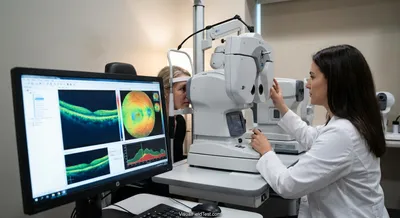
Each OCT result comes with color-coded maps and numbers. Green usually means “within normal limits,” yellow means “borderline,” and red indicates...

In a rabbit glaucoma model (pressure raised by a gel in the eye), researchers injected NGF around the eye (retrobulbar) before damage. Those rabbits...
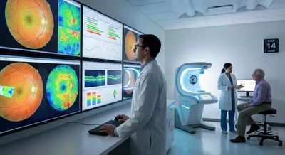
Doctors don’t judge an OCT scan in isolation. Instead, the scan machine compares your eye measurements to a built-in reference database of healthy...

NAD<sup>+</sup> is a ubiquitous coenzyme that facilitates ATP production via glycolysis and oxidative phosphorylation, and serves as a substrate for...
Our visual field test is inspired by the perimetry methods eye care professionals use. Check for blind spots and track changes over time.
Test Your VisionOCT stands for optical coherence tomography, a noninvasive imaging test that uses light waves to create cross-sectional pictures of the back of the eye. It produces detailed, almost microscopic views of layers in the retina and the optic nerve head, so doctors can see structures that are invisible with ordinary examination. The machine scans across the eye and measures the echo time delay of reflected light, translating that information into thin slices that together form a 3D picture. Because it is quick and painless, OCT is widely used in eye clinics for diagnosis and routine monitoring. In conditions that affect retinal cells or optic nerve fibers, OCT can measure thickness and detect early thinning before vision loss is obvious. This ability helps clinicians decide when to start or change treatment and to track whether a disease is getting better or worse over time. OCT is also useful for spotting fluid, swelling, or other structural changes that guide procedures and medication choices. However, image quality can be affected by cataracts, eye movements, or poor fixation, and the test is one piece of information used alongside vision tests and exams. Advances in OCT continue to improve resolution and speed, making it an essential tool for preserving sight and detecting eye disease early.