Introduction
Glaucoma is a complex eye disease in which no single test can provide a definitive answer. Instead, a battery of tests is needed to build a full picture of your eyes: measuring pressure, examining the drainage angle, evaluating the optic nerve head, and mapping your visual field. Each test provides one piece of the puzzle. When you understand what each test does and what the numbers mean, you become an active partner in your care – not just a passive patient sitting in the dark. This guide will explain why multiple tests are necessary and how each one contributes unique information about your eye pressure, anatomy, nerve health, and vision, with clear explanations of the results you’ll receive.
Why Multiple Tests Matter
Glaucoma is defined by damage to the optic nerve, often associated with high eye pressure (intraocular pressure or IOP), but it can occur even with “normal” pressure. For example, many glaucoma patients actually have relatively low measured IOP because they have thin corneas, which can make pressure readings appear falsely low (pmc.ncbi.nlm.nih.gov). Conversely, a very thick cornea can make IOP look higher than it really is. On the other hand, some eyes with higher pressure never develop glaucoma. Therefore, doctors must look at eye anatomy and function in addition to pressure. This means examining the drainage angle (to see if fluid can escape properly), inspecting the optic nerve for damage, and testing your peripheral vision. In practice, this requires a comprehensive evaluation with complementary tests (pmc.ncbi.nlm.nih.gov). One review of international guidelines notes that general screening is of “limited clinical utility,” and no single test has both the sensitivity and specificity needed (pmc.ncbi.nlm.nih.gov). The takeaway is that a combination of pressure measurement, imaging, and visual field testing is used to confirm or rule out glaucoma.
Empowering you as a patient means explaining each test and result. When you walk out of the exam room, you should know, for example, “The average IOP measured 18 mmHg and the pachymetry showed my cornea is thin, which means my true IOP is probably higher,” or “My OCT shows red areas where my nerve fiber layer is thinner than normal.” Armed with this knowledge and the actual printouts of your tests, you can track trends over time and ask informed questions.
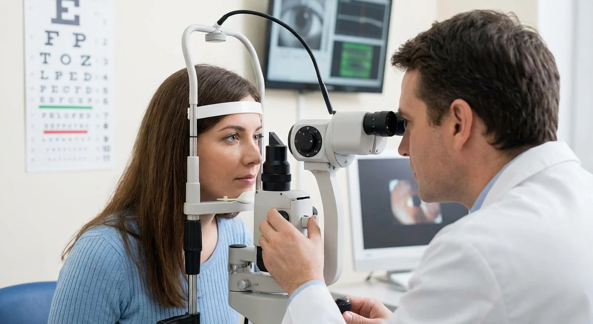
Measuring Intraocular Pressure (Tonometry and Pachymetry)
The only modifiable risk factor for glaucoma is high eye pressure. Measuring IOP is therefore a crucial first step, but even this has nuances.
-
Goldmann Applanation Tonometry (GAT) is the gold standard for IOP measurement. (pmc.ncbi.nlm.nih.gov) In this test, a tiny probe gently flattens (“applanates”) the cornea, using a slit-lamp microscope. GAT has been used for decades and is very well validated. (pmc.ncbi.nlm.nih.gov) It requires anesthetic eye drops and careful technique. Most clinical trials and glaucoma treatment thresholds are based on Goldmann IOP values. Because GAT relies on flattening the cornea, its reading is accurate for an “average” cornea (about 520 microns thick (eugs.bitblox.eu)). But if your cornea is much thinner or thicker, the reading can be off (more on that below).
-
Non-Contact (Air-Puff) Tonometry is the familiar–and more comfortable–test that blows a brief puff of air onto your eye. It also measures pressure by analyzing how the cornea flattens under the air impulse. Modern air-puff devices have shown very strong correlation with Goldmann measurements (pmc.ncbi.nlm.nih.gov). In one study, air-puff readings were within about 1–2 mmHg of GAT (with correlation coefficient >0.8) (pmc.ncbi.nlm.nih.gov). In other words, if your GAT reading is 18 mmHg, the air-puff might measure roughly 19–20, which is clinically negligible. Air-puff tonometry is quick, no-touch, and does not require anesthetic drops (pmc.ncbi.nlm.nih.gov). Many doctors use it for initial screening or quick checks. However, it can sometimes overestimate pressures in eyes with very high IOP or other corneal issues, so elevated readings are often re-checked with Goldmann for confirmation.
-
Rebound Tonometers (e.g. iCare) are portable devices you can use in a clinic (and in some cases at home) without drops. A tiny probe literally bounces off the cornea, and the speed of rebound indicates pressure. iCare tonometers are increasingly used for quick screening or for patients who can’t tolerate Goldmann (children, uncooperative patients, or when anesthesia is not given). Studies show iCare measurements strongly agree with Goldmann in the low-to-moderate pressure range (pmc.ncbi.nlm.nih.gov). For example, in one trial the average IOP by iCare and Goldmann was almost identical (18.3 vs 18.5 mmHg) with excellent correlation (pmc.ncbi.nlm.nih.gov). However, iCare tends to slightly underestimate very high pressures (for eyes over ~23 mmHg) and is more influenced by corneal thickness (pmc.ncbi.nlm.nih.gov). Its big advantage is convenience: no drops needed and minimal training to use. Some clinics loan iCare units for home use (“iCare HOME”) so patients can track their own pressures.
-
Dynamic Contour Tonometry (Pascal DCT) uses a special concave sensor tip that matches the shape of the cornea. It measures pressure continuously and is designed to be less dependent on corneal properties. Studies have found that DCT readings tend to be slightly higher on average than Goldmann readings (pmc.ncbi.nlm.nih.gov). Importantly, DCT is least affected by corneal thickness (pmc.ncbi.nlm.nih.gov). In a comparative study, adding 45 microns of corneal thickness increased GAT and air-puff readings substantially but had minimal effect on DCT readings (pmc.ncbi.nlm.nih.gov). In other words, if you have an unusually thick or thin cornea, DCT may give a truer IOP value. However, DCT devices are bulkier and less common in routine practice.
-
Central Corneal Thickness (Pachymetry) is measured in millimeters by a pachymeter device (ultrasound or optical) and is critical context for tonometry. Because tonometers flatten the cornea, a thinner-than-normal cornea will be pushed flatter by the same force than an average cornea, resulting in a falsely low pressure reading (eugs.bitblox.eu). Conversely, a thick cornea resists flattening and produces a falsely high reading. For example, studies note that Goldmann applanation is only accurate near 520 µm; far thinner values grossly underestimate true pressure (eugs.bitblox.eu), while very thick corneas can overestimate pressure. This is why pachymetry is routinely done alongside IOP: if your cornea is significantly thin, your doc will know that a “normal” IOP might actually mask risk, and if it’s thick, a high reading may not be as alarming (pmc.ncbi.nlm.nih.gov). In fact, nearly half of glaucoma patients have corneas thinner than average, which partly explains why they had “normal” pressure despite disease (pmc.ncbi.nlm.nih.gov).
-
Diurnal and Fluctuation Effects. Any single pressure measurement during an office visit is just a snapshot. We now know that IOP fluctuates throughout the day and night, and these swings can be an independent risk factor for glaucoma progress. A meta-analysis found that long-term IOP fluctuation itself raises the risk of field loss (hazard ratio ~1.43 for those with large long-term swings) (pmc.ncbi.nlm.nih.gov). In other words, an eye that varies between 18 and 26 mmHg over days or weeks may be at higher risk than one that stays around 18 all the time (pmc.ncbi.nlm.nih.gov). Because of this, some specialists order measurements at different times of day or use 24-hour monitoring (like a contact lens sensor or home tonometry) to catch unseen peaks. It also underscores recording several measurements over time rather than relying on a lone reading.
By understanding tonometry and pachymetry, you’ll know how your IOP is being measured and corrected. For instance, you can ask: “My reading was 18 mmHg but my corneas are thin – what’s the adjusted pressure?” or “My pressure spiked in the morning – should I be trying to capture more readings throughout the day?”
Examining the Drainage Angle (Gonioscopy)
Glaucoma isn’t just about pressure – it’s also about how fluid drains from your eye. The fluid (aqueous humor) leaves through a tiny angle between the iris and cornea at the front of the eye. In open-angle glaucoma, this drainage angle is wide open but the outflow mechanism fails somehow. In angle-closure glaucoma, the angle is narrow or blocked, preventing fluid outflow and causing pressure to rise. Gonioscopy is the only way to directly see and grade this angle.
-
Van Herick Estimate. Before gonioscopy, an ophthalmologist can do a quick slit-lamp estimate called the Van Herick test. By shining a narrow slit of light at the edge of the cornea and iris, the examiner can compare the peripheral chamber depth to corneal thickness. If the space is narrow (less than one-quarter of corneal thickness), it suggests a narrow angle that merits formal gonioscopy (eugs.bitblox.eu). This is a non-contact, screening step but is not definitive.
-
Gonioscopy With a Lens. For a true angle view, your doctor uses a special gonioscopy lens on the eye (like a small contact lens with mirrors). This lets light enter the eye without total internal reflection, so the doctor can see angle structures through the lens. The angle is graded by how wide it appears. Common grading systems include Shaffer (Graded 0 to 4 by angle width in degrees; 0 = closed, 4 = very open) and Spaeth (a more detailed system considering iris insertion and angle depth). These tell us if the angle is physiologically open or dangerously narrow.
-
Appositional vs. Synechial Closure. During gonioscopy, your doctor may notice the iris touching the trabecular meshwork (angle wall). If the iris is simply pressing against the angle (appositional closure), it may open once pressure is reduced or after a laser iridotomy. But if there are actual adhesions (peripheral anterior synechiae) gluing the iris over the meshwork, this indicates chronic closure that may not fully reverse. If synechiae are present, an iridotomy alone might not eliminate blockage, and more advanced treatments like surgical goniosynechialysis may be needed.
-
Underutilization Warning. Gonioscopy is critical but often skipped. A 2024 U.S. study found that over 70% of patients had no recorded gonioscopy at all during their initial glaucoma evaluation (pmc.ncbi.nlm.nih.gov). This is alarming because missing a narrow-angle eye can lead to a sudden angle-closure crisis later. In fact, both the American Academy of Ophthalmology and the World Glaucoma Association recommend gonioscopy at the first glaucoma visit (and periodically thereafter, e.g. every 5 years) to check the angles (pmc.ncbi.nlm.nih.gov). In practice, if you haven’t had gonioscopy, you should ask for it – especially if you have any symptoms (blurry vision, halos) or risk factors (very farsighted, family history of angle-closure, Asian or Arctic ancestry). Knowing your angle anatomy turns your glaucoma evaluation from guesswork into precision.
Evaluating the Optic Nerve and Retina
The optic nerve head (the “eye’s cable”) is the central site of damage in glaucoma. A careful examination and imaging of the nerve head and surrounding retina is essential for diagnosis and tracking.
-
Clinical Optic Nerve Exam. The doctor will look through a slit lamp or ophthalmoscope and assess the optic disc (the circular spot where nerve fibers exit). Key features include: the cup-to-disc ratio (the size of the central “cup” compared to the full disc) and the neuroretinal rim thickness. In glaucoma, the cup typically enlarges as rim tissue is lost. A normal cup-to-disc ratio is often around 0.3 (cup is 30% of disc) but varies with disc size. Ratios above ~0.6 or asymmetry greater than ~0.2 between eyes are suspicious. The doctor also checks the rim shape: normally the rim is thicker at the bottom and top (“ISNT rule”), but glaucoma often thins the rim first at the superior and inferior poles. Other findings include disc hemorrhages (tiny flame-shaped bleeds on the disc surface) and retinal nerve fiber layer (RNFL) defects (wedge-shaped patterns of missing nerve fibers visible with a red-free filter). Research shows disc hemorrhages are relatively rare in healthy eyes (<2% prevalence) but occur more often in glaucoma (up to 10-15% of glaucomatous eyes) (pmc.ncbi.nlm.nih.gov). Importantly, a disc hemorrhage is a red flag – it often precedes further nerve damage. One analysis found eyes with hemorrhages had significantly faster visual field loss during follow-up (pmc.ncbi.nlm.nih.gov). Examining the RNFL is also valuable: loss of the bright NFL striations on the retina can be seen as dark “notches” extending from the disc. In fact, studies note that careful RNFL inspection can reveal damage before a visual field defect appears (pmc.ncbi.nlm.nih.gov). All these observations (cup size, rim thinning, hemorrhages, RNFL defects, and any asymmetry between eyes) collectively inform the diagnosis and staging of glaucoma.
-
Optic Disc Photography. High-quality color photos of the optic nerve are often taken to establish a baseline. These stereoscopic disc photos preserve the 3D view of the nerve head. Comparing future photos to baseline allows the doctor to see subtle changes over time (e.g. new rim thinning or hemorrhages). It’s like having a “snapshot” of your optic nerve on file. In clinical trials, disc photos are a key method for detecting progression. Patients should ask for disc photos to be done early in their care and requested for personal records.
-
Optical Coherence Tomography (OCT). OCT has revolutionized glaucoma care by providing quantitative, cross-sectional imaging of the optic nerve and retina. An OCT scan (noninvasive and painless) produces a map of the retinal nerve fiber layer (RNFL) thickness around the optic nerve, as well as thickness of the ganglion cell–inner plexiform layer (GCL-IPL) in the macula. These layers contain the nerve fibers and cell bodies that are lost in glaucoma. The OCT device compares your eye’s values to a built-in normative database of healthy eyes (matched for age, etc.). On the OCT printout you’ll see color-coded maps and graphs:
-
Color-Coded Thickness Map: This shows cross-sectional scans or thickness maps. Typically, warm colors (green/yellow/red) indicate thicker normal tissue, whereas cool colors (blue/green) indicate thinner areas (journals.lww.com). For example, on an RNFL thickness map, a long green arc is normal, but any red zones may indicate thinning. The “RNFL deviation map” is often green for normal areas and yellow/red to flag points outside normal limits (journals.lww.com).
-
TSNIT or Profile Graph: This stands for Temporal–Superior–Nasal–Inferior–Temporal (a circle around the nerve). It is often graphed as two lines (one for each eye) showing RNFL thickness versus clock hour. The reference range (normal) is shown as a green band. If your line (often one solid, one dashed for each eye) dips into the yellow/red zone, that point is abnormally thin. Comparing both eyes on the same plot highlights asymmetry (journals.lww.com).
-
Ganglion Cell Analysis: Many OCTs also provide a map of the macular ganglion cell layer. This usually appears as an elliptical or oval map across the central retina, again with green/yellow/red coding. Damage in glaucoma often first shows up nasally or inferiorly in the macula on these maps.
-
Numerical Summaries: The OCT will also give average RNFL thickness (global, and by quadrant), numerical comparison to normal (in standard deviations or percentile), and possibly a “glaucoma probability score.” These ease interpretation but you can cross-check the visual maps.
-
Progression Analysis: If you have multiple OCTs over time, many devices can display a trend graph or event analysis to see if nerve thickness is decreasing. The software may flag points of significant loss across visits.
-
Understanding your OCT printout can feel complex at first, but remember: green = good, yellow = borderline, red = likely abnormal. If your average RNFL thickness is shown in red in the report, that means it is thinner than 99% of normals at that age. If a ruled trend line in a graph shows a downward slope, that means thinning is progressing. Ask your doctor to review these reports with you. For example, if you see a new red (abnormal) sector on the RNFL map compared to last year’s green map, that’s important to catch early.
Visual Field Testing
Visual field (perimetry) tests measure how well you see in all directions (especially peripheral vision). Since glaucoma typically causes “patchy” loss of side vision, automated perimetry is indispensable. The Humphrey Field Analyzer (HFA) is the standard instrument. Here’s what to know:
-
24-2 vs. 10-2 Testing: The common screening program is the HFA 24-2 test, which examines 54 points in the central 24 degrees of the visual field (on a 6-degree grid). This allows detection of the early classic arcuate scotomas of glaucoma. However, it has relatively few points within the central 10°. The 10-2 test covers a finer 68-point grid over the central 10°, useful for detecting paracentral defects near fixation that standard fields miss. Current advice is that if any significant defect appears near the center on a 24-2 test, the eye should be retested with a 10-2 field (www.ncbi.nlm.nih.gov). A newer “24-2C” test adds extra points in the central 10° to the 24-2 grid, improving detection of central loss (www.ncbi.nlm.nih.gov). These tests are done one eye at a time; you press a button whenever you see a tiny light flash while staring at a fixed target.
-
Reliability Indices: Each field printout comes with reliability scores. “Fixation losses” measure how often you looked away from the target (tracking the blind spot), and false positives/negatives gauge if you were pressing incorrectly. High false positives (spurious clicks) or false negatives (missing obvious lights) mean the test results may be unreliable. It often takes two tries to get a reliable baseline. In fact, the first visual field test almost always has learning artifacts – it’s common to see you “miss” many points due to inexperience (pmc.ncbi.nlm.nih.gov). Thus, your doctor will typically establish a baseline with two good fields done a few weeks apart before drawing any conclusions about progression. Always be well-rested and properly corrected (prescription glasses on) before a field test, and try to respond consistently.
-
Reading the Field Printout: The main part of the report shows your sensitivity at each point. A grayscale map uses darker shades to indicate lower sensitivity (a “black hole” means very poor vision at that spot). Below that, total deviation (TD) numbers show how many decibels each point is below the age-normal average. The pattern deviation (PD) map adjusts for any overall dimming (for example, if you had a cataract, it removes that general shift to highlight localized losses). Key indices include: Mean Deviation (MD) – the average difference from normal across the field (0 dB is normal; negative MD means overall loss) – and Visual Field Index (VFI) – a percentage score (100% is a full-scene, 0% is near blind). The VFI is especially useful for tracking progression over time (steeper downward slopes mean faster loss). When you get your printout, focus on whether key clusters of points are darkened or flagged in PD, and keep an eye on the MD/VFI trends in serial tests.
-
Staging & Progression: Together with OCT and optic nerve exam, fields tell us if glaucoma is stable or worsening. For example, if your VFI drops from 90% to 80% over two years, or if new defects appear in a pattern block of 3 points at <5% level, that indicates progression. By law (and by many guidelines), patients detected with early glaucoma should have fields done at least once a year, more often (every 6 months or less) if there’s faster change.
-
Emerging Field Tests: Aside from Humphrey, there are home or tablet-based perimeters like Melbourne Rapid Fields (MRF). One trial found MRF to be “cost-effective, time-saving, and user-friendly,” with results generally comparable to the Humphrey 24-2 (pmc.ncbi.nlm.nih.gov). Such home perimetry devices can now supplement clinic visits, especially for patients in remote areas or with mobility issues. Always ensure any home test is done in a dark, quiet room and follow instructions carefully.
Advanced and Emerging Tests
Eye care is at the cutting edge of technology. In addition to the standard exams, some advanced tests and new technologies are being developed or rolled out to catch glaucoma even earlier and monitor it more precisely:
-
Optical Coherence Tomography Angiography (OCT-A): This is a newer OCT mode that visualizes blood flow in the tiny capillaries around the optic nerve and macula, without dye. Studies show that OCT-A can detect reduced perfusion before structural nerve thinning is obvious. For example, patients with glaucoma have been found to have lower capillary density around the optic nerve head “beginning temporally even before RNFL thinning” is measurable (eyewiki.org). Similarly, glaucoma and even ocular hypertensive eyes have reduced macular capillary density compared to normals (pmc.ncbi.nlm.nih.gov). OCT-A is not yet standard in all clinics, but it hints at diagnosing glaucoma via its vascular changes rather than anatomy alone.
-
Electroretinography (ERG) for Ganglion Cells: The Pattern ERG (PERG) is an electrical test that specifically probes retinal ganglion cell (RGC) function. In glaucoma, RGCs are damaged, so PERG can be abnormal even before visual field loss. Clinically, one finds that the PERG “N95” wave often has longer latency (delay) and reduced amplitude in early glaucoma. In fact, studies have shown that glaucoma suspect eyes often already have a delayed N95, whereas eyes with manifest glaucoma show reduced N95 amplitude (pmc.ncbi.nlm.nih.gov). This means PERG can detect RGC dysfunction at a stage not visible on OCT or fields. PERG requires special equipment and trained staff, so it’s mainly used in research or specialist centers, but it’s a promising early biomarker.
-
Adaptive Optics Imaging: This cutting-edge imaging technology corrects for the eye’s aberrations and can resolve microscopic detail on the retina. Adaptive optics scanning laser ophthalmoscopes (AO-SLO) and adaptive optics OCT have already been used to image individual photoreceptors and even see retinal capillaries†. In research settings, people are experimenting with AO to visualize individual retinal ganglion cells in vivo. In theory, this could directly count nerve cells or spot early cell death before functional loss. It’s not something you’ll get today in clinic, but it’s an area of active investigation.
-
Artificial Intelligence (AI) and Machine Learning: Powerful AI algorithms are now being applied to fundus photos, OCT scans, and even visual field data to assist diagnosis and predict progression. These systems can detect subtle patterns invisible to the human eye. For example, deep learning models have been trained to forecast who will develop glaucoma. One study using sequential optic disc images reported ~88% accuracy in predicting glaucoma onset 1–3 years before it happened (pmc.ncbi.nlm.nih.gov). Another used advanced models to flag fields likely to worsen. In practice, AI can serve as a second set of eyes: flagging suspicious OCT changes, quantifying rate of RNFL loss, and guiding physicians on when to intensify treatment.
-
Home Tonometry and Portable Perimetry: We already mentioned the iCare HOME rebounding tonometer. In clinical trials, home IOP monitoring has proven very useful. In one series, patients measured their own IOP at waking, midday, evening, and bedtime for several days. The home measurements captured many high peaks that were missed in the office. For example, mean peak IOP at home was 21.3 mmHg compared to 17.4 in-clinic (pmc.ncbi.nlm.nih.gov). This information led doctors to change treatment in 55% of those eyes (pmc.ncbi.nlm.nih.gov). Other innovations include 24-hour contact lens sensors (like the Triggerfish) and smartphone/tablet perimetry (MRF and others mentioned above). These tools move glaucoma management from occasional snapshots toward continuous monitoring, catching changes earlier.
Together, these technologies aim to detect glaucoma as early as possible and to quantify change more precisely. As these tools become more widely available, patients can gain even more information about their disease in a less invasive or time-consuming way (“tele-glaucoma” clinics, anyone?).
Practical Patient Considerations
-
Testing Schedule: How often should these tests be done? There is no one-size-fits-all answer. Your follow-up schedule depends on your glaucoma stage and risk factors. Guidelines now emphasize individualized monitoring (www.reviewofoptometry.com). As a rough idea:
- If you are simply a glaucoma suspect (raised pressure or suspicious nerve but no damage yet), you might have a full work-up (IOP, gonioscopy, OCT, visual field) every 12 months.
- If you have mild glaucoma under good control, many doctors do OCT and visual fields about once a year, or every 6–12 months if uncertainty exists.
- For moderate to advanced glaucoma, testing is more frequent – often every 3–6 months – because we need to catch any progression quickly.
- After any significant change (like a surgery or medication change), the first 3–6 months usually involve more frequent checks (every few weeks or months) to see how the pressure and fields respond.
- People with high-risk traits (African or Inuit ancestry, strong family history, thin corneas) may be monitored more aggressively as well.
The updated AAO Preferred Practices include tables with recommended intervals based on risk and severity (www.reviewofoptometry.com). It’s reasonable to review this schedule with your doctor and ask why each test is done at a given interval. If you are stable, your ophthalmologist might space out visits; if you are fast-progressing, they may want more frequent data points.
-
Ask for Your Results: Be proactive. Ask for copies of your OCT printouts and visual field reports at each visit. Keep them in a safe place (or electronically) so you can track your own trends. Learning to read the basics (as we have summarized above) helps you notice things like a worsening VFI on fields or new red sectors on OCT. It empowers you to ask, “My last 3 fields all show growth of the dark area in the lower right; are we seeing field loss despite stable pressure?” or “The OCT RNFL average thickness in my right eye went from 80µm last year to 75µm now – is that significant?”
-
Test Preparation: Proper preparation can improve test accuracy. For tonometry, remove contact lenses or eye makeup before pressure measurement. For OCT, pupils should be fully dilated if your doctor requests retinal imaging (dilation drops are common before photography or wide scans). For visual fields, ensure you are comfortable, not sleep-deprived, and have had your usual medications (unless told otherwise) so that energy levels are normal. Don’t drink excessive coffee or other stimulants right before tests, as caffeine can raise IOP slightly (pmc.ncbi.nlm.nih.gov). (One study showed a single strong coffee raised IOP by about 1 mmHg over the next hour (pmc.ncbi.nlm.nih.gov); for someone at the threshold, even a 1–2 mmHg rise could matter.)
-
Medication Effects: Remember that glaucoma drops can influence test results. Ideally do IOP measurements without recent drops if assessing baseline pressure, or always account for medication timing. For example, if you took latanoprost last night, your morning IOP may be lower than it would be without medication. Communicate all eye drops you use, and ask if you should omit a dose before testing. In addition, systemic medications can affect tests: certain steroid eye drops (or even oral steroids) can raise IOP, whereas antihypertensives usually do not. Also warn your doctor of any caffeine or supplements you’ve taken.
-
Out-of-Pocket Costs: In many countries, glaucoma tests are covered by insurance, but in the U.S. there are copays and deductibles. A typical glaucoma work-up (doctor exam with dilated fundus exam, IOP, and gonioscopy) might be around $150 on a cash basis. Each additional diagnostic test (OCT, visual field) can add another $100–$250 without insurance. If you have Medicare or private insurance, much of this is usually covered (often only a small copay or 10-20%). If you have high deductible plans or no insurance, the costs can add up. It’s worthwhile to check with your provider or billing department. Many clinics also prioritize which tests are essential: e.g. they may alternate OCT and fields or skip gonioscopy if not indicated, to reduce costs. If money is a concern, discuss it openly – there are sometimes lower-cost options (like non-contact tonometry instead of GAT, or fewer field tests). Community screening programs may offer free or reduced exams for at-risk individuals.
-
Red Flags and When to See a Specialist: Watch your results for warning signs. Sudden spikes or very high IOP (above 30–35 mmHg) at any visit should prompt urgent evaluation. A disc hemorrhage seen on exam or OCT is a poor prognostic sign (pmc.ncbi.nlm.nih.gov) and often triggers closer follow-up. Rapid visual field worsening (for example, losing more than 2–3 dB of VF Index in a year) should lead to immediate discussion of treatment change. If you have glaucoma or even borderline findings, keeping an up-to-date chart of your own MD and VFI trends, along with image printouts, helps spot trends.
Typically, optometrists and general ophthalmologists manage most glaucoma, but you should ask for a glaucoma specialist (an ophthalmologist fellowship-trained in glaucoma) if any of the following apply: worsening despite treatment, very high-pressure readings, or very narrow/closed angles on gonioscopy. Specialists will have experience with complex cases and can perform laser or surgery if needed.
Screening and Advocacy
So who should be tested for glaucoma? And when? Screening guidelines vary around the world.
- The American Academy of Ophthalmology (AAO) recommends that all adults have a glaucoma screening exam by age 40 (journals.lww.com). People with glaucoma risk factors should be evaluated earlier. Risk factors include older age (especially over 60), African or Inuit ancestry, strong family history of glaucoma, very high myopia, and systemic conditions like diabetes or hypertension. The AAO notes that as part of a routine eye exam at 40 you should have an IOP check, a nerve exam, and at least one comprehensive eye exam including glancing at the angles (journals.lww.com).
- In contrast, the European Glaucoma Society (EGS) and many international bodies do not currently recommend population-wide screening, citing insufficient evidence that it improves outcomes (journals.lww.com) (pmc.ncbi.nlm.nih.gov). They emphasize testing people who present to any eye-care provider with risk factors rather than inviting everyone in. For example, the Pan-American Ophthalmology Association recommends targeting people over 65, those with a strong family history, or those with ocular hypertension (journals.lww.com).
- The World Glaucoma Association (WGA) consensus statements similarly focus on risk evaluation rather than blanket screening. “Glaucoma meets some criteria for screening” (it is common, asymptomatic early, and treatable), but its low prevalence in the general population and the imperfect nature of tests mean indiscriminate screening can lead to many false referrals.
- Indeed, research shows screening programs can work best when integrated into other eye-health services. For example, adding a glaucoma check during diabetic retinopathy screening or eye camps can raise detection in underserved communities. Community initiatives like the Michigan “MI-SIGHT” program provide free glaucoma screening and follow-up in Spanish and other languages for vulnerable populations (pmc.ncbi.nlm.nih.gov). These efforts help close the detection gap: common barriers were language and cost of care, and facilitators were interpreters and affordable eye exams (pmc.ncbi.nlm.nih.gov). Telemedicine screening (remote OCT or field testing with digital uploads) is promising as well.
In practice, present these points to your eye doctor: if you’re turning 40 (or older) and have not had a glaucoma exam, ask for one. If you have any risk factors or symptoms (even mild), insist on gonioscopy and a nerve exam. Stay informed of guidelines: for example, the AAO now suggests baseline OCT of the RNFL and ganglion cell layers in glaucoma patients (www.reviewofoptometry.com). And if you live in a region with higher glaucoma rates (for example, people of Asian descent for angle-closure risk), consider more proactive screening (some guidelines suggest checking all patients over 50 in high-risk areas).
A personalized testing timeline can help. Here is a sample schedule that you could bring to your provider:
- Age 20–39: One routine eye exam by 30, with IOP check each visit. If you have very high risk (family history, very high myopia), have a glaucoma specialist exam by age 30–35, including at least one visual field and optic nerve photo.
- Age 40–49: Baseline comprehensive glaucoma evaluation by 40 (IOP + gonioscopy + optic nerve exam + OCT + visual field). If all is normal, routine maintenance exams every 2 years with pressure checks.
- Age 50–59: If over 50, have a dilated exam by an ophthalmologist every 1–2 years. Anyone with risk factors (African/Inuit ancestry, family history, thin corneas) should have a full glaucoma workup at least annually.
- Age 60+: Exams with gonioscopy, IOP, and visual fields every 1–2 years for everyone; more often if you have known glaucoma or thick/thin corneas.
These are general suggestions – your doctor will tailor the exact intervals. But discussing your personal risk and a plan helps ensure nothing is overlooked.
Conclusion
Glaucoma testing is not just a black-box mystery – it’s a suite of complementary tools that together keep your optic nerve safe. By knowing why each test is done, what it measures, and what changes in your results mean, you transform from a worried patient into an informed partner in your care. Always feel free to ask your eye doctor: “What did that number mean? Why do we do this test next time?” Over time, tracking your own OCT and visual field printouts (as well as IOP and disc photos) can alert you to trends. Glaucoma is often indolent, so early detection and vigilant follow-up are key. Use the guidelines as a roadmap (screen by 40, test high-risk eyes earlier, follow up according to disease severity) and take advantage of new technologies like home tonometry and app-based fields when available. Finally, remember community resources and telehealth can help expand access: if cost or distance is a barrier, look for local screening initiatives or eye camps that offer glaucoma checks.
Being proactive with these tests is the best defense against glaucoma’s stealthy damage. With knowledge and regular monitoring, you and your doctor can catch progression early and tailor treatment – preserving your vision for years to come.