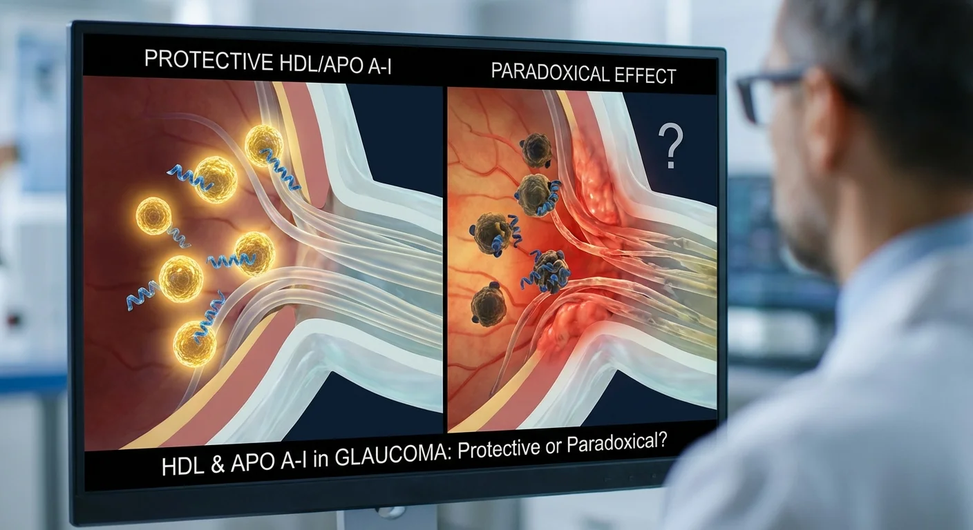
HDL Cholesterol, ApoA-I and Glaucoma – Friend or Foe?
Glaucoma – a leading cause of vision loss – is influenced not only by eye pressure but also by blood flow and inflammation in the eye. Some researchers are asking whether HDL cholesterol (often called “good cholesterol”) and its main protein component, apolipoprotein A-I (ApoA-I), help protect the optic nerve, or if under certain conditions they work in a paradoxical way.
In general health, HDL carries cholesterol from tissues back to the liver and has anti-inflammatory and antioxidant actions. For example, HDL stimulates blood vessel cells to make nitric oxide (NO), a molecule that relaxes vessels and improves blood flow (pmc.ncbi.nlm.nih.gov). HDL particles also carry enzymes like paraoxonase-1 (PON1) that break down harmful oxidized fats. In fact, research in eye disease notes that low PON1 activity (and thus dysfunctional HDL) has been seen in glaucoma patients (pmc.ncbi.nlm.nih.gov). ApoA-I, the main protein on HDL, itself has strong anti-inflammatory effects: its levels drop sharply during acute inflammation, and it can suppress key inflammatory signals like TNF-α and IL-1 (pmc.ncbi.nlm.nih.gov). In short, under healthy conditions, HDL and ApoA-I tend to support blood vessel health and tame inflammation – which should, in theory, help keep the tiny retinal and optic nerve blood vessels open.
The “HDL Paradox” in Chronic Disease
However, the story gets more complex in chronic diseases. Studies have found that in persistently inflamed states (like arthritis, diabetes, or heart disease), HDL can become “dysfunctional” (pubmed.ncbi.nlm.nih.gov). Instead of protecting vessels, it may carry abnormal proteins or lose its beneficial enzymes. One review notes that during atherosclerosis or chronic inflammation, HDL can even take on pro-inflammatory properties (pubmed.ncbi.nlm.nih.gov). In rheumatic diseases, a so-called “lipid paradox” is seen: patients often have low cholesterol but higher heart risk, because inflammation both lowers HDL levels and makes the remaining HDL work poorly (pmc.ncbi.nlm.nih.gov).
Even in the general population, valuing very high HDL can be misleading. A large Copenhagen study found that people with extremely high HDL had higher mortality, resulting in a U-shaped risk curve (pubmed.ncbi.nlm.nih.gov) (pubmed.ncbi.nlm.nih.gov). In other words, too much HDL was paradoxically linked to worse outcomes. This does not mean HDL is bad per se, but it highlights that the simple HDL cholesterol number does not always capture its true function.
HDL, ApoA-I and Eye Blood Flow
How might this apply to the eye? Glaucoma involves loss of retinal nerve cells and optic nerve damage. Good optic nerve health likely depends on steady blood supply. Optical Coherence Tomography Angiography (OCTA) is a scan that lets doctors see tiny blood vessels in the eye. Studies show that glaucoma patients often have reduced vessel density on OCTA – especially around the optic nerve and macula – and worse visual field if blood flow is poorer. For example, one study found that each 1% drop in optic nerve head capillary density on OCTA doubled the risk of glaucoma visual field worsening (pmc.ncbi.nlm.nih.gov). In other words, better ocular perfusion (blood flow) appears to slow disease progression.
Given HDL’s role in healthy vessels, we might expect higher HDL or ApoA-I to support ocular perfusion. Indeed, some groups have found higher HDL or ApoA-I is linked to healthier eye measures. A molecular-vision study of 282 normal-tension glaucoma patients reported that higher HDL was associated with less optic nerve cupping and thicker nerve fiber layers (both signs of milder glaucoma damage) (pmc.ncbi.nlm.nih.gov) (pmc.ncbi.nlm.nih.gov). Another meta-analysis across 7196 glaucoma patients found that, on average, glaucoma was associated with slightly lower HDL-C than in people without glaucoma (pubmed.ncbi.nlm.nih.gov). In some Chinese glaucoma patients, HDL and ApoA-I levels inversely correlated with eye pressure – more HDL linked to lower intraocular pressure (possibly via blood vessel and drainage effects) (pmc.ncbi.nlm.nih.gov).
On the other hand, there is evidence of a paradox in eye disease. One eye-fluid study found higher ApoA-I levels in glaucoma patients compared to controls. The authors suggested this might reflect ongoing inflammation, since ApoA-I can rise in some damaged tissues (pmc.ncbi.nlm.nih.gov). In blood studies, results are mixed: some detect no HDL difference, others like the normal-tension glaucoma study noted, higher HDL seemed protective. Overall, the pattern hints that good HDL function may benefit the eye, but only if HDL really works well.
Inflammation (hs-CRP) as a Key Moderator
A critical factor is systemic inflammation, often measured by high-sensitivity C-reactive protein (hs-CRP). When inflammation is low (normal hs-CRP), HDL usually works as expected. But when hs-CRP is high, HDL appears to lose its punch. For instance, a study of heart disease patients found that higher hs-CRP was strongly linked to lower cholesterol efflux capacity – a lab measure of HDL’s ability to remove cholesterol (pmc.ncbi.nlm.nih.gov). In those inflamed patients, HDL-C no longer predicted efflux capacity. Translated to glaucoma, this suggests: if a glaucoma patient has high chronic inflammation, their HDL might not help much with eye blood flow or protection.
We might predict that in low-CRP patients, higher HDL/ApoA-I would associate with better OCTA perfusion and slower glaucoma progression, whereas in high-CRP patients this benefit could be blunted or reversed. This theory mirrors findings in cardiovascular research: CRP can “paralyze” HDL’s normal powers (pmc.ncbi.nlm.nih.gov). It also fits glaucoma’s link with neuro-inflammation.
Adjusting for Lifestyle, Medications, and Liver Health
Interpreting HDL levels requires care. Some common factors can raise or lower HDL, so studies must adjust for them. For example, moderate alcohol intake typically raises HDL (pmc.ncbi.nlm.nih.gov), so alcohol users often have higher HDL-C. Certain drugs—especially statins or niacin—can also raise HDL, while others may alter lipid profiles. Liver health is crucial: the liver makes most HDL components, so chronic liver disease often leads to lower HDL-C and dysfunctional HDL particles (pmc.ncbi.nlm.nih.gov). In advanced liver disease, researchers observed very low HDL-C and impaired HDL enzymes (pmc.ncbi.nlm.nih.gov). Because of this, any analysis of HDL with glaucoma needs to adjust for alcohol use, lipid medications, and liver function tests (pmc.ncbi.nlm.nih.gov) (pmc.ncbi.nlm.nih.gov). Doing so helps isolate whether HDL itself (and not these other factors) is linked to eye blood flow or disease.
Measuring HDL Function – Possible in Clinics?
Measuring raw HDL cholesterol and ApoA-I content is routine: most patients can get these by standard blood tests. Apolipoprotein A-I is often available on extended lipid panels (immunoassays quantify it routinely (pmc.ncbi.nlm.nih.gov)). However, these measures only tell us quantity, not quality. The best tests of HDL function (such as cholesterol efflux capacity or HDL inflammatory index) are complex and experimental. For instance, the classic cholesterol efflux assay (using cultured cells and radio-labeled cholesterol) provides insight into HDL function, but it is time-consuming and not available in routine practice (pmc.ncbi.nlm.nih.gov). Likewise, directly measuring oxidized HDL or PON1 activity requires specialized labs.
Some intermediate proxies exist. Nuclear magnetic resonance (NMR) labs can count HDL particles or classify HDL “sub-fractions” – these are mainly research tools. ApoA-I level itself can be seen as a crude proxy (high ApoA-I usually means functional HDL is present), but it is influenced by the issues above (inflammation, etc.). The take-home point: Right now, doctors mainly get HDL-C and ApoA-I from standard tests. Truly functional assays remain research-only.
In the future, we might see simpler proxies for HDL health – for example, ratios of apolipoproteins or novel blood tests – but they are not part of routine eye care today. Instead, eye doctors could consider existing markers: for example, a glaucoma patient with very high CRP might remind us that even if their HDL-C is good, the HDL particles may not be protecting the eye.
Conclusion
In summary, HDL cholesterol and ApoA-I have many vascular and anti-inflammatory roles that should help protect the optic nerve by supporting retinal perfusion (pmc.ncbi.nlm.nih.gov) (pmc.ncbi.nlm.nih.gov). For most people, raising HDL through healthy lifestyle or treatment could be beneficial. However, in chronic disease or high-inflammation states, HDL can become dysfunctional, and very high HDL can even signal trouble (pubmed.ncbi.nlm.nih.gov) (pubmed.ncbi.nlm.nih.gov). In glaucoma specifically, lower HDL-ApoA often goes along with worse disease, suggesting a protective trend (pmc.ncbi.nlm.nih.gov) (pmc.ncbi.nlm.nih.gov), but some studies find higher ApoA-I in glaucoma eyes – possibly reflecting inflammation (pmc.ncbi.nlm.nih.gov).
Current evidence suggests that optimal eye health likely requires not just high HDL levels, but well-functioning HDL. Monitoring inflammation (hs-CRP) and controlling it may be as important as watching HDL levels. Future eye exams may include measuring inflammation or more advanced lipid markers. For now, routine tests (HDL-C, ApoA-I) give some clues, but researchers are still working on practical ways to measure HDL function.
What patients can do today: Focus on overall vascular health. Exercise regularly, eat a balanced diet, avoid smoking, and manage weight – all of which tend to improve HDL quality. If you have chronic inflammation (high CRP) or liver issues, work with your doctor to address those, as they can impair HDL’s protective effects. While we await new HDL-function tests in clinics, it remains wise to keep HDL in a healthy range and keep systemic inflammation low to support eye health and potentially slow glaucoma’s advance.