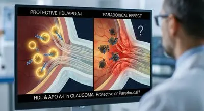
HDL cholesterol and apolipoprotein A-I: protective or paradoxical in glaucoma?
In general health, HDL carries cholesterol from tissues back to the liver and has anti-inflammatory and antioxidant actions. For example, HDL...
Deep research and expert guides on maintaining your visual health.
In general health, HDL carries cholesterol from tissues back to the liver and has anti-inflammatory and antioxidant actions. For example, HDL...

However, combining many compounds also has pitfalls. Overlapping mechanisms can lead to diminishing returns. The so-called “antioxidant paradox”...
Visual field loss from conditions like glaucoma can go unnoticed. Start a free trial and screen for potential blind spots in minutes.
Optical coherence tomography angiography is an imaging technique that shows blood flow in the tiny vessels of the eye without needing injected dye. It works by taking many rapid cross-sectional scans and detecting subtle differences caused by moving blood cells, then building a map of the vascular networks. The result is a detailed picture of the superficial and deeper capillary layers in the retina and around the optic nerve. Because it does not require contrast dye, the test is faster and safer for many patients and can be repeated often to track changes. Clinicians use it to spot early signs of diseases that affect blood vessels in the eye, such as diabetic retinopathy, age-related macular degeneration, and glaucoma. Seeing the microvasculature helps doctors understand disease activity, decide on treatments, and monitor how well therapies are working. The images can reveal vessel loss, abnormal new vessel growth, and regions with poor blood flow that might not be obvious on other scans. However, the technique can produce artifacts from eye movement, blinking, or poor image quality, so results must be interpreted carefully. It also has limited ability to show leakage, which some other tests can demonstrate more directly. Despite its limitations, this technology has become an important, noninvasive tool for eye care and research.