Glaucoma and Intraocular Pressure: The Role of the Outflow Pathway
Glaucoma is a group of eye diseases that can cause vision loss by damaging the optic nerve. High intraocular pressure (IOP) – the fluid pressure inside the eye – is a major risk factor for glaucoma. Normally, fluid made inside the eye (aqueous humor) drains out through the trabecular meshwork (TM) and Schlemm’s canal (SC) at the front (anterior segment) of the eye. When this drainage becomes blocked or restricted, fluid builds up and pressure rises. In many forms of glaucoma, doctors see extra extracellular matrix (ECM) – the network of proteins and structural components outside cells – accumulating in the TM and SC. This thickened ECM acts like extra “debris” in the drainage channels, making it harder for fluid to exit. Over time, this increased resistance to outflow causes IOP to climb, which can damage the optic nerve and lead to loss of vision (pmc.ncbi.nlm.nih.gov).
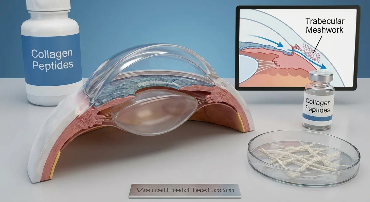
In a healthy eye, the TM and SC work together like a plumbing system. The TM is a spongy, porous tissue lined by endothelial cells, and it sits just in front of Schlemm’s canal (see illustration below). Fluid flows out through pores in the TM and the inner wall of SC into a blood vessel-like channel (Schlemm’s canal) to exit the eye. Research shows that most of the normal resistance to fluid outflow comes from the juxtacanalicular TM region (the deepest part of the TM right next to Schlemm’s canal) and from the basement membrane of the inner wall of Schlemm’s canal (pmc.ncbi.nlm.nih.gov). In glaucoma, the TM and SC basement membrane become abnormally thick and stiff, filled with extra collagen, fibronectin, and other ECM proteins (pmc.ncbi.nlm.nih.gov). These changes make the outflow paths narrower, like clogging a drain, which raises IOP.
(pmc.ncbi.nlm.nih.gov) Figure: Fluid drains from the anterior chamber through the trabecular meshwork (TM) and inner wall of Schlemm’s canal (SC). Most outflow resistance – the “bottleneck” – is in the deep TM and inner SC wall (pmc.ncbi.nlm.nih.gov).
ECM Remodeling in the Trabecular Meshwork
In glaucoma, the TM cells (which behave somewhat like fibroblasts, the connective tissue cells found in skin and other organs) produce extra matrix and fail to break it down properly. The balance of matrix metalloproteinases (MMPs) and their inhibitors (TIMPs) shifts so that more ECM is deposited. At the same time, powerful signaling proteins are at play. A key culprit is transforming growth factor-beta (TGF-β). Both TGF-β1 and TGF-β2 are growth factors that normally help tissues heal and regulate ECM, but in glaucoma the level of TGF-β2 in the eye’s fluid (aqueous humor) is abnormally high (pmc.ncbi.nlm.nih.gov). Experiments show that TGF-β2 stimulates TM cells to make more collagen and other matrix molecules, and to cross-link the fibers (via lysyl oxidase enzymes) (pmc.ncbi.nlm.nih.gov). This creates a fibrotic phenotype (like a scar) where the TM is filled with solid matrix and becomes stiffer.
Another important factor is connective tissue growth factor (CTGF), also called CCN2. CTGF is induced by TGF-β and further promotes matrix production. Studies in human TM cells found that TGF-β increases CTGF, and that adding CTGF to TM cells causes them to deposit much more ECM (pmc.ncbi.nlm.nih.gov). Blocking CTGF (for example with an antibody) prevents these fibrosis-like changes (pmc.ncbi.nlm.nih.gov). In glaucoma patients, CTGF levels are elevated in the TM, and research suggests CTGF may create a positive feedback loop: as collagen builds up, CTGF drives even more collagen to be made (pmc.ncbi.nlm.nih.gov). In other words, thinner, normal TM becomes thicker and scarred.
Integrins are surface receptors that let TM cells sense and bind to the ECM around them. When integrins bind to collagens or fibronectin, they send signals inside the cell that affect its shape, survival, and function. In the TM and Schlemm’s canal cells, many integrins connect to ECM proteins like collagen and laminin (pmc.ncbi.nlm.nih.gov). This “outside-in” signaling can, for example, activate enzymes like FAK (focal adhesion kinase) that influence the actin cytoskeleton. Abnormal ECM (like extra fibronectin or collagen) can therefore trigger inside-out signals too. For instance, when fibronectin is high in glaucoma, it may bind to RGD-recognizing integrins on TM cells, altering their behavior (pmc.ncbi.nlm.nih.gov). However, how collagen fragments or peptides might directly affect integrins in eye cells specifically is still being studied.
Overall, the TM and Schlemm’s canal become more fibrotic in glaucoma due to a combination of excess ECM, increased cross-linking, and profibrotic signals (TGF-β, CTGF, cytokines) (pmc.ncbi.nlm.nih.gov) (pmc.ncbi.nlm.nih.gov). This fibrotic remodeling raises outflow resistance and IOP. (For more details on TM pathophysiology, see reviews by Vranka et al. and others (pmc.ncbi.nlm.nih.gov) (pmc.ncbi.nlm.nih.gov).)
Collagen Peptides: Effects on Fibroblasts and ECM
Collagen peptides are short chains of amino acids (small protein fragments) derived from collagen. They are commonly taken as dietary supplements for skin, joint, or bone health. In the lab, scientists have tested collagen peptides on various cell types (especially skin fibroblasts) to see what they do at the molecular level. Recent studies suggest that collagen peptides can stimulate fibroblasts and influence key pathways like integrins, TGF-β, CTGF, and MMPs. While data on ocular cells is limited, findings from skin and other tissues provide clues.
-
Fibroblast proliferation and matrix production. Multiple studies have found that collagen peptides can make skin fibroblasts multiply and produce more collagen. For example, Brandão-Rangel et al. (2022) showed that adding collagen peptides to human dermal fibroblasts caused a significant increase in cell proliferation and in the expression of pro-collagen type I (the main collagen of skin) (pmc.ncbi.nlm.nih.gov). Similarly, another in vitro study found that collagen peptides at moderate concentrations boosted the genes for collagen type I (COL1A1), elastin (ELN), and proteoglycan versican (VCAN) in dermal fibroblasts (pmc.ncbi.nlm.nih.gov). In both cases, fibroblasts made more of the building blocks of the connective tissue matrix. A systematic review of studies on hydrolyzed collagen reported that doses of about 50–500 µg/mL of collagen peptides are enough to stimulate fibroblast activity and collagen synthesis in human cells (pmc.ncbi.nlm.nih.gov). In short, collagen peptides appear to help rebuild and strengthen the extracellular scaffolding by prompting fibroblasts to grow and make more matrix.
-
Anti-inflammatory effects and TGF-β. Surprisingly, collagen peptides also have anti-inflammatory actions. In the Brandão-Rangel study, collagen peptides not only spurred collagen production but also suppressed inflammatory markers. When skin cells were exposed to a bacterial toxin (LPS), adding collagen peptides greatly lowered the induced levels of cytokines IL-6, IL-8, TNF-α and others (pmc.ncbi.nlm.nih.gov). At the same time, the peptides raised the levels of TGF-β (and VEGF) in the fibroblasts (pmc.ncbi.nlm.nih.gov). In other words, collagen peptides acted like a signal to calm inflammation and shift cells into a growth/repair mode. Because TGF-β is both anti-inflammatory and pro-fibrotic, this could be a double-edged sword: more TGF-β may help healing, but it could also drive fibrosis if unchecked. Indeed, in the same study the highest dose of collagen peptides (10 mg/mL) was needed to upregulate pro-collagen and TGF-β (pmc.ncbi.nlm.nih.gov). Another report in skin cells found that certain collagen-derived dipeptides (like ile-hydroxyproline) activated the TGF-β/Smad pathway, promoting collagen synthesis (documentsdelivered.com). Thus, collagen peptides can engage the very pathways (TGF-β signaling, Smad) that normally control ECM production.
-
Integrin signaling. Collagen is a natural ligand for certain integrins (notably α2β1 integrin binds collagen). Recent work in skin models shows that collagen peptides can increase the expression of collagen-binding integrins and activate associated signals. Mistry et al. (2024) found that porcine collagen peptides applied to skin cells significantly raised integrin α2β1 levels and triggered downstream signaling via ERK and FAK pathways (eprints.ncl.ac.uk). (These pathways normally respond to the cell binding to the ECM.) In those experiments, blocking the β1 integrin subunit prevented the collagen peptide effects in keratinocytes, although fibroblasts still responded, suggesting multiple routes of activation (eprints.ncl.ac.uk). The take-home is that collagen peptides can “prime” cells to sense and adhere to collagen. In a trabecular meshwork context, integrin α2β1 is present and mediates collagen binding (pmc.ncbi.nlm.nih.gov). If collagen peptides similarly boost α2β1 on TM cells, that might increase adhesion to the surrounding matrix, potentially influencing outflow.
-
MMPs and TIMPs (matrix remodeling). The matrix metalloproteinases (MMPs) and their inhibitors (TIMPs) control how fast the ECM is broken down. Excess MMP activity leads to ECM degradation, while too much TIMP can preserve ECM and lead to fibrosis. In skin models, collagen peptides seem to reduce the expression of some MMPs. Liu et al. (2019) showed that certain collagen peptide metabolites in culture suppressed activation of AP-1, lowered the protein levels of MMP-1 and MMP-3, and thereby depressed collagen degradation (documentsdelivered.com). Another study noted that increased collagen accumulation in fibroblasts was linked not only to more collagen synthesis but also to less degradation, with collagen peptides inhibiting MMP-1 and MMP-2 activity (pmc.ncbi.nlm.nih.gov). In summary, collagen peptides tend to tip the balance toward matrix buildup by breaking down less collagen. If TIMPs were also affected, research is limited, but one could imagine peptides might also influence TIMP production or activity as part of regulating matrix.
Comparing Eye Cells to Skin, Tendon, and Lung Fibrosis Models
How do these collagen-peptide effects compare between eye cells and other fibrotic tissues? In all these tissues, TGF-β and CTGF are known drivers of fibrosis. For example, in the skin and lung, chronic injury triggers persistent TGF-β signaling, activating fibroblasts (or myofibroblasts) to make excess collagen and ECM (as reviewed by Grafanaki et al.) (pmc.ncbi.nlm.nih.gov). In lung diseases like idiopathic pulmonary fibrosis, collagen-producing alveolar cells and fibroblasts ramp up collagen I and III production under TGF-β influence. In tendon disorders, TGF-β and CTGF similarly jumpstart fibrotic matrix deposition (pmc.ncbi.nlm.nih.gov). In these systems, collagen fragments and crosslinks often accumulate, and the stiffness of tissue increases.
There are no direct studies of dietary collagen peptides on trabecular meshwork cells to date. But we can draw parallels: Skin dermal fibroblasts and TM cells are both mesenchymal cells that respond to TGF-β. In both, TGF-β induces collagen, fibronectin, and proteoglycan genes. CTGF is likewise a common mediator. For instance, a corneal fibroblast study (an eye cell type related to TM cells) showed that TGF-β induced much more CTGF+collagen production when cells were grown on collagen matrix (pmc.ncbi.nlm.nih.gov). That suggests collagen-rich environments prime eye fibroblasts for fibrosis, not unlike skin. Similarly, tendon fibroblasts secrete CTGF and collagen under TGF-β signaling (pmc.ncbi.nlm.nih.gov), and lung fibroblasts do the same (Pulmonary fibrosis is treated by anti-TGF-β in experimental models).
In short, fibrotic injury pathways are conserved: TGF-β and CTGF upregulate matrix genes and downregulate MMPs (often via AP-1 pathway), while integrin signaling can further activate TGF-β and matrix production (pmc.ncbi.nlm.nih.gov) (pmc.ncbi.nlm.nih.gov). Collagen peptides in skin and other models tend to reinforce these pro-healing/pro-fibrotic signals (more TGF-β, more collagen, less MMPs (pmc.ncbi.nlm.nih.gov) (pmc.ncbi.nlm.nih.gov)). This suggests that what collagen peptides do in the skin (promote ECM build-up) might be similar in other connective tissues. However, the eye’s TM/SC has unique fluid dynamics, so we must be cautious in extrapolating.
Could Collagen Peptides Raise or Lower IOP?
Given this information, could taking collagen peptide supplements affect eye pressure? We can sketch two opposing hypotheses:
-
Increase outflow resistance (raise IOP): Collagen peptides clearly drive collagen production and fibroblast proliferation in other tissues. If TM cells responded similarly, they might produce extra ECM in the meshwork, effectively clogging it further. Peptide-induced TGF-β signaling and CTGF release (as seen in skin cells) could worsen the fibrotic TM changes already present in glaucoma. Also, by suppressing MMPs (as some studies show), collagen peptides might reduce matrix turnover, letting ECM accumulate. In skin wound healing contexts, CTGF causes scarring; by analogy, more CTGF in the TM could increase the “scarring” of the outflow pathway. Therefore, one plausible outcome is that collagen supplements could increase outflow resistance by thickening the TM/SC matrix, thereby raising IOP. This could be especially relevant for people predisposed to glaucoma or ocular hypertension.
-
Decrease outflow resistance (lower IOP): On the other hand, collagen peptides have anti-inflammatory effects and might promote healthy tissue remodeling. If collagen peptides helped TM cells maintain a normal ECM (for example, by enhancing regeneration of a properly organized matrix), they might improve outflow. Increasing integrin signaling (α2β1) could potentially help TM cells reorganize collagen fibrils in a way that eases flow (since cell-matrix adhesion and cytoskeletal tension can affect pore size). Moreover, by boosting VEGF and healing pathways, there is a theoretical chance that peptides enhance TM repair and fluid clearance. Finally, collagen peptides sometimes carry small fragments that can bind and neutralize TGF-β or CTGF (some research on endostatin peptides shows anti-fibrotic effects). In tendon models, for instance, controlled delivery of CTGF (with collagen) improved healing without excessive scarring (pmc.ncbi.nlm.nih.gov). If collagen supplements in the eye led to more normal TM structure or counteracted pathological fibrosis, they might decrease resistance.
Realistically, the net effect of collagen peptides on the eye is not known and could be complex. No clinical trial has tested collagen supplements for eye pressure. The evidence from other tissues leans toward a pro-fibrotic action (more matrix, more collagen), which would suggest collagen peptides might tend to raise IOP in susceptible eyes. But because peptides also modulate inflammation and growth signals, the outcome could vary. It might depend on dose, peptide size, and individual eye condition.
In conclusion, the scientific evidence shows that collagen-derived peptides influence fibroblasts to proliferate and produce more matrix (via integrin, TGF-β, CTGF signaling) while at the same time reducing inflammatory MMP activity. In glaucoma, where TM fibrosis already elevates IOP, such actions could exacerbate outflow blockage. However, a balanced view must note the anti-inflammatory and repair aspects of these peptides as well. Until direct studies are done in ocular cells or patients, we can only hypothesize. For now, anyone concerned about glaucoma should use caution and consult an eye doctor before taking supplements intended to affect connective tissue, as they might theoretically impact eye pressure.