Uric Acid: Antioxidant Versus Pro-oxidant in Glaucoma
Introduction: Glaucoma is a progressive optic nerve disease in which oxidative stress and vascular dysfunction are thought to contribute to retinal ganglion cell loss. Uric acid (UA), the end product of purine metabolism, is of growing interest because it circulates at high levels in humans and has complex redox effects. In blood, UA is a potent antioxidant (scavenging radicals in plasma) (pmc.ncbi.nlm.nih.gov). However, inside cells or as crystals, UA can promote inflammation and oxidative stress. Recent studies on glaucoma have reported conflicting findings: some suggest higher serum UA correlates with worse glaucoma, whereas others suggest the opposite. We review these data, and explore how UA interrelates with autonomic (heart rate variability), endothelial, and kidney factors. We also consider common gout medications (which lower UA) and their potential eye effects. Personalized analyses by sex, kidney health, and metabolic status are warranted. Finally, we outline simple urine/blood tests (e.g. serum UA and kidney panels) a person can obtain and interpret to gauge risk factors.
Uric Acid and Glaucoma: Conflicting Clinical Evidence
Studies of serum UA in glaucoma patients have yielded mixed results. A 2023 systematic review and meta-analysis (1,221 glaucoma patients vs. 1,342 controls) found that mean serum UA was slightly higher in glaucoma cases by about 0.13 mg/dL – not statistically significant (pmc.ncbi.nlm.nih.gov). In that review, three of six studies actually found lower UA in glaucoma patients (suggesting a protective antioxidant role), while three found higher UA in glaucoma (suggesting UA as a risk marker) (pmc.ncbi.nlm.nih.gov). For example, one report in primary open-angle glaucoma (POAG) noted significantly lower UA levels in patients than in controls, with the lowest UA in those with the most severe visual field loss (pmc.ncbi.nlm.nih.gov). That study even showed the UA–glaucoma trend was strongest in men (pmc.ncbi.nlm.nih.gov). In contrast, other studies found higher UA in glaucoma. Elisaf et al. reported that POAG subjects (without diabetes) had higher UA (≈6.2 mg/dL) than age-matched non-glaucoma controls (≈5.0 mg/dL, P=0.006) (www.sciencedirect.com). Another study found normal-tension glaucoma (NTG) patients had higher UA than controls (5.8 vs. 4.9 mg/dL) (www.sciencedirect.com). These discrepancies may relate to glaucoma subtypes (e.g. NTG vs. angle-closure) or population differences. For instance, several Chinese cohorts found lower UA in acute angle-closure glaucoma and slower glaucoma progression in those with higher UA (www.sciencedirect.com) (www.sciencedirect.com).
In summary, some data suggest a protective role (lower UA in worse glaucoma) (pmc.ncbi.nlm.nih.gov), while others imply UA is a risk factor (higher UA in glaucoma cases) (www.sciencedirect.com) (www.sciencedirect.com). This mirror-opposite pattern underlies the “antioxidant vs. pro-oxidant” paradox. Because human studies differ by design, glaucoma definition, and populations, consensus is lacking. Physicians and patients should note that evidence is inconclusive: UA could reflect either inadequate antioxidant defense (if low) or systemic metabolic stress (if high).
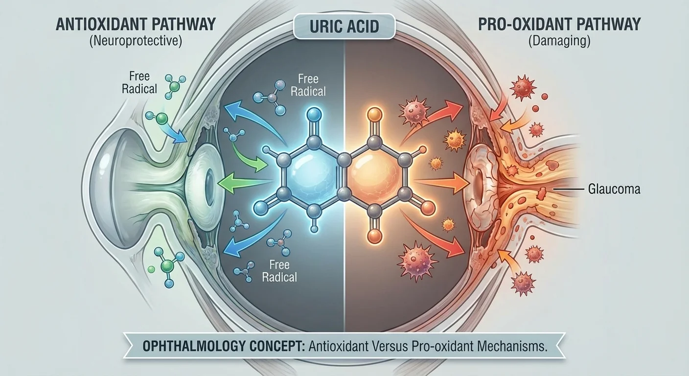
Biochemistry of Uric Acid: Antioxidant vs. Pro-oxidant
Biochemically, UA has a classic dual nature. In the blood, urate is actually one of the major antioxidants. For example, it can scavenge singlet oxygen, peroxyl and hydroxyl radicals (pmc.ncbi.nlm.nih.gov). Evolutionarily, humans lost the enzyme uricase so that circulating UA (~300–400 μM) significantly contributes to plasma antioxidant capacity. In the central nervous system, this may protect neurons: animal studies show UA administration protects hippocampal neurons from oxidative injury (pmc.ncbi.nlm.nih.gov). Thus acute rises in UA (e.g. after ischemia) have sometimes been neuroprotective.
Yet paradoxically, chronically high UA is epidemiologically linked to conditions of oxidative stress: obesity, hypertension, metabolic syndrome, and kidney disease (pmc.ncbi.nlm.nih.gov). How can a strong antioxidant correlate with oxidative diseases? The resolution is that UA’s effects depend on context. Inside cells or when interacting with other molecules, UA can become a pro-oxidant. For example, urate can react with peroxynitrite to form radicals that oxidize lipids (including LDL) and damage membranes (pmc.ncbi.nlm.nih.gov) (pmc.ncbi.nlm.nih.gov). In endothelial and adipose cells, high UA triggers oxidative stress pathways (pmc.ncbi.nlm.nih.gov). Indeed, experimental studies show that adding UA to cultured adipocytes or vascular cells increases intracellular reactive oxygen and entry into inflammatory states (pmc.ncbi.nlm.nih.gov). In summary: UA is antioxidative in the bloodstream but can generate ROS and inflammation within tissues (pmc.ncbi.nlm.nih.gov) (pmc.ncbi.nlm.nih.gov).
In the eye, UA can form needle-like monosodium urate crystals that induce inflammation. Case reports describe “ocular tophaceous gout,” where GH deposits in ocular structures trigger uveitis and high intraocular pressure (IOP) (pmc.ncbi.nlm.nih.gov). In animal models, blocking UA production reduced ocular inflammation: for example, a sustained-release ocular film of febuxostat (a xanthine oxidase inhibitor) lowered IOP and inflammation in rabbits with experimentally induced ocular gout (pmc.ncbi.nlm.nih.gov). Although rare, these findings highlight that UA-driven inflammation can occur in ocular tissues.
More broadly, the paradox suggests that moderate UA may be beneficial (antioxidant), but excessive or crystallizing UA is harmful (pro-oxidant). In glaucoma research, this means both interpretations are plausible: low UA might mean lack of a needed free-radical scavenger, while high UA could reflect vascular/renal comorbidity that exacerbates optic nerve damage.
Heart Rate Variability, Autonomic Dysfunction, and Uric Acid
Beyond direct oxidative effects, UA may link to glaucoma through systemic autonomic and cardiovascular factors. Heart rate variability (HRV) is a noninvasive marker of autonomic balance. Low HRV (indicating sympathetic overactivity) has been associated with glaucoma progression in several studies. Separately, elevated UA is tied to cardiac arrhythmias and autonomic dysregulation. In a Korean population survey of ~10,800 adults, hyperuricemia (UA ≥7 mg/dL in men, ≥6 in women) nearly tripled the odds of heart rate irregularity (overall arrhythmia risk) (pmc.ncbi.nlm.nih.gov). This hyperuricemia–arrhythmia link persisted after adjusting for age, sex, hypertension, diabetes, CKD, and smoking (pmc.ncbi.nlm.nih.gov). In chronic kidney disease patients on dialysis, those with hyperuricemia had smaller increases in HRV after dialysis, again reflecting impaired autonomic recovery (pmc.ncbi.nlm.nih.gov).
Because glaucoma (especially normal-tension types) has also been associated with autonomic dysfunction, it is plausible that high UA could worsen glaucoma indirectly by affecting blood pressure and heart rate patterns. For example, if hyperuricemia drives sympathetic tone, ocular perfusion could be compromised. Data directly linking UA to HRV in glaucoma is still emerging, but the broader pattern suggests UA and ANS function are connected.
Endothelial Dysfunction and Uric Acid
Endothelial function (ability of blood vessels to dilate via nitric oxide) is crucial for healthy ocular blood flow. Chronic hyperuricemia has been shown to impair endothelial function systemically. In a large Japanese cohort study (n=1000), higher serum UA was strongly associated with impaired flow-mediated dilation (FMD), a measure of endothelial health (pmc.ncbi.nlm.nih.gov). The association was especially evident in women and in individuals not on antihypertensive drugs (pmc.ncbi.nlm.nih.gov). In other words, people with higher UA had stiffer vessels and reduced NO-mediated dilation. Even in healthy adults, UA accumulation is thought to reduce NO and increase pro-inflammatory signals. By analogy, compromised endothelial function could diminish optic nerve head perfusion and resilience.
Conversely, some smaller studies found no association between UA and endothelial markers in healthy subjects, so the effect may need existing metabolic stress. Nevertheless, given that many glaucoma patients (especially with normal-tension glaucoma or exfoliation syndrome) have coexisting vascular risk factors, hyperuricemia could tip the balance toward dysfunction. This underscores that UA’s vascular impact – particularly on microcirculation – might influence glaucoma risk or progression.
Metabolic Syndrome, Kidney Function, and Uric Acid
High uric acid often occurs in metabolic syndrome and precedes or predicts diabetes. Insulin resistance itself can raise UA by reducing renal excretion. One review noted that even in people without overt gout, higher UA levels were independently linked to features of metabolic syndrome and prediabetes (pubmed.ncbi.nlm.nih.gov). Hyperinsulinemia reduces kidney urate excretion, creating a vicious cycle: more UA impairs endothelial NO and worsens insulin resistance (pubmed.ncbi.nlm.nih.gov). In other words, UA and metabolic factors (obesity, hypertension, lipids, glucose) fuel each other. Since metabolic syndrome is associated with glaucoma, UA may be one shared element. Stratified analyses should therefore adjust for obesity, blood sugar, and lipid levels when assessing UA’s effect on glaucoma risk.
Chronic kidney disease (CKD) is another key comorbidity. The kidney normally clears most UA, so impaired renal function causes UA to rise. UA itself may also contribute to CKD progression. In fact, “elevated serum uric acid is a marker for decreased renal function” and may play a causal role in CKD and hypertension (pmc.ncbi.nlm.nih.gov). Large population studies show higher UA predicts faster kidney decline and higher risk of end-stage renal disease. Importantly, several epidemiologic studies have found that glaucoma patients have a markedly higher incidence of CKD. For example, a Korean national cohort (>478,000 subjects) found primary open-angle glaucoma increased the hazard of developing CKD by over 7-fold (HR ≈7.6) (www.sciencedirect.com). Newly diagnosed glaucoma patients also had much higher acute kidney injury and kidney failure rates than non-glaucoma patients (www.sciencedirect.com). This co-occurrence suggests shared pathophysiology – possibly via microvascular damage or pressure regulation – and implicates UA as a common link. Indeed, UA is called a “key player in kidney disease pathophysiology” and a marker of CKD (www.sciencedirect.com) (pmc.ncbi.nlm.nih.gov). In summary, renal health mediates UA levels and glaucoma risk: poor kidneys raise UA and also can independently impact intraocular and cerebrovascular dynamics.
Gout Therapies and Ocular Effects
Given the interplay of UA with glaucoma-related factors, one might ask whether urate-lowering therapies influence eye health. Common gout medications include xanthine oxidase inhibitors (allopurinol, febuxostat) and anti-inflammatories (colchicine, NSAIDs).
-
Allopurinol: A decades-old XO inhibitor, allopurinol can rarely cause ocular side effects secondary to hypersensitivity (e.g. Stevens–Johnson syndrome with conjunctivitis), though these are very rare. Interestingly, in a comprehensive review of systemic drugs, allopurinol was listed as having an association with cataract formation (pmc.ncbi.nlm.nih.gov). The evidence for this is not strongly causative, but patients on long-term allopurinol might mention it during ophthalmic exams. On the other hand, animal models suggest allopurinol can protect the retina: in diabetic rats, allopurinol reduced retinal inflammation and vascular leakage by lowering UA and oxidative stress (pmc.ncbi.nlm.nih.gov). There is also speculation that protecting retinal ganglion cells with antioxidant therapy might be advantageous, though no human trials have tested allopurinol for glaucoma specifically.
-
Febuxostat: A newer XO inhibitor, febuxostat has a different safety profile. A large population study (Korea, n>200,000) found no difference in the risk of retinal microvascular complications between new users of febuxostat vs. allopurinol over ~200 days follow-up (www.nature.com). This suggests neither drug uniquely predisposes to (or protects from) ischemic retinal disease in diabetics or gout. Intriguingly, a recent experimental approach delivered febuxostat in an eye-drop film and achieved sustained lowering of IOP and intraocular inflammation in a gouty eye model (pmc.ncbi.nlm.nih.gov). That suggests urate-lowering locally might ease crystalline inflammation, but clinical relevance is uncertain.
-
Colchicine and others: Colchicine is used for gout flares; its ocular side effects are not well-documented. We did not identify specific glaucoma risks from colchicine. General anti-inflammatory gout treatments (steroids, NSAIDs) are known to raise IOP or cause cataracts, but these are systemic steroid side-effects rather than specific urate effects.
In practice, glaucoma patients with gout should continue essential gout therapy. There is no clear evidence that allopurinol or febuxostat worsens glaucoma or that they can halt it. However, since high UA may contribute to oxidative/metabolic harm, some clinicians argue it is prudent to optimize urate levels within normal range. Any patient on gout drugs should have routine eye examinations as part of general health monitoring.
Sex Differences and Stratified Analyses
Sex (biological gender) influences UA and vascular risk. Men naturally have higher normal UA levels than premenopausal women. In many studies of vascular disease, elevated UA tends to be a stronger risk marker in women (pmc.ncbi.nlm.nih.gov) (pmc.ncbi.nlm.nih.gov). For example, the Japanese endothelial study found the UA–endothelial dysfunction link was more pronounced in females (pmc.ncbi.nlm.nih.gov). Accordingly, analyses of UA in glaucoma should stratify by sex. It’s possible that the same UA level might represent higher relative risk in women.
Metabolic factors also influence UA differently by sex. Women with metabolic syndrome may have higher relative UA increases. Age is relevant too: UA rises after menopause.
Renal function stratification is also important. Since CKD drastically alters UA, studies must adjust or stratify by kidney health (eGFR or albuminuria). A mild UA elevation in someone with CKD may be less concerning (since GFR is low) than the same UA in a healthy kidney. Conversely, high UA in a person with normal GFR suggests overproduction and might signal other risk.
Finally, metabolic syndrome (obesity, diabetes, hypertension, dyslipidemia) underlies both UA elevation and glaucoma risk. Future research should analyze subgroups: e.g. glaucoma patients with vs. without metabolic syndrome, or by HbA1c levels, to see if UA’s effect on glaucoma is modified by these factors (pubmed.ncbi.nlm.nih.gov) (pmc.ncbi.nlm.nih.gov).
Accessible Tests and How to Interpret Them
Individuals interested in monitoring UA-related risk can request several routine lab tests. These can be ordered by a physician or through direct-access lab services. Key tests include:
-
Serum Uric Acid: A simple blood test. Normal ranges are roughly 4.0–8.5 mg/dL in adult men and 2.7–7.3 mg/dL in adult women (emedicine.medscape.com). (Values vary slightly by lab.) A reading above the upper range is termed hyperuricemia. For example, a man with 9 mg/dL or a woman with 7.5 mg/dL would be above normal. High values suggest increased risk of gout and can reflect reduced kidney clearance or high purine turnover (emedicine.medscape.com). Extremely low UA (e.g. <2 mg/dL) is uncommon and usually of no concern outside rare genetic conditions. In general:
- UA in upper-normal range (e.g. 6–7 mg/dL) can be seen in healthy people, but if accompanied by other risk factors (obesity, kidney disease, hypertension) it may warrant lifestyle modification and follow-up.
- UA above normal should prompt evaluation for gout symptoms and kidney function (see below).
-
Basic Metabolic Panel (BMP) / Kidney Function: This blood test includes serum creatinine and estimated glomerular filtration rate (eGFR). Normal creatinine is roughly 0.6–1.2 mg/dL (higher end in men, lower end in women depending on muscle mass) (emedicine.medscape.com). More importantly, labs automatically calculate eGFR (which adjusts for age, sex, race). An eGFR > 60 mL/min/1.73m² is considered acceptable kidney function; values persistently below 60 indicate chronic kidney disease (CKD). If eGFR is low, the kidneys’ ability to clear UA is reduced, so an elevated UA in that setting may be explained by CKD. Clinically, if eGFR ≥90 you have normal function; 60–89 is mildly reduced; under 60 suggests moderate-to-severe CKD.
-
Urinalysis / Urine Albumin: A urine test can detect microalbuminuria, an early sign of kidney microvascular damage. Although not directly about UA, it signals renal endothelial health. Normal urinary albumin-to-creatinine ratio (ACR) is <17 mg/g in men and <25 mg/g in women (emedicine.medscape.com). An ACR above 30 mg/g (30–300 mg/g) defines microalbuminuria (emedicine.medscape.com). Elevated urine albumin suggests kidney involvement (e.g. hypertension or early diabetic kidney disease). If such tests are high-normal or elevated, the same UA value should be interpreted cautiously – even normal-range UA might be excessive if the kidneys are partly impaired.
-
Blood Glucose and Lipids: Since UA is tightly linked to metabolic syndrome, checking fasting glucose, A1C, and a lipid panel is wise. Elevated glucose or A1C (>5.6%) indicates impaired glucose metabolism; high triglycerides or low HDL are also metabolic signs. These tests are widely available in direct-access labs. Interpretation follows usual guidelines (e.g. FPG & A1C for diabetes risk, LDL for cholesterol management). Even prediabetes raises concern for metabolic syndrome, which often accompanies hyperuricemia and vascular risk.
-
Others: Blood pressure monitoring, while not a blood test, is important – both hypertension and UA synergistically harm vessels (pmc.ncbi.nlm.nih.gov). Home or pharmacy BP checks can be included in risk evaluation. (Devices like Fitbit do HRV, but that is far less standardized for self-use.)
All these tests can often be ordered by a primary care physician or via consumer labs (Quest/ LabCorp direct access, etc.). Results should be discussed with a doctor. As a rule of thumb:
- High UA or low eGFR warrants further evaluation. Lifestyle measures (reducing red meat, alcohol; controlling weight) can lower UA. Medications (like allopurinol) are prescribed when gout flares or very high UA occur.
- Borderline UA with normal sugars and lipids is usually watched.
- Microalbuminuria or reduced eGFR flags kidney protection.
- Any abnormal should trigger a holistic approach (diet, exercise, blood pressure, glycemic control), since UA is one piece of metabolic and vascular health.
Regular monitoring (e.g. annual checks) can track changes. Importantly, results near thresholds (e.g. UA 7.2 mg/dL in a woman or 8.5 in a man) may prompt preventive steps even if technically “normal.”
Conclusion
In summary, serum urate occupies a complex place in glaucoma biology. It is theoretically protective as a powerful antioxidant, yet epidemiologically suspect as a marker of vascular and metabolic stress. Human data on glaucoma are inconclusive – studies show both higher and lower UA in patients. The dual role makes sense biochemically: UA combats free radicals in plasma (pmc.ncbi.nlm.nih.gov) but can promote oxidative injury in tissues (pmc.ncbi.nlm.nih.gov). Its impact on heart rate variability and endothelial function suggests that systemic hyperuricemia could amplify glaucomatous damage via blood flow dysregulation (pmc.ncbi.nlm.nih.gov) (pmc.ncbi.nlm.nih.gov). Renal dysfunction ties the knot tighter, since poor kidney clearance raises UA and separately affects eye health (www.sciencedirect.com) (pmc.ncbi.nlm.nih.gov). Given the ambiguities, future glaucoma studies should stratify by sex, renal status, and metabolic syndrome.
For clinicians and patients, the practical takeaway is that UA is a modifiable risk factor. While we do not recommend UA-lowering specifically to treat glaucoma, controlling high urate (through diet or medication) benefits overall vascular health and prevents gout. Patients concerned about glaucoma risk may consider checking their uric acid and related labs and addressing anything abnormal. Continued research will clarify whether chronically optimal UA (neither too high nor too low) protects vision in susceptible individuals.