Introduction
Imagine running a race and later noticing rainbow-like halos around lights, a brief blurring of vision, or a dull ache in your eyes. For most people this would sound alarming – yet such symptoms are often painless and temporary in a hidden condition called Pigment Dispersion Syndrome (PDS). PDS tends to strike healthy, young, myopic (nearsighted) adults, especially men in their 20s–40s. These individuals are often active and otherwise feel fine. Yet their eyes carry a concealed risk: tiny dust-like pigment granules rubbing off the back of the iris and clogging the eye’s drainage system. Over time this can raise eye pressure and lead to Pigmentary Glaucoma (PG), a form of glaucomatous nerve damage. (pmc.ncbi.nlm.nih.gov) (pmc.ncbi.nlm.nih.gov).
This article dives deep into what PDS is, how it can progress, and what it means for athletes and fitness enthusiasts. We’ll explain the eye’s anatomy in plain language, give clear examples from real studies, and outline the latest evidence (up to 2026) on exercise and PDS. You’ll learn why PDS often feels “silent,” how doctors spot it, and – most importantly – how people with PDS/PG can safely stay active.
What is Pigment Dispersion Syndrome?
The Eye’s Pigment “Dust”
The iris (the colored part of the eye) has a back layer called the iris pigment epithelium, rich in dark melanin granules. In a normal eye, these pigment cells stay put. In PDS, however, the iris is slightly bowed backward and rubs repeatedly against the lens zonules (tiny fibers holding the lens). This rubbing releases pigment particles into the eye’s fluid (the aqueous humor) (pmc.ncbi.nlm.nih.gov) (doctorlib.org).
Reverse Pupillary Block: One big factor is a “reverse pupillary block” mechanism. Normally fluid flows from behind the iris, through the pupil, to the front of the eye and drains out. In PDS eyes, however, the iris bows back like a sail (often in myopic eyes with deep anterior chambers (pmc.ncbi.nlm.nih.gov) (doctorlib.org)). This can create a one-way “ball-valve” effect: fluid struggles to flow forward, causing pressure behind the iris and pushing the iris even more backward. This iris concavity greatly increases rubbing between the iris pigment and the underlying structures (pmc.ncbi.nlm.nih.gov) (bjo.bmj.com). The result is repeated bouts of pigment shedding – think of it like dust collecting on a car’s windshield wipers.
Where Do the Pigments Go?
Once free in the eye’s aqueous fluid, the pigment granules float around and deposit on various tissues in the front of the eye (pmc.ncbi.nlm.nih.gov). The most important deposit is in the trabecular meshwork (TM) – the eye’s drainage grate. Pigment accumulates in the meshwork, clogging it and reducing fluid outflow (pmc.ncbi.nlm.nih.gov) (bjo.bmj.com). Over time this backs up fluid and raises intraocular pressure (IOP).
Other classic signs (often seen by doctors, not by patients) include:
- Krukenberg Spindle: A vertical spindle-shaped band of pigment on the central corneal endothelium (the inner lining of the clear cornea) (pmc.ncbi.nlm.nih.gov). Convection currents in the eye cause the pigment to line up like a spindle.
- Iris Trans-illumination Defects: The iris develops spoke-like, radial defects that look like little gaps in a wheel when light shines through (pmc.ncbi.nlm.nih.gov). These are where the iris pigment cells have been stripped away.
- Zentmayer (Scheie) Line: A line of pigment on the back surface of the lens equator (near the top/bottom of vision).
- Sampaolesi Line: Pigment just in front of the Schwalbe’s line (the edge of the drainage angle).
- Homogeneous Angle Pigmentation: On gonioscopic exam (special mirror view of the angle), the entire trabecular meshwork is stained darkly with pigment (pmc.ncbi.nlm.nih.gov) (bjo.bmj.com).
These findings – pigment showering the cornea, iris defects, and a heavily pigmented drainage angle – form the classic triad of PDS/PG (pmc.ncbi.nlm.nih.gov) (pmc.ncbi.nlm.nih.gov).
Analogy: Imagine your eye’s drainage as a sponge filter. Pigment granules are like fine sand tossing into the water you pour through. Over time, the sand clogs the sponge, slowing drainage (outflow) and causing pressure to build up behind the faucet.
If the outflow obstruction is significant and chronic, eye pressure rises (ocular hypertension). When this pressure damages the optic nerve (seen as thinning of nerve fibers and vision field loss), it becomes Pigmentary Glaucoma (PG) (pmc.ncbi.nlm.nih.gov) (bjo.bmj.com). In the disease spectrum, PDS is the early stage (pigment release and high pressure risk) and PG is the later stage (actual glaucoma damage) (pmc.ncbi.nlm.nih.gov).
PDS to Pigmentary Glaucoma: Risk and Progression
How Likely is PDS to Become Glaucoma?
Fortunately, most people with PDS do not immediately go blind. Estimates vary, but current evidence suggests only a subset progress to true glaucoma. In clinic-based studies, about 10–50% of PDS patients eventually develop PG (bjo.bmj.com) (pmc.ncbi.nlm.nih.gov). A recent 2026 review summarized one large observation: about 10% of PDS eyes converted to PG by 5 years, and 15% by 15 years (pmc.ncbi.nlm.nih.gov). Earlier reviews even cited up to 50%, but those older numbers likely come from biased samples (people already in eye clinics) (bjo.bmj.com) (pmc.ncbi.nlm.nih.gov). In the general population, progression is likely at the lower end of that range, roughly 10–20% over one or two decades (pmc.ncbi.nlm.nih.gov) (pmc.ncbi.nlm.nih.gov).
The key risk factors for progressing from PDS to glaucoma are well documented (pmc.ncbi.nlm.nih.gov):
- High Trabecular Pigment: Eyes with a very dark, crowded trabecular meshwork (seen on exam) are at greatest risk (the “filter is nearly plugged”).
- Elevated Pressure from the Start: Higher baseline IOP in a PDS eye means more stress on the nerve.
- Younger Age: Paradoxically, younger patients may have more vigorous pigment shedding, so PDS often appears in youth and can progress more quickly.
- Male Sex: Men with PDS convert more often than women (pmc.ncbi.nlm.nih.gov) (pmc.ncbi.nlm.nih.gov).
- Myopia (Nearsightedness): Moderate myopes have deeper anterior chambers and more iris-lens contact, predisposing to PDS and PG (pmc.ncbi.nlm.nih.gov) (bjo.bmj.com).
- Race: PG is much more common in Caucasians than in darker-pigmented eyes (bjo.bmj.com) (pmc.ncbi.nlm.nih.gov). (Many African-American or Asian patients do not show the iris transillumination defects because their eyes produce less visible pigment release, though the risk patterns are less well studied outside white populations (pmc.ncbi.nlm.nih.gov).)
- Family History: A family history suggests a genetic susceptibility.
- Visible Signs: Detecting a Krukenberg spindle or other pigment signs in both eyes raises the odds that glaucoma may follow (pmc.ncbi.nlm.nih.gov).
- Chronicity: A longstanding PDS (multiple years) increases odds, as pigment has more time to accumulate.
The European Glaucoma Society notes that overall PDS accounts for only about 1–1.5% of all glaucoma cases, underscoring that it’s a minority form of glaucoma (pmc.ncbi.nlm.nih.gov) (bjo.bmj.com). Nonetheless, for each PDS patient, vigilance is crucial. PG tends to affect a younger population (often diagnosed in the 30–50 year range (pmc.ncbi.nlm.nih.gov) (bjo.bmj.com)) and any vision loss at that age is significant, even if total blindness is rare (pmc.ncbi.nlm.nih.gov).
Statistics to Note: PDS appears in about 1–2% of people (pmc.ncbi.nlm.nih.gov), whereas typical open-angle glaucoma is 3–4% in older adults. Of those with PDS, roughly 10–20% may develop glaucoma over time (pmc.ncbi.nlm.nih.gov) (pmc.ncbi.nlm.nih.gov).
There is also an age-related “burn-out” phenomenon described: as patients grow older (past 50–60), the iris often becomes less concave and sheds less pigment (pmc.ncbi.nlm.nih.gov) (pmc.ncbi.nlm.nih.gov). This means PDS may slow or even abate with age. Studies have observed that older PDS patients tend to have lower IOP and slower progression (pmc.ncbi.nlm.nih.gov). However, any nerve damage already done is permanent, so earlier cases must be managed proactively.
The Exercise Connection: What Does the Research Say?
Jogging and Jumping: Triggering Pigment Release
A striking theme in PDS research is the effect of physical activity. Since the 1980s, doctors have noted that jarring or high-impact exercise can provoke pigment showers and IOP spikes in PDS eyes. In a classic 1992 study, Haynes et al. had 14 PDS patients, 10 PG patients, and 10 healthy controls all do 45 minutes of jogging. They found that eyes with PDS/PG were significantly more likely to spit out pigment into the front chamber after exercise, compared to controls (www.aaojournal.org). Some PDS eyes had suddenly clouded aqueous with pigment granules immediately post-run. The pressure often rose as a result, though in that small study it was modest. Interestingly, eyes on the miotic drug pilocarpine (which constricts pupils and pulls the iris taut) showed much less pigment release: in fact, pre-treatment with pilocarpine “appeared to inhibit exercise-induced pigment dispersion” (pubmed.ncbi.nlm.nih.gov). Based on these findings, the authors concluded that not all PDS patients need to avoid exercise, but anyone who jogs or does similarly strenuous activity should get checked before and after. If heavy pigment release occurs, one strategy is starting pilocarpine drops rather than giving up the exercise (pubmed.ncbi.nlm.nih.gov).
Earlier, in 1980, Schenker et al. reported two cases of PDS patients who each had sudden painful IOP spikes after vigorous exercise (in one case, heavy lifting triggered a painful “attack” (pubmed.ncbi.nlm.nih.gov)). These were isolated case reports, but they raised the alarm that exercise can aggravate PDS. In the late 1980s, a larger study by Smith et al. deliberately tested exercise in 10 PG patients using movements meant to jostle the lens-iris. Surprisingly, on average these glaucoma patients did not show a significant IOP rise over the two hours after exercise (pubmed.ncbi.nlm.nih.gov). Only 2 eyes (out of 100+) had a 6–7 mmHg spike at 15 minutes, which then fell back to baseline by 30 minutes (pubmed.ncbi.nlm.nih.gov). The authors suggested that, overall, exercise may not reliably raise IOP in PG eyes as much as feared.
What Causes This?
Why would exercise release pigment? The theory is that vigorous motion and pupil movements cause momentary reverse block events:
- High-Impact/Bouncing Activities: Running, basketball, high-impact aerobics, jumping rope or on a trampoline – all of these involve repeated accelerations and jolts. Each bounce might briefly shift the iris position or pulse fluid, scraping more pigment off the back of the iris pigment epithelium. Patients often report more pigment in their eye after a hard run (kingvision.org) (pmc.ncbi.nlm.nih.gov).
- Rapid Head Movements: Turning the head quickly (e.g. checking lanes while cycling) can similarly agitate the fluid and iris.
- Pupil Dilation Events: Many intense activities happen in dim light or stress, causing pupils to dilate. A large pupil lets the peripheral iris bow back more easily.
- Straining/Valsalva: Heavy lifting or certain exercises involve holding breath and straining, which spikes thoracic pressure. This can transiently raise venous pressure and thus eye pressure. (Pilocarpine use in one small study suggests that preventing dilation and iris bowing is helpful, since pilocarpine reduced dispersion in Haynes’ jogging study (pubmed.ncbi.nlm.nih.gov).)
- Inverted Positions: Being upside-down (e.g. handstands or some yoga poses) pushes blood and fluid differently in the eye. Although not studied in PDS specifically, experts warn that inversion can temporarily raise IOP (kingvision.org).
In fact, an optometrist’s blog in 2026 summarized exercise tips for PG patients: “High-impact or bouncing activities may increase pigment liberation. Straining or breath-holding can raise eye pressure. Inverted positions may temporarily elevate IOP” (kingvision.org). Conversely, it recommended low-impact cardio (walking, swimming, cycling) as safer (kingvision.org).
Case Examples
Consider these concrete scenarios (anecdotal but illustrative):
- Runner’s halos: A 35-year-old marathon runner with known PDS notices occasional halos and mild blurring after long runs. His ophthalmologist finds that after he jogs, his eye has many pigment cells floating and the pressure is 8–10 mmHg higher. Yet at rest it’s normal.
- Weightlifter’s pressure: Another patient, a competitive weightlifter, experienced a red eye and eye pain after a powerlifting session. On exam she had an IOP of 42 mmHg (very high) and fresh corneal pigment deposits. This suggests acute pigment repulsion from repeated pupil dilation and straining.
- Scuba diver/flip-turner: Sometimes even a forceful acrobatic move can matter. There are reports of swimmers with PDS noticing visual disturbances after diving and quickly turning (rapid head movement under water).
These examples highlight that exercise can be a trigger in PDS/PG, but they are not universally experienced by all patients. Indeed, the evidence is limited. The strongest research (Haynes 1992) shows exercise can cause pigment dispersal (www.aaojournal.org); a smaller study (Smith 1989) found little average effect (pubmed.ncbi.nlm.nih.gov). That mixed data suggests individual differences.
Strength of evidence: These findings come from small studies and case reports (1992, 1989, 1980) and a couple of literature reviews, not from large clinical trials (www.aaojournal.org) (pubmed.ncbi.nlm.nih.gov). So the exercise connection is well-documented in ophthalmology literature but not extremely common-knowledge. More research is needed, but eye doctors agree it’s worth mentioning.
Who is Affected? The Athlete’s Demographic
PDS/PG tends to hit an unusual profile of patients – young, apparently healthy adults. Key demographic patterns are:
- Age: Usually 20s to 40s (occasionally into 50s). EGS guidelines note PG is typically diagnosed in the 30–50 year range (bjo.bmj.com). This is much younger than typical open-angle glaucoma (which peaks after age 60).
- Sex: More common in men than women – roughly a 3:1 ratio (pmc.ncbi.nlm.nih.gov) (pmc.ncbi.nlm.nih.gov). It’s not completely clear why, but the theory is that men often have deeper eyes or engage in more high-impact activity (though both sexes can get it).
- Refractive Error: High prevalence of myopia (nearsightedness). Myopic eyes are longer and typically have more room behind the iris, favoring that posterior bows (pmc.ncbi.nlm.nih.gov) (bjo.bmj.com). Many patients notice red or irritated eyes early in life and may have worn glasses since childhood.
- General Health: Patients are otherwise generally healthy. They do not have diabetes or stroke risk like older glaucoma patients.
- Race: Predominantly Caucasian (white) individuals. Classic PDS findings (spoke-like iris defects, Krukenberg spindle) are seen most often in lighter-colored irides (pmc.ncbi.nlm.nih.gov). In darkly pigmented eyes (e.g. many African or Asian patients) those iris defects may not show, so PDS is under-recognized in those groups. But any ethnicity with those characteristic findings should be considered.
- Family History: Often a family history of glaucoma or even incidental known PDS/PG can be present, indicating genetic factors.
This profile – a young, nearsighted, athletic Caucasian man – is classic. For an athlete in that group (e.g. a 28-year-old male marathoner who’s worn glasses since college), PDS may lurk unnoticed. On routine eye checks, his doctor might spot the pigment deposits. Without symptoms, he’d likely be surprised to learn that vigorous exercise is connected to that hidden eye risk.
The condition is sometimes called “the cyclic hemorrhagic glaucoma” (old term) or “silent glaucoma” because of the demographics and symptoms. It often strikes people who otherwise look after their health and have no obvious risk factors. That’s exactly why athletes should know about it.
Symptoms and Detection
Why PDS is Called “Silent”
One of the challenges with PDS is that it is often asymptomatic until it causes trouble. Many people have no idea anything is wrong with their eyes. Doctors frequently discover PDS incidentally during routine exams. In fact, it is often detected when measuring pressure at an eyeglass or contact lens check-up.
When symptoms do occur, they tend to be temporary and vague, especially tied to triggering events like exercise or bright lights (pmc.ncbi.nlm.nih.gov) (pmc.ncbi.nlm.nih.gov). Examples of reported symptoms include:
- Curved halo or rainbow around lights (especially at night). Blood or fluid pressure behind the cornea can cause mild corneal edema, producing colored rings or halos.
- Migraine-like headache or brow ache after vigorous activity, related to short-lived high eye pressure.
- Eye pain or redness, usually mild and transient, following an IOP spike (for instance, after a hard workout).
- Sudden blurring of vision that comes and goes. This can happen if a lot of pigment or fluid is released into the pump, momentarily clouding vision.
- Photophobia (light sensitivity) during the episode, again due to corneal changes.
In many cases, these symptoms resolve after the activity and leave no permanent complaint, which is why people often shrug them off.
Key point: Because of such fleeting signs, PDS is often called a “sleeping” or “silent” condition. Aircraft separated from vision loss by a thin thread.
Consistent hints that should prompt evaluation include recurrent halos, intermittent blurring after workouts, or periodic eye discomfort. If an athlete ever notices these, it’s worth an eye exam.
How Doctors Diagnose PDS/PG
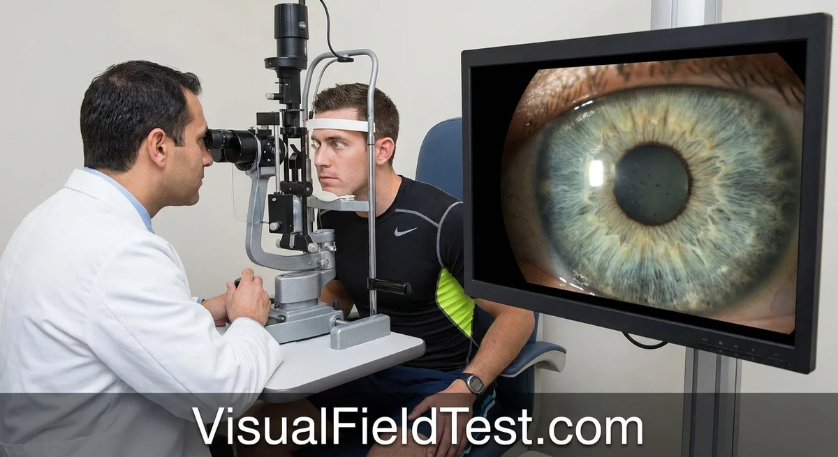
An eye doctor will use several tests, many of which you’ve likely seen:
-
Tonometry (Pressure Test): A puff-of-air test or a gentle pressure on the eye (using fluorescein dye or a tonometer) measures intraocular pressure. Many PDS patients will have pressures above normal range (>21 mmHg), but about half may have “normal” pressure at any given time (doctorlib.org). Because PDS causes fluctuating pressure, a single reading can miss the spikes.
-
Slit-lamp Exam: Under the microscope, the doctor looks for characteristic signs:
- Krukenberg spindle (linear pigment on the cornea).
- Iris transillumination defects (seen when light is shone through the iris).
- Pigment floating in the anterior chamber.
- Pigment on the lens (Zentmayer line), if visible.
-
Gonioscopy: This is a key test. The doctor places a special contact lens with a mirror on the eye to view the angle where fluid drains. In PDS, the entire trabecular meshwork often looks deeply and uniformly pigmented, sometimes with a thick band of pigment all around (360 degrees) (pmc.ncbi.nlm.nih.gov). You may also see the bowed iris.
Note: Gonioscopy confirms the angle is open (ruling out angle-closure) and shows the pigment deposition (“homogeneous for 360 degrees”) typical of PG (doctorlib.org).
-
Optical Coherence Tomography (OCT): This imaging scans the appearance of the optic nerve and retinal nerve fiber layer. Thinning of the nerve fibers would indicate glaucoma damage (PG stage). OCT of the anterior segment can also image the iris bowing in research settings, but it’s not routine in clinics.
-
Visual Field Test: A computerized field test (Humphrey visual field) checks for any loss of peripheral vision, a hallmark of glaucoma damage. If PDS has progressed to PG, subtle patterns like nasal steps or arcuate defects may appear.
-
Other: Low-tech clues include a history of eye rubbing (which can distribute pigment) or a glowing exam where the pupils are dilated (sometimes revealing transillumination defects).
In practice, combining these findings yields the diagnosis: pigment deposits + angle findings + (possibly) elevated IOP. Only with optic nerve or field damage does one call it pigmentary glaucoma (pmc.ncbi.nlm.nih.gov) (bjo.bmj.com).
Because PDS can lie hidden, regular eye screenings (especially for myopes) are crucial. The simplest first clue might be a thorough eye exam by an ophthalmologist or optometrist stating “suspicious pigment” or “classic Krukenberg spindle” when you have no other symptoms.
Management and Treatment
The treatment goals for PDS/PG are the same as for any glaucoma: lower eye pressure and protect the optic nerve. The range of options includes watchful waiting, medicines, lasers, and surgery. Here’s what the evidence and experts advise:
Observation and Medications
-
Observation: If someone has PDS but no ocular hypertension or nerve damage, doctors may simply observe closely (especially if few symptoms). This involves: treating any transient spikes, avoiding known triggers, and re-checking IOP and fields regularly (often every 6–12 months). There is no “cure” to reverse the pigment; the strategy is to guard against pressure buildup.
-
Eye Drops: If IOP is elevated or if there are early signs of nerve stress, first-line treatment is usually glaucoma eye drops. Prostaglandin analogs (like latanoprost) are often the first choice because they are potent and dosed once daily (bjo.bmj.com). Other classes include beta-blockers (e.g. timolol), carbonic anhydrase inhibitors (e.g. dorzolamide), and alpha agonists. Each works either to decrease fluid production or improve outflow. Occasionally, a miotic like pilocarpine may be prescribed on a trial basis since it can pull the iris taut and reduce pigment liberation (as seen in the jogging study (pubmed.ncbi.nlm.nih.gov)). However, pilocarpine causes a constricted pupil (which can blur vision and cause headaches) and is poorly tolerated by many young patients (www.ncbi.nlm.nih.gov). In practice, doctors often avoid it unless absolutely needed.
-
Evidence: There is no specific medication proven uniquely for PDS; the strategy follows standard glaucoma guidelines. The Cochrane Review (2016) on pigmentary glaucoma treatments confirms that we rely on general glaucoma therapies, and that all evidence for special PDS treatments is limited (pmc.ncbi.nlm.nih.gov).
Laser Treatments
-
Laser Peripheral Iridotomy (LPI): This laser creates a small hole in the peripheral iris to equalize pressure between the front and back chambers, theoretically eliminating the “reverse block” and iris concavity. For a time, many doctors performed LPI in PDS to prevent pigment release. However, multiple long-term studies and a Cochrane review have not shown a clear benefit in preventing IOP rises or glaucoma progression (pmc.ncbi.nlm.nih.gov) (bjo.bmj.com). Recent guidelines call iridotomy “controversial” and recommend it only in selected cases: for instance, very young patients with clear evidence of iris-lens apposition and because they can tolerate the procedure (pmc.ncbi.nlm.nih.gov) (bjo.bmj.com). (The evidence here is weak/low-quality. (pmc.ncbi.nlm.nih.gov)) In plain terms, LPI may help a few individuals but it’s not a guaranteed fix and isn’t routine.
-
Laser Trabeculoplasty: In open-angle glaucoma, lasers that target the trabecular meshwork – either Argon Laser Trabeculoplasty (ALT) or Selective Laser Trabeculoplasty (SLT) – can improve outflow. For pigmentary glaucoma, the meshwork is already bathed in pigment, making lasers tricky. Still, SLT has been used. One trial showed SLT was at least as effective as med drops in general open-angle cases (bjo.bmj.com), but there is no strong evidence specifically in pigmentary cases. Some clinicians use SLT if medications fail to control IOP. Notably, ALT/SLT can themselves cause a transient IOP spike in heavily pigmented eyes, so they must be done with care.
Surgery
If eye drops (and possibly lasers) cannot keep pressure at a safe target, glaucoma surgery is considered. Options include:
- Trabeculectomy (creating an alternate drainage route under a flap in the sclera).
- Tube shunt implants (placing a small tube/graft to drain fluid).
- Minimally-Invasive Glaucoma Surgeries (MIGS): These are newer micro-stents or gels to improve outflow. The experience with MIGS in PG is limited, but they might be considered in mild cases as a safer option.
- Canaloplasty: A non-penetrating surgery mentioned in newer PDS gradation papers; it dilates the natural canal of Schlemm.
Classic glaucoma surgery outcomes in PG patients are usually good “on paper,” since these patients are younger and have healthy tissues. One review noted pigmentary glaucoma patients often do well after filtering surgery, though surgeons watch for a higher risk of bleb infection and occasional hypotony (too low pressure) in young male eyes (pmc.ncbi.nlm.nih.gov) (bjo.bmj.com). The bottom line: if PDS has already caused significant optic nerve damage, the treatment strategy is the same as any open-angle glaucoma – aggressively lower the IOP to halt further loss (pmc.ncbi.nlm.nih.gov).
Should Athletes Stop or Modify Exercise?
This is the million-dollar question for active patients. The take-home message from experts and the limited evidence is: do not stop exercise completely, but be smart and individualized. Here’s what current thinking suggests:
-
Don’t assume you must quit sports. The removed message from 1992 and subsequent commentary is that not every PDS patient needs to give up running or sports (pubmed.ncbi.nlm.nih.gov). On the contrary, general exercise is beneficial for overall health and often lowers baseline IOP in normal glaucoma patients (pmc.ncbi.nlm.nih.gov).
-
Monitor your response. Dr. Haynes and colleagues recommend monitoring eye pressure before and after a typical workout the patient does regularly (pubmed.ncbi.nlm.nih.gov). If your pressure or pigment release skyrockets every time you jog or do jumping jacks, you can tailor your activities. If it’s modest, you may continue with simple precautions.
-
Choose lower-impact if needed. Based on physiologic understanding, experts often advise swapping out very bouncy exercises for gentler ones. For example, walking, swimming, cycling, or using an elliptical are generally considered safer (kingvision.org) (pmc.ncbi.nlm.nih.gov) because they raise fitness without constant head movement or jolting. On the other hand, activities like running, jumping rope, high-intensity interval training, or contact sports (basketball, soccer with heading) are the ones to approach with caution (kingvision.org).
-
Avoid Valsalva maneuvers. When lifting weights, be careful not to do a tight breath-hold. Breathing steadily helps keep eye pressure from spiking. If weight training is important to you, try lighter weights with higher reps, or exercise machines that stabilize the head.
-
No need for extreme restrictions. Even inverted yoga poses or head-down positions only cause temporary IOP rises (kingvision.org). If you enjoy yoga, just be aware (and maybe avoid the most dogged headstands for now).
-
Regular check-ups. If you have PDS/PG, regular ophthalmology visits (every 3–6 months) are crucial. If you plan to change your workout routine significantly, tell your doctor. They might check your IOP before/after a gym session to see what effect it has.
In short, current guidance is not to impose a blanket exercise ban. The British Glaucoma Society (EGS) nomenclature even comments that aside from PDS, most glaucoma patients are actually encouraged to exercise. Exercise can have positive effects on eye health and blood flow (pmc.ncbi.nlm.nih.gov). For PDS users, it’s a matter of balancing benefits with the one-time risks of pigment storms.
Example recommendation: One clinician’s blog summarized: “Exercise remains an important part of glaucoma management – even with pigmentary glaucoma. The goal is not restriction, but intentional movement choices. By selecting low-impact activities and maintaining consistent follow-up care, patients can confidently support both their overall wellness and their visual health.” (kingvision.org).
Controversies and Uncertainties
Several aspects of PDS/PG remain under debate:
- LPI Effectiveness: As mentioned, iridotomy was once routine in PDS, but evidence for its benefit is currently lacking (pmc.ncbi.nlm.nih.gov) (bjo.bmj.com). Some doctors still use it in special cases (young eyes with big iris concavity). Research is ongoing; a key point to stress is that routine LPI for all PDS patients is not evidence-based practice.
- Exercise Guidelines: Almost no formal guidelines exist on exercise for PDS – no large trials on sports and eye pressure. Current advice is largely expert opinion from small studies. We don’t know, for example, how a thousand meter row (with head dips) compares to stair-climbing. Athletes should work with doctors to figure out what’s safe for them.
- Prognosis Variability: Why do some PDS patients never progress while others do? Genetic factors (like mutations in GPNMB or others (pmc.ncbi.nlm.nih.gov)) may play a role, but we lack predictive tests. The concept of a “burn-out” stage in older age is still just a hypothesis (though supported by clinical observation (pmc.ncbi.nlm.nih.gov)).
- Non-Caucasian Populations: Most studies are on white patients. PDS may behave differently in other ethnic groups (less obvious pigment dispersion, maybe different risk factors). We need more data on race and PDS.
- Optimizing IOP Spikes: It’s unknown whether long-term intermittent spikes (e.g. after exercise) cause more harm than a slower, steady pressure. In classic glaucoma, large fluctuations are known to be bad (pmc.ncbi.nlm.nih.gov). It seems prudent to avoid repeat spikes, but how aggressive to be is unsettled.
Prognosis and Living with PDS/PG
If you have PDS/PG, what does the future look like? Here are the reassuring and cautionary aspects:
- Visual prognosis: Overall, pigmentary glaucoma tends to have a better outlook than typical open-angle glaucoma. Total blindness from PG is rare (pmc.ncbi.nlm.nih.gov). However, any glaucoma at a young age should be taken seriously because damage (once done) is irreversible.
- Monitoring: Frequent eye check-ups are essential. Regular IOP checks, optic nerve exams, and visual field tests help detect any progression early. Even if your pressure is normal at one visit, it could spike another day, so consistent follow-up (every 3–6 months in the early years) is the norm.
- Lifestyle: Beyond exercise, other factors matter. For example, sleeping slightly elevated (head of bed 20–30°) can slightly reduce nighttime pressure (this is true for many glaucomas, including PDS) (pmc.ncbi.nlm.nih.gov). Good hydration is advised, and avoid caffeine right before an exam if possible since caffeine can raise pressure a bit.
- Education: Patients do best when they understand their condition. Be aware that coloring contacts, long-term steroids, and eye rubbing could worsen pigment release. Tell any other doctors you see (like for migraines) that you have a form of glaucoma so they avoid dilating your eyes unnecessarily.
With modern treatments, most PDS patients with glaucoma maintain good vision. The main task is to catch it early and prevent damage. As one review summarizes: “With appropriate monitoring and timely pressure-lowering treatment, many individuals with PDS can live full lives without significant vision loss.” (pmc.ncbi.nlm.nih.gov) The important part is monitoring and taking action promptly if signs worsen.
Conclusion
Pigment Dispersion Syndrome is a stealthy eye condition that often lurks in active, healthy-looking individuals. By understanding the biology – tiny pigment granules being abraded off the iris and slowing the eye’s drainage – we see why it can raise pressure and cause pigmentary glaucoma. We now know (from decades of studies) that vigorous exercise can stimulate these pigment “dust storms” in the eye, especially in susceptible people. However, exercise itself is not a forbidden fruit; with smart precautions and monitoring, most patients can stay fit and preserve vision.
In summary:
- What matters: PDS is diagnosed by eye exam signs (Krukenberg spindle, iris defects, angle pigment) and managed by keeping eye pressure low.
- Who to watch: Young, myopic, physically active patients (often men) should be aware of this risk.
- Key advice: Keep exercising for overall health, but prefer low-impact activities if you have PDS/PG. Always follow your eye doctor’s guidance and reporting any new vision symptoms immediately.
- Outlook: With regular care and modern treatments, most people with PDS/PG do well and maintain good sight. Awareness and adherence to exam schedules are crucial.
In the end, the hidden eye risk in athletes can be managed. Regular ophthalmic check-ups and a dialogue about your sports routine will help you protect your vision without giving up the activities you love.