
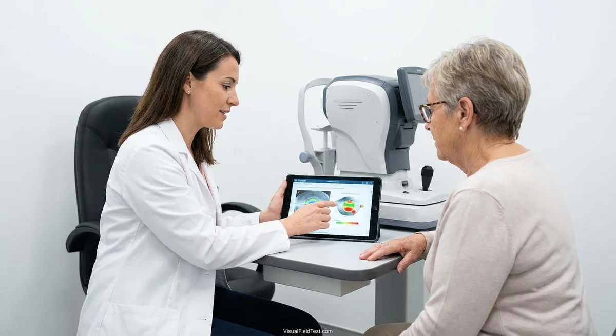
The Future of Glaucoma Care May Be Personal: Matching Treatment to Each Patient’s Risk
Glaucoma is a chronic optic nerve disease and a leading cause of irreversible blindness. Traditionally, doctors have focused on one main factor – eye pressure – to diagnose and treat glaucoma. But in recent years experts have realized that glaucoma behaves very differently from person to person. In fact, two patients with the same eye pressure can have very different outcomes. For example, one patient might slowly lose vision despite moderate pressure, while another with high pressure stays stable for years. This is because many hidden factors – genetic traits, eye anatomy, blood flow, lifestyle habits and more – all influence glaucoma risk (pmc.ncbi.nlm.nih.gov) (pmc.ncbi.nlm.nih.gov).
Today we are on the brink of truly personalized glaucoma care, where doctors will tailor follow-up plans and treatments to each person’s unique risk profile. In this article we’ll explore how clinicians estimate glaucoma risk now, and how future tools like advanced imaging, genetics and artificial intelligence (AI) may change things. We’ll give examples of different patient profiles and imagine what glaucoma care might look like in 2030. We’ll also consider possible pitfalls, like too many tests or unequal access to new technology.
Why Two Patients with the Same Pressure Can Have Different Outcomes
A key reason is that glaucoma is multifactorial. High eye pressure (intraocular pressure, IOP) is the best-known risk factor, but it is far from the only one. Some people’s optic nerves are simply more vulnerable than others’. For example, one large study (the Ocular Hypertension Treatment Study) found that people who went on to develop glaucoma tended to be older, already have larger “cup-to-disc” ratios in their optic nerve, and have thinner corneas than those who did not (ohts.wustl.edu). In other words, an older person with a fragile optic nerve and a very thin cornea might suffer damage at a given pressure level that a younger person with a robust nerve might tolerate. Similarly, about half of glaucoma patients never have very high pressure – so-called normal-tension glaucoma – but still lose vision because of other problems like poor blood flow or genetic factors (pmc.ncbi.nlm.nih.gov). The European Glaucoma Society even emphasizes that “IOP is not the only factor” in glaucoma risk (pmc.ncbi.nlm.nih.gov).
To put it another way: imagine two people, both with an eye pressure of 25 mmHg. Patient A has a thin cornea (which actually masks higher true pressure) and a family history of glaucoma. Patient B has a thick cornea and no family history. Patient A’s optic nerve may already be stressed from years of even slightly elevated pressure and blood flow issues, so glaucoma damage can progress more quickly. Patient B’s healthier eyes and strong corneas might tolerate that pressure without harm for much longer. In short, each eye is different – like a unique machine with its own weak points – so identical pressures don’t guarantee identical outcomes (ohts.wustl.edu) (glaucomatoday.com).
How Doctors Estimate Glaucoma Progression Risk Today
Currently, eye doctors (ophthalmologists) piece together many clues to judge each patient’s risk of vision loss. There’s no single “glaucoma paint-by-numbers” formula used for everyone, but clinicians pay attention to known risk factors and test results. Some key elements include:
- Baseline eye pressure (IOP): Even if pressure isn’t the whole story, higher IOP generally raises glaucoma risk. Yet doctors also consider pressure fluctuations over time, not just one reading (pmc.ncbi.nlm.nih.gov).
- Optic nerve appearance: A large or asymmetric cup-to-disc ratio (the hollow in the optic nerve head) suggests more damage or susceptibility (pmc.ncbi.nlm.nih.gov). If one eye’s nerve shows more cupping, that eye may need stricter control.
- Visual field tests: A standard visual field test maps what areas a person can see. Early loss in these tests indicates glaucoma onset. Doctors look at field results over time – a faster rate of field loss means higher risk.
- Retinal imaging (OCT): Technologies like Optical Coherence Tomography (OCT) give high-resolution scans of the optic nerve and its retinal nerve fiber layer. Thin or thinning fiber layers can signal higher progression risk even before fields are affected.
- Corneal thickness (pachymetry): The central cornea’s thickness is measured because it affects pressure readings. A thin cornea not only underestimates true IOP, it also independently correlates with nerve vulnerability (glaucomatoday.com). In fact, the Ocular Hypertension Study found people with corneas ≤555 µm had three times the risk of glaucoma compared to those with thicker corneas (glaucomatoday.com).
- Age: Older patients generally have higher risk. Each additional decade of age slightly increases the odds of progression.
- Myopia (nearsightedness): Being very nearsighted stretches the eye and optic nerve, raising glaucoma risk (pmc.ncbi.nlm.nih.gov).
- Family history: A strong clue – a first-degree relative (parent, sibling) with glaucoma boosts risk dramatically. One review found relatives of glaucoma patients had a 22% lifetime risk, versus only about 2–3% for relatives of people without glaucoma (pmc.ncbi.nlm.nih.gov).
- Race/ethnicity: People of African descent have higher rates of open-angle glaucoma, and those of Asian descent have more angle-closure forms (pmc.ncbi.nlm.nih.gov). Certain genetic backgrounds color risks.
- Systemic health: Conditions like diabetes and high or low blood pressure [L557–560] can worsen optic nerve health. For instance, very low blood pressure at night (“nocturnal hypotension”) or sleep apnea may starve the eye of blood, adding risk (pmc.ncbi.nlm.nih.gov) (pmc.ncbi.nlm.nih.gov).
- Lifestyle factors: Smoking, for example, damages tiny blood vessels and is linked to glaucoma progression (pmc.ncbi.nlm.nih.gov). Migraine and systemic vasospastic issues can also hint at vulnerable optic nerve perfusion (pmc.ncbi.nlm.nih.gov).
- Medication adherence: Known modifiable factor – if a patient doesn’t stick to treatments, risk climbs.
Often, doctors will use risk calculators or scoring systems. For example, the Ocular Hypertension Treatment Study (OHTS) provided a calculator for patients with high pressure but no glaucoma. It combines age, pressure, corneal thickness, optic disc measurements and more to estimate a 5-year glaucoma risk (ohts.wustl.edu) (glaucomatoday.com). Such tools quantify how multiple factors interplay.
In practice, doctors integrate all these clues. If most signs point to low risk (thick corneas, no family history, only slight optic changes), a patient might only need mild treatment or routine monitoring. But high-risk patients – say, an older person with very cupped optic nerves and thin corneas – would likely get aggressive treatment to lower pressure promptly (ohts.wustl.edu) (pmc.ncbi.nlm.nih.gov).
The Role of Key Tests: OCT, Visual Fields, Pachymetry and More
Two tests are especially important today:
-
Visual Field Testing: This functional test charts a person’s field of vision (often using a computerized device). It detects visual field loss from glaucoma – for example, small scotomas (blind spots) that develop in peripheral vision. Tracking changes in the field over months or years lets doctors calculate how fast vision is worsening. Faster loss means a higher risk profile and need for stronger therapy.
-
Optical Coherence Tomography (OCT): This is an imaging “CAT scan” of the eye. OCT gives a high-resolution cross-section of the retina and optic nerve. It measures the thickness of retinal nerve fibers and shows structural damage. Thinning on OCT often precedes visible field loss. By comparing OCT images over time, doctors spot subtle nerve fiber decline. This helps them catch progression earlier and tailor treatment. (Emerging OCT angiography can even image blood flow around the optic nerve.)
Other measurements round out the picture:
- Pachymetry for corneal thickness, as noted.
- Gonioscopy to check the iris and angle (to rule out angle-closure threat).
- Photography of the optic nerve to record appearance.
- Intraocular Pressure Checks (often at different times of day or after posture changes).
Together, these tests help classify each patient. One might say: “Our patient has moderately damaged fields and moderately thin nerve fiber layers, with IOP usually in the mid-20s. Given her thin corneas and a family history of glaucoma, her risk is above average.” Another patient with similar pressures but normal OCT and no family risk might be classified as lower risk.
AI for Tailoring Follow-Up and Treatment
Artificial Intelligence (AI) is starting to enter glaucoma care, promising to personalize decisions further. Advanced AI systems can analyze large amounts of data – images, test histories, even genetics – to spot patterns a human might miss.
For example, a recent review of over 150 studies found that deep-learning AI on fundus photos or OCT scans can match or even exceed specialist accuracy for glaucoma detection (pmc.ncbi.nlm.nih.gov). More impressively, some sequence-based AI models could detect subtle worsening of visual fields up to 1.7 years earlier than traditional trend analysis (pmc.ncbi.nlm.nih.gov). In other words, an AI algorithm looking at a series of fields and OCTs could warn a doctor long before visual acuity worsens visibly. Other AI models have been trained to predict which patients are likely to need surgery – one multi-modal network combining OCT, field tests and clinical data achieved an accuracy (ROC AUC ~0.92) in forecasting eventual need for incisional surgery (pmc.ncbi.nlm.nih.gov).
In practical terms, AI could customize monitoring schedules. Instead of a fixed “every 6 months”, an AI-guided system might say, “This patient’s data suggest a high chance of rapid change, so check in 3 months. That one looks stable; checking in 9–12 months is fine.” AI can also help triage: smartphone-based programs might let patients do preliminary vision or photo tests at home or in a clinic kiosk, flagging only high-risk cases to see a specialist (pmc.ncbi.nlm.nih.gov).
By 2030, many expect clinical decision support tools – essentially AI-backed “second opinions” – to be routine. These tools will integrate each person’s OCT scans, field history, genetics and even daily eye pressures (from implants or wearable sensors) into a risk score. The doctor and patient could then use that score to pick treatment intensity. For instance, an AI might combine age, genetic markers and OCT data to recommend a lower pressure goal for a patient who, say, has an OPTN gene variant that makes nerves fragile (pmc.ncbi.nlm.nih.gov) (pmc.ncbi.nlm.nih.gov). We are not quite at that level yet, but research suggests it's coming: one study’s AI even answered patient case questions as accurately as a glaucoma specialist, hinting at future clinical-assistant tools (pmc.ncbi.nlm.nih.gov).
However, caution is needed. AI systems must be carefully validated and free from bias. For example, recent work has shown that some AI glaucoma-screening models perform worse in racial minority groups unless specifically calibrated (www.nature.com). This underlines the importance of building AI that works for all eyes, not just a subset.
Advanced Treatments: Tailoring Therapy to Risk
Personalized care means matching the type of treatment to the patient’s needs. New technologies are giving doctors more options beyond just “drops or surgery”.
-
Sustained-release drug implants: These are tiny devices or gels that release glaucoma medications gradually over months, removing the need for daily eye drops. The first FDA-approved example was a bimatoprost implant (brand name Durysta) that slowly releases a prostaglandin drug inside the eye. Studies have shown such implants can keep pressure down for 3–4 months per injection, with efficacy comparable to daily drops (pmc.ncbi.nlm.nih.gov). Other implants are in trials (e.g. travoprost intracameral implants) (pmc.ncbi.nlm.nih.gov). In future, a patient who fires glaucoma drops might instead get a quarterly implant. This is especially good for someone who struggles with drop compliance or who needs very steady pressure control. In practice, your doctor might personalize this: “Because you have moderate risk and difficulty using drops, let’s try an implant that lasts 6 months” vs. “You have higher risk, so an implant plus drops might be needed.”
-
Laser therapies: Selective laser trabeculoplasty (SLT) is already a popular first-line or adjunctive treatment. It can be seen as a personalized step-up for mild cases. Some doctors now use SLT upfront for many open-angle glaucoma patients so they need fewer drops. Others are studying intermittent very-low-power laser pulses (micropulse laser) aimed at neuroprotection. In the future, whether and when to use laser may be personalized – for example, family history of aggressive glaucoma might trigger early laser use.
-
Minimally Invasive Glaucoma Surgery (MIGS): These techniques involve tiny stents or shunts placed in the eye’s drainage angle through a small incision. They generally have fewer risks than traditional surgery but also different pressure-lowering power. A patient with moderately high risk (e.g. needing modest IOP reduction with low complications) might be offered MIGS. Someone with very advanced disease might go straight to more potent surgeries (below). By 2030 surgeons are likely to pick among many MIGS devices depending on the eye’s anatomy – for instance, one type of stent may work better for a certain angle shape or stage of disease.
-
Filter/trabeculectomy and shunts: Classic glaucoma surgeries like trabeculectomy or tube shunts remain the most powerful ways to drop pressure. Typically reserved for high-risk cases today, they will still be used for those who need big pressure drops or have failed other treatments. But even selection of filters could become more personalized: for someone with a high risk gene or very “frail” nerves, a doctor might lower the target pressure to the low teens and perform surgery earlier rather than after multiple drops have failed.
-
Neuroprotective/neuroregenerative treatments: These aim to protect or heal the optic nerve itself, not just lower pressure. Currently no reliably proven neuroprotective drug exists for glaucoma, but many are in research. Examples include brimonidine (some evidence of nerve protection beyond pressure), antioxidants, and investigational agents delivering growth factors to retinal cells (pmc.ncbi.nlm.nih.gov) (pmc.ncbi.nlm.nih.gov). By 2030, we may see at least one therapy approved to directly guard optic nerve cells, especially for patients whose risk seems driven by vascular or genetic susceptibility. (For instance, a patient with normal-pressure glaucoma might receive an extra medication targeting neuroprotection.) Gene therapy is also on the horizon for rare inherited glaucoma: if a patient has a known mutation (like MYOC), future treatments might alter the gene or supply a missing factor.
Each treatment choice would be personalized. For a high-risk patient, a doctor might propose starting with a sustained-release implant plus drops and a laser. For a low-risk patient, just monitoring or a single drug might suffice.
A Glimpse at 2030: Personalized Glaucoma Care in Action
Imagine a patient named Maria, age 60, who comes in for her glaucoma check-up in 2030. Maria’s data has been compiled into a digital record for years: baseline scans of her optic nerve, yearly visual fields, her DNA risk profile (from a genetic test done in 2025), and even a smart contact lens that has recorded her nighttime eye pressures. An AI system crunches all this information into a personalized risk report. It finds that Maria has early optic nerve changes, a family history, a moderate genetic risk score, and a pattern of slightly lower nighttime blood pressure. Her predicted 5-year vision loss risk is high despite currently acceptable pressure.
Given this risk profile, her doctor recommends an aggressive yet tailored plan:
- Immediate therapy: Instead of just prescribing more eyedrops, the doctor discusses a sustained-release glaucoma implant now to ensure steady pressure control without Maria worrying about dosing every day. The plan anticipates a possible second implant in one year.
- Laser treatment: Because Maria’s optic nerves are vulnerable and family history strong, the doctor also performs a quick outpatient laser procedure to boost drainage.
- Follow-up schedule: The AI schedules Maria to return in 3 months (rather than the usual 6) for an exam and OCT scan review. The frequency can be adjusted by the system if things look stable.
- Lifestyle tweaks: Knowing Maria has mild sleep apnea from her history, the team arranges for a sleep specialist consult, since controlling that might help her eyes.
- Digital monitoring: Maria can use a home OCT device or a smartphone app (FDA-approved) to do a quick check of her vision field every month. If the app detects any worrying change, it will alert her doctor even before her scheduled visit.
Now compare John, age 50, whose risk factors are few: moderate glaucoma in one eye, good eye exam, thick corneas, and normal blood pressure at all times. His personalized risk report shows a very low chance of fast progression. At his visit, he and his doctor agree on a more relaxed plan: an eye pressure drop, and routine check-ups every year. He won’t need invasive implants or extra appointments.
By 2030, this kind of stratified approach – high-risk patients getting early interventions, low-risk patients avoiding unnecessary treatments – could become standard. Glaucoma clinics might routinely use apps and algorithms to guide who needs what level of care.
Risks of Overtesting, Overtreatment, and Unequal Access
While personalization promises better care, it also raises concerns. Overtesting can lead to patient anxiety, extra costs, and false alarms. For example, if a high-risk algorithm flags every tiny change as dangerous, a patient might undergo needless procedures or frequent exams. We’ve already seen in medicine that too much screening can sometimes do more harm than good. Doctors will have to balance vigilance with pragmatism.
Overtreatment is another worry. Lowering pressure always has side effects (medications can irritate eyes, surgeries have risks). If an algorithm seems to predict vision loss, will every patient be offered surgery just in case? We must avoid a one-size-fits-all “treat everything” mindset. Even with better risk scores, doctors should still consider each patient’s overall health, life expectancy, and preferences. Not every marginal risk increase justifies aggressive therapy.
Health equity is a final big issue. Right now, nearly half of people with glaucoma worldwide don’t even know they have it, especially in underserved communities (www.nature.com). Advanced tools – genetic tests, AI clinics, high-end imaging – may be available first in wealthy settings. There is a danger that only affluent patients benefit from personalized glaucoma care while others fall further behind. For example, a recent study noted that Black and Hispanic patients often first present with more severe visual loss, largely because of limited access to eye care (www.nature.com). We must ensure new technologies help bridge, not widen, this gap. Innovations like smartphone screening tools or low-cost AI could help reach under-served areas, but this will require deliberate effort, training and resources.
Finally, AI algorithms themselves can be biased if trained on limited data. As one group re-trained an OCT-based glaucoma AI, they found initial models performed worse in non-white patients. They had to specifically adjust the AI (“fair identity normalization”) to equalize accuracy (www.nature.com). This highlights how careful development and regulation are needed. The future of glaucoma care should include rules and standards (like those in development for medical AI) to safeguard patients everywhere.
Conclusion
Glaucoma care is moving beyond the old “one size fits all” model. We now understand that an individual’s genetic makeup, eye anatomy, lifestyle and health factors all combine to make his or her glaucoma unique. By pooling all this information, from OCT scans and family history to AI-driven risk scores, doctors can tailor monitoring and treatment to each patient.
In the next decade, many routine glaucoma visits may feel more like personalized consultations. High-risk patients might get early implants or combined therapies; low-risk patients might enjoy longer intervals between visits and fewer medications. Tools from tomorrow – AI analysis, smart sensors, gene panels – will sharpen our predictions and choices.
At the same time, we must tread carefully. More data does not automatically mean better outcomes; it can also mean more confusion if not handled wisely. Patients and doctors alike should remember that even the best algorithms are guides, not oracles. And society must seek to make these advances available to all, not just a fortunate few.
With thoughtful use, personalized glaucoma care could help prevent many cases of needless vision loss. By 2030 and beyond, matching treatment intensity to individual risk could turn the tide on glaucoma’s historic reputation as the “sneak thief of sight.” The future may indeed be personal – a future where each patient’s care plan is as unique as their own risk profile.