Myopia and Glaucoma: When Nearsightedness Raises the Stakes
Myopia (nearsightedness) is becoming very common worldwide. In fact, by 2050 about half of people will likely be nearsighted (pmc.ncbi.nlm.nih.gov). Most kids and adults know myopia means trouble seeing far away, but people often forget that high myopia can also lead to serious eye problems like retinal disease and glaucoma (pmc.ncbi.nlm.nih.gov). Glaucoma is a leading cause of irreversible blindness. In glaucoma, pressure in the eye (called intraocular pressure) damages the optic nerve – the cable in the back of the eye that sends sight to the brain. Many studies find people with moderate to severe myopia have higher rates of glaucoma than people with normal vision (pmc.ncbi.nlm.nih.gov). For example, one large study (the Blue Mountains Eye Study) found mild myopia roughly doubled glaucoma risk, while stronger myopia increased it about three-fold (pmc.ncbi.nlm.nih.gov). In simple terms, an eye stretched by myopia is more likely to develop glaucoma, so doctors may watch nearsighted patients more carefully.
Diagnosing glaucoma in myopic eyes is tricky. Moderate-to-high myopia changes the eye’s shape and anatomy, making standard glaucoma tests harder to interpret (pmc.ncbi.nlm.nih.gov) (pmc.ncbi.nlm.nih.gov). We will explain how a long eye changes the optic nerve, why that makes visual field tests and OCT scans (advanced imaging) harder to read, and what this means for patients. We also cover how to slow myopia in children and why nearsighted adults need regular glaucoma screening. Finally, we’ll clarify the difference between ocular hypertension (just high eye pressure) and true glaucoma (actual nerve damage).
How Myopia Changes the Eye’s Shape
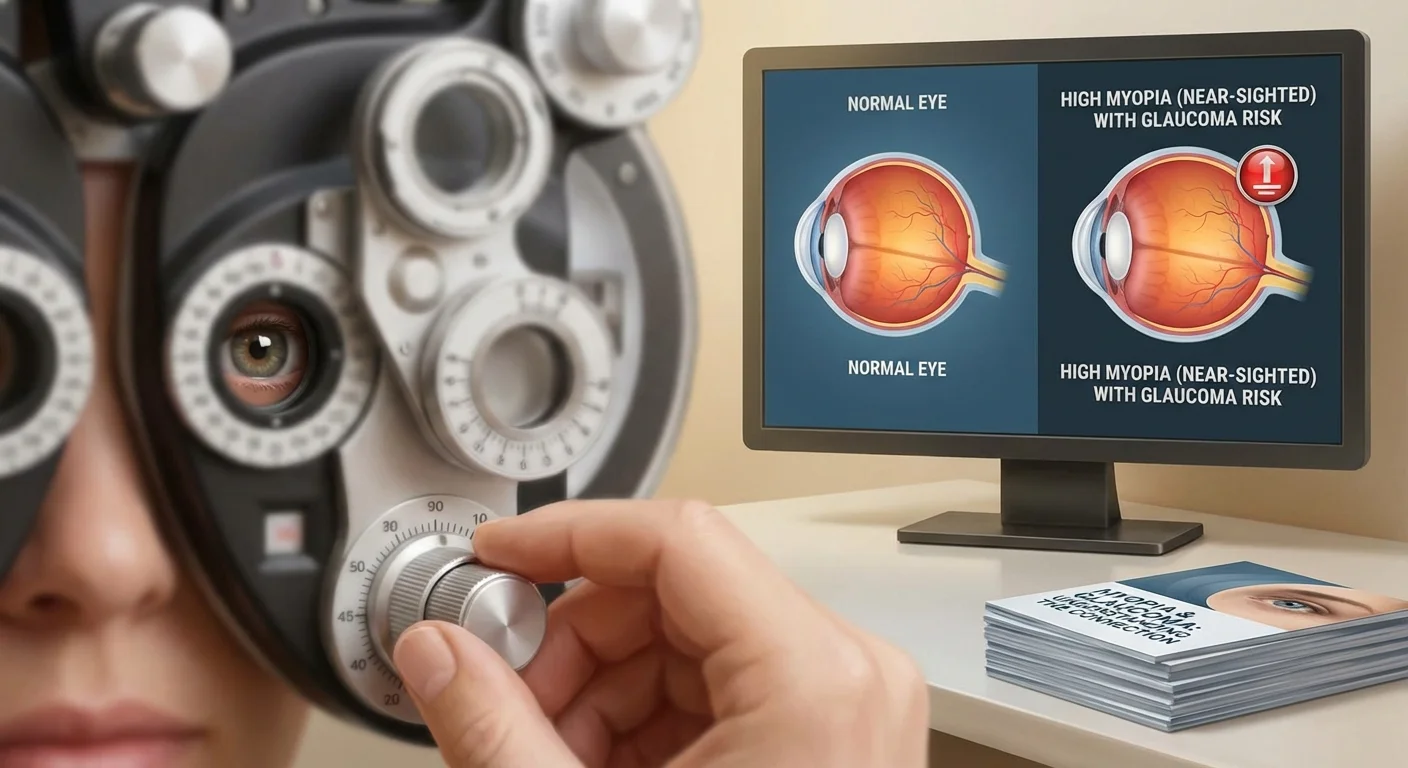
Nearsighted eyes are longer than normal from front to back (an increased axial length). Imagine blowing up a balloon – as it expands it thins and stretches the material. Similarly, an elongated myopic eyeball stretches the retina (the light-sensitive tissue) and optic nerve tissues. These changes include:
- Tilted outlook: The optic nerve head (also called the optic disc, where nerve fibers leave the eye) often appears tilted or slanted in high myopes (pmc.ncbi.nlm.nih.gov) (pmc.ncbi.nlm.nih.gov).
- Larger disc area: High myopic eyes usually have a bigger optic disc that may appear blurry or oval (pmc.ncbi.nlm.nih.gov) (pmc.ncbi.nlm.nih.gov).
- Peripapillary atrophy (PPA): You may see pale, scalloped patches (areas of thinning) around the optic nerve (pmc.ncbi.nlm.nih.gov) (pmc.ncbi.nlm.nih.gov).
- Shallow cupping: In glaucoma, the optic disc “cupping” (a hollowing-out) is a sign of nerve loss. Myopic discs tend to be flatter and shallower, which can mask or mimic the cupping seen in glaucoma (pmc.ncbi.nlm.nih.gov) (pmc.ncbi.nlm.nih.gov).
- Retinal stretching: The retina and its nerve fibers become thinner and more loosely packed, even in a normal myopic eye (pmc.ncbi.nlm.nih.gov) (pmc.ncbi.nlm.nih.gov).
These structural changes mean that a nearsighted eye’s optic nerve can look quite different from a normal eye’s, even without any disease. In short, doctors have to tell apart “normal myopic appearance” from “glaucoma damage,” and that can be hard. For instance, an expert review notes that disc tilt, eye elongation, and PPA in myopic eyes make it difficult to spot glaucomatous changes (pmc.ncbi.nlm.nih.gov). High myopic glaucoma often shows a large, elongated optic disc with a broad, shallow cup and big areas of peripapillary atrophy (pmc.ncbi.nlm.nih.gov). Because these patterns can look like or hide signs of glaucoma, careful comparison with past exams and both eyes’ appearance is important.
Why Myopia Raises Glaucoma Risk
Many large studies confirm that nearsighted people get glaucoma more often than non-mypopic people (pmc.ncbi.nlm.nih.gov). In fact, there is a dose-response relationship[^1]: the more severe the myopia, the higher the chance of glaucoma (pmc.ncbi.nlm.nih.gov) (pmc.ncbi.nlm.nih.gov). For example, the Blue Mountains study (an Australian population study) found that people with mild myopia (–1.0 to –3.0 diopters) had about twice the glaucoma risk of normal vision adults, and those with myopia worse than –3.0 had three times the risk (pmc.ncbi.nlm.nih.gov). Other studies in diverse populations (Latino, Asian, etc.) showed that increasing axial length (longer eyeball) goes hand-in-hand with higher open-angle glaucoma rates (pmc.ncbi.nlm.nih.gov). A recent review summarized that “numerous large population-based studies have demonstrated an increase in glaucoma prevalence with increasing myopia” (pmc.ncbi.nlm.nih.gov).
Why do myopic eyes develop glaucoma more often? The stretched retina and nerve fiber layer are less robust, so they may be more vulnerable to pressure damage. Also, myopic eyes often have blood flow or structural changes that can predispose to optic nerve damage. In any case, experts now treat moderate-to-high myopia as a glaucoma risk factor (pmc.ncbi.nlm.nih.gov) (pmc.ncbi.nlm.nih.gov). If you have –6 diopters or more of myopia, your eye pressure and optic nerve should be checked regularly, as though you have another risk factor like family history or diabetes.
Challenges in Glaucoma Tests for Myopic Eyes
Diagnosing glaucoma usually involves measuring eye pressure, looking at the optic nerve, and testing peripheral vision (visual field). In myopic eyes, each of these steps can give false alarms or hide problems.
Interpreting Visual Field Tests
A visual field test checks your side vision by making you press a button when you see lights in your blind spot or edge of vision. Many nearsighted people – especially younger high myopes – will show spots of reduced vision on this test even if they don’t have true glaucoma (pmc.ncbi.nlm.nih.gov). Studies report that a significant number of high-myopia patients have “glaucoma-like” field defects despite never progressing to real damage (pmc.ncbi.nlm.nih.gov). For example, in one large registry of young highly myopic patients, about 16% already showed visual field patterns that look like glaucoma (pmc.ncbi.nlm.nih.gov). Another series of myopic patients labeled “glaucoma suspects” were followed for years and showed no worsening in fields or optic nerve cupping (pmc.ncbi.nlm.nih.gov).
What does this mean? In plain terms, myopia can cause spots or scotomas on a visual field test that mimic early glaucoma. It might be due to subtle stretching or retinal changes that don’t actually mean the nerve is dying. Researchers caution that “a proportion of myopic patients may be misdiagnosed as having glaucoma and be taking unnecessary treatment” (pmc.ncbi.nlm.nih.gov). The takeaway is that a single visual field test in a myopic eye is less definitive. Eye doctors will compare fields over time (looking for change) and use other clues (like a disc hemorrhage or thinning nerve fiber on one side) to decide if it’s true glaucoma (pmc.ncbi.nlm.nih.gov) (pmc.ncbi.nlm.nih.gov).
Interpreting OCT and Imaging Scans
OCT (Optical Coherence Tomography) is like an ultrasound but with light: it creates cross-section images of the retina and optic nerve and measures the thickness of layers like the retinal nerve fiber layer. In a normal eye, OCT can detect early nerve fiber loss by noticing thinning. However, in a high myope, the baseline (starting) thickness of nerve fibers is already low due to stretching (pmc.ncbi.nlm.nih.gov) (pmc.ncbi.nlm.nih.gov). Worse, the reference “normals” in the machine’s database are usually people with normal eye shapes. So a myopic eye may be flagged as “abnormal” on OCT charts even when it’s healthy (pmc.ncbi.nlm.nih.gov).
Specifically, experts note that in highly elongated eyes, the nerve fiber bundles get crowded and pulled toward the temple side. This causes common artifacts: the OCT may show a patch of missing nerve fibers (a pseudodefect) where there is none (pmc.ncbi.nlm.nih.gov). Large areas of peripapillary atrophy and a tilted disc can also fool the software. The review by Park summarized: “RNFL measurements by OCT in high myopia may exhibit variations owing to disc tilting, large atrophy, and segmentation error... convergence of RNFL bundles temporally ... causing artifacts” (pmc.ncbi.nlm.nih.gov).
What can be done? Some OCT machines now offer a “myopia” mode or expanded database, which can reduce false positives (pmc.ncbi.nlm.nih.gov). Clinicians also look at the raw thickness curves rather than only the color-coded map, to decide if the thinning is real or an artifact (pmc.ncbi.nlm.nih.gov). In addition, focusing on the macula (center of vision) and the ganglion cell layer there can help, because glaucoma often affects certain areas. In short, OCT in a very nearsighted eye should be interpreted with caution, knowing that the “norm” might not apply.
Summary of Diagnosis Challenges
Because of these issues, diagnosing glaucoma in myopic patients often rests on looking for change over time and on combining many signs. One review states that careful longitudinal follow-up (repeating exams over months or years) is “the most critical factor in facilitating the detection of early glaucomatous changes” in high myopes (pmc.ncbi.nlm.nih.gov). In practical terms, an eye doctor might watch a slightly suspicious myopic eye rather than jump to treatment, unless the changes clearly progress. Finding an optic disc hemorrhage (a tiny bleeding spot on the nerve) or onset of a typical nerve defect can tip the balance toward a glaucoma diagnosis.
Preventing Myopia Progression in Children
Because high myopia is a risk factor for glaucoma (and other problems), a lot of effort goes into slowing myopia as kids grow. Modern research has identified several proven strategies to slow axial elongation in children (pmc.ncbi.nlm.nih.gov):
- Outdoor time: Encourage children to spend time outdoors every day (bright natural light seems to slow eye growth). In fact, clinical trials and systematic reviews show that schools that add an extra 40–60 minutes of outdoor recess see significantly lower new cases of myopia (pmc.ncbi.nlm.nih.gov) (pmc.ncbi.nlm.nih.gov).
- Atropine eye drops: These are very low-dose eyedrops (often 0.01–0.05%) given nightly. Even tiny doses of atropine can reduce the rate of myopia progression by 30–60% in schoolchildren (pmc.ncbi.nlm.nih.gov). (Atropine is an older drug, but at low concentrations it is safe and minimally blurs near vision.)
- Special lenses or glasses: Certain multifocal or dual-focus contact lenses and spectacle lenses are designed to slow myopia (for example, by having different power zones or special defocus zones). These are approved in some countries and show fitting benefit in trials (pmc.ncbi.nlm.nih.gov).
- Orthokeratology: This is hard contact lenses worn overnight to gently reshape the cornea. Kids wake up with better vision and the lens effect also slows eye growth. Studies find it can reduce progression by about 30–50%.
- Good visual habits: Even simple steps help. Limit nearby screen/reading time without breaks. Ensure good lighting when reading. Regular outdoor play is one of the easiest changes for families to start.
A recent expert review notes that “evidence is growing regarding the potential benefits of applying these interventions in children with myopia” (pmc.ncbi.nlm.nih.gov) (pmc.ncbi.nlm.nih.gov). None of these methods stops myopia completely, but they can make a meaningful difference over the years of growth. The goal is to end up with less severe nearsightedness in adulthood, which in turn lowers risks of glaucoma and retinal problems later.
What Nearsighted Adults Should Know About Glaucoma
If you are an adult with myopia (particularly moderate to high), you should be aware that you have a higher-than-average risk for glaucoma. Here is what to keep in mind:
- Regular eye exams: Get comprehensive exams as often as your eye doctor recommends (often yearly). This should include a check of eye pressure, examination of the optic nerve (with a dilated pupil eye exam), and possibly imaging or visual field tests. The Cleveland Clinic advises that people with high pressure or risk factors “see their provider and follow suggestions about... regular eye exams” (my.clevelandclinic.org). Early detection is key.
- Know your ocular pressure: On each visit, make sure your eye pressure is measured. Eyes run at different pressures, but above about 21 mmHg is considered high. Even normal or “low-tension” pressures in high myopes should be tracked carefully if there are nerve changes.
- Watch for nerve health: Ask if your doctor sees any optic disc changes or thinned areas on an OCT. But also remember the OCT caveats above: if you are very myopic, a single abnormal scan isn’t definitive. Trends over time matter more.
- Family history matters: If glaucoma runs in your family or you’ve had eye injury, be extra vigilant. Myopia plus family history may warrant preventive treatment (like pressure-lowering drops) even before damage appears.
- Discuss ocular hypertension vs glaucoma: Ocular hypertension (OHT) is a term for when eye pressure is high but there is no detectable nerve damage (my.clevelandclinic.org). By definition, glaucoma means there is damage to the optic nerve (often seen as specific nerve thinning or visual field loss). Precisely, Cleveland Clinic explains: “Ocular hypertension can cause glaucoma. Glaucoma happens when high IOP damages the optic nerve” (my.clevelandclinic.org). In practice, an eye with high pressure but no field loss or nerve cupping is labeled “ocular hypertension.” That eye still needs close monitoring because it is at risk. If consistent optic nerve changes or field loss appear, then it becomes glaucoma.
With proper care, many people with ocular hypertension or early glaucoma can prevent vision loss. In fact, the Cleveland Clinic notes, “It’s important to make and keep appointments for regular eye examinations. Finding problems early is best” (my.clevelandclinic.org). If a doctor prescribes pressure-lowering drops or recommends laser treatment for your eye pressure, it’s because reducing pressure is the best way to prevent glaucoma damage.
Ocular Hypertension vs. Glaucoma
To be very clear: Ocular hypertension means only high eye pressure, with no signs of nerve damage yet (my.clevelandclinic.org). It is a risk factor for glaucoma (premonitory state), but not everyone with ocular hypertension develops glaucoma (my.clevelandclinic.org) (my.clevelandclinic.org). For instance, one person with pressure of 24 mmHg but a completely healthy optic nerve and fields is generally told “We’ll watch closely” because their risk is elevated. On the other hand, if that person’s optic nerve shows characteristic thinning and the visual field also begins to fail, we call that glaucoma – the disease of nerve damage.
In some eye exams, the difference can be subtle. Doctors rely on all evidence together: pressure readings, nerve photos, OCT layers, and visual fields, to label it correct. Importantly, controlling pressure is the main way to protect the nerve. If you have ocular hypertension, you may be started on drops that lower pressure to prevent the hammer of glaucoma from striking.
Conclusion
Nearsightedness is not just about blurry distance vision – moderate and high myopia actually changes eye anatomy in ways that raise the risk of glaucoma. We now know that people with strong myopia have significantly higher chances of developing glaucoma (pmc.ncbi.nlm.nih.gov) (pmc.ncbi.nlm.nih.gov). Unfortunately, the same changes that raise the risk (elongated eye, tilted disc, thinner retina) also make it harder to detect glaucoma early using standard tests. That is why eye doctors emphasize careful, repeated exams and a complete look at the whole picture when examining myopic patients (pmc.ncbi.nlm.nih.gov) (pmc.ncbi.nlm.nih.gov).
For parents of children who are becoming myopic, the message is: use the proven tools to slow progression – outdoor time, atropine drops, special lenses – so that the child’s final prescription is as small as possible (pmc.ncbi.nlm.nih.gov). For adults with significant myopia, the take-home is: get glaucoma screenings regularly. Ask about your optic nerve, ask if any test results look suspicious, and if you have ocular hypertension, do not ignore it. With modern treatments, glaucoma progression can be slowed or stopped if caught early.
In summary, being nearsighted means you should take extra steps to protect your vision (pmc.ncbi.nlm.nih.gov) (my.clevelandclinic.org). Schedule regular eye exams, follow up on any worrisome findings, and control any high pressure with your doctor’s guidance. Understanding that nearsighted eyes are at higher risk will help you and your eye care team keep your vision safe for years to come.