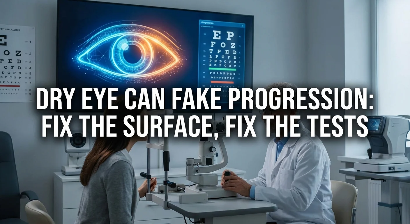
Dry Eye & Glaucoma: Fix the Surface, Fix the Tests
Persistent dryness and irritation of the eye surface—often called dry eye disease or ocular surface disease—is very common in people with glaucoma (especially those using eye drops). This surface problem can blur vision, make you blink more, and change the way your eyes see light. That in turn can throw off glaucoma tests. For example, a frustrated, burning eye may cause more false-positive or tracking failures on a visual field test (pmc.ncbi.nlm.nih.gov) (pubmed.ncbi.nlm.nih.gov), or a rough tear film can cause the OCT scan light to scatter and “miss” part of the nerve fiber layer. In short, dry eye can fake glaucoma progression by making tests look worse even when the nerve fibers haven’t changed.
Before declaring your glaucoma is getting worse, doctors must check and optimize the ocular surface. Treating the dryness and lids first will give more reliable test results. In this article we explain why, and give a step-by-step guide to “fixing the surface.”
How Ocular Surface Disease Skews Glaucoma Tests
-
Visual Field Tests (Perimetry): If your eyes are irritated or your vision fluctuations, it’s hard to concentrate during a perimetry test (when you press a button for each light you see). Studies have shown that patients with more severe dry eye symptoms tend to have more tracking failures and unreliable field tests (pmc.ncbi.nlm.nih.gov). In one study, giving just one drop of artificial tears before testing noticeably improved visual field quality – lowering false negatives and giving a better average result (pubmed.ncbi.nlm.nih.gov). In practice, unresolved dryness can make it seem like your field is worse.
-
OCT Scans: Optical coherence tomography (OCT) uses light to map the retina and nerve layers. A healthy tear film and clear cornea are needed for crisp images. Dry or irregular tears can cause segmentation errors – the software might misidentify layer boundaries when the images are foggy or have debris. (pmc.ncbi.nlm.nih.gov). These errors often underestimate nerve fiber thickness by a few microns (pmc.ncbi.nlm.nih.gov). In other words, an OCT scan taken through a bad tear film can look falsely thin, mimicking glaucoma damage. In fact, a classic study found that about 20–50% of automated OCT RNFL scans had some error, especially when image quality was lower (pmc.ncbi.nlm.nih.gov). (Always ask your doctor if they checked scan quality or redid an OCT if the results looked surprising.)
-
Why It Matters: If dryness causes a test to look worse on one visit (and better the next), the glaucoma doctor might misread this as disease progression. By contrast, if the surface is healthy and stable, any change is more likely real. We want to remove the “fake” changes caused by the surface.
Bold and bright perfectly healthy eyes will give much more consistent test results. So treat first, test second.
Improving the Ocular Surface: A Step-by-Step Regimen
Your eye doctor or optometrist may suggest a stepwise approach to restore a smooth, hydrated surface. Here’s a typical plan:
-
Use Frequent Preservative-Free Artificial Tears: Instill a preservative-free lubricating eye drop several times a day, before testing and in between glaucoma meds. Preservatives in many eye drops (like benzalkonium chloride) can worsen dryness and irritation over time (pmc.ncbi.nlm.nih.gov). Opting for preservative-free formulations (which many pharmacies carry) helps prevent further damage. Artificial tears re-establish a smooth tear film and clear vision. Research shows that even one drop immediately before a field test can improve the result (pubmed.ncbi.nlm.nih.gov). In practice, try to keep your tears topped up all day (for example, 4–6 times daily or as directed).
-
Lid Hygiene (Warm Compresses + Cleaning): Many glaucoma patients have oily gland dysfunction or blepharitis, which make tears unstable. Warm compresses soften the dried oils clogging the lids. Evidence suggests that applying a warmed (around 104°F/40°C) moist compress to each eyelid for about 10 minutes daily significantly improves tear quality and dry eye symptoms (pmc.ncbi.nlm.nih.gov) (pmc.ncbi.nlm.nih.gov). After the compress, gently massage your lids toward the lashes to express the oils. Then clean the lid margins with a mild lid scrub (baby shampoo on a cotton swab or commercial lid wipes). Regular lid hygiene (warming, massaging, and cleaning) is the cornerstone of treating dry eye and blepharitis (pmc.ncbi.nlm.nih.gov). In MGD (meibomian gland disease), good lid hygiene has been shown to “re-establish tear film stability, improve gland secretion, and decrease dry eye symptoms” (pmc.ncbi.nlm.nih.gov).
-
Protect From Irritants: Air conditioning, fans, wind, smoke or low humidity can aggravate dry eyes. During the treatment phase, avoid these triggers if possible. Take breaks during long screen use to blink normally. A humidifier at home or day glasses can also help keep tears from evaporating.
-
Short Course Anti-Inflammatory Treatment: If the ocular surface is quite inflamed (red, painful, or heavily stained) and not improving with tears alone, your doctor might prescribe a short course of mild steroid eye drops (for example, loteprednol or fluorometholone) or a non-steroidal anti-inflammatory drop. Topical steroids are powerful anti-inflammatories and have been shown to significantly reduce dry eye symptoms and corneal staining (pmc.ncbi.nlm.nih.gov), even more than artificial tears alone. (Their effect is usually seen in 1–2 weeks.) Caution: Steroids can raise eye pressure in some people, so this must be used under supervision and only briefly. For long-term improvement, the doctor may also consider immunomodulators like cyclosporine or lifitegrast, but these take weeks to act. For immediate test accuracy, even a week or two of a safe steroid ointment (under doctor guidance) can calm the surface. Some combination drops (steroid+antibiotic) are also used for blepharitis.
-
Recheck & Repeat Before “Progression”: After a few weeks or a month of these surface treatments, repeat the glaucoma tests (visual field, OCT) with the surface optimized. In many cases, findings that looked concerning at the first visit are actually stable once the eye is lubricated and comfortable. For example, one study showed that after using artificial tears, patients’ visual field mean deviation improved and false defect points decreased (pubmed.ncbi.nlm.nih.gov). Similarly, an OCT scan done through a clear tear film will suffer fewer segmentation errors (pmc.ncbi.nlm.nih.gov). If needed, keep the regimen going and re-test again, rather than rushing to escalate glaucoma therapy based on a possibly spurious change.
In summary, a typical “dry eye regimen” before repeat testing might include:
- Artificial tears (preservative-free) throughout the day
- Daily warm compress (10–15 min) + gentle lid massage and cleansing
- Short-term prescription anti-inflammatory if significant inflammation remains
- Continue for several weeks, then repeat glaucoma tests.
Before-and-After Case Examples
Below are illustrative examples of how optimizing the surface can change test results. (Note: these are representative cases, not your actual data. Any test results after treatment are shown without personal patient identifiers.)
-
Visual Field Improvement: A 70-year-old man with stable glaucoma had a sudden worsening on his visual field – his mean deviation (MD) dropped and pattern defects grew. He also admitted he hadn’t been using artificial tears. After committing to preservative-free artificial tears 4×/day and warm compresses for 4 weeks, he returned for a repeat field test. This time his MD was back to baseline and the false defects vanished, showing no true progression. (Studies confirm that giving tears immediately before a visual field can yield a better test and higher MD (pubmed.ncbi.nlm.nih.gov).)
-
OCT Artifacts Cleared: A 60-year-old woman’s routine OCT RNFL scan showed a suspicious thinning in one quadrant that looked worrisome. On exam, she had marked eyelid crusting and punctate staining on her cornea (signs of dryness/blepharitis). She was treated with lids cleansers and eye drops. A week later the OCT was repeated. The new scan showed a smooth RNFL contour with no apparent thinning – confirming that the initial anomaly had been an artifact of the bad surface, not true nerve loss.
(Ideally, doctors would capture "before and after" scan images or field printouts in these scenarios to illustrate the point. It underscores why it’s crucial to treat dryness first.)
Quick Dry Eye Screening in Glaucoma Visits
Because dry eye is so common in glaucoma patients (up to 60% on drops develop it (pmc.ncbi.nlm.nih.gov)), a little screening at the start of each visit can save confusion later. Here are some fast screening “tools”:
-
Symptom Questions: Ask about burning, scratchiness, or fluctuating vision. A simple question like “Do your eyes often feel dry, gritty, or tired?” or using a short questionnaire (for example, the DEQ-5 or SPEED questionnaire) can quickly identify people with significant dry eye symptoms (pmc.ncbi.nlm.nih.gov) (pubmed.ncbi.nlm.nih.gov). The DEQ-5 is a 5-question survey used in studies to gauge dryness before testing (pmc.ncbi.nlm.nih.gov). Even asking patients “How often do you need to blink or use drops when you read?” provides a clue.
-
Blink Test: A quick self-test is to see how long the patient can comfortably keep their eyes open looking at a target without blinking. A time under ~10 seconds may indicate tear film instability. (One study found that requiring patients to hold eyes open without blinking predicted the presence of dry eye with good accuracy (pubmed.ncbi.nlm.nih.gov).) If someone blinks very frequently or complains of immediate discomfort, that suggests their tears need rehydration.
-
Quick Exam Clues: Observe the eyes at the slit lamp: look for red lids, flakes/crust on lashes, or uneven tear breakup after a blink. A quick instillation of fluorescein dye (if your office has time) can reveal short tear breakup time. Notice if the tear meniscus (pool of tears on the lower lid) is low. Also evaluate eyelid margin for excess oil or debris. These signs mean the surface is abnormal.
-
Action on the Spot: If screening suggests dryness, don’t wait. You can often have the patient place a drop of artificial tears in each eye before performing any glaucoma pressure measurement or visual field test. This simple step may improve test accuracy immediately (pubmed.ncbi.nlm.nih.gov). Likewise, remind patients to follow the preservative-free tear or lid hygiene plan you’ll recommend.
By integrating these brief checks into your clinic visit, glaucoma progression can be distinguished from test error. One insightful study concluded that dry eye symptom scores should be considered when interpreting visual field reliability metrics (pmc.ncbi.nlm.nih.gov). In practice, if a patient is very dry or on lots of drops, be more cautious about calling a field “true loss” until the surface is treated.
Conclusion
Dry eye disease is common in glaucoma, but its effects on testing can be easily overlooked. An irritated, unstable ocular surface can mimic disease progression by making visual fields inconsistent and OCT scans inaccurate. The cure is simple in principle: “Fix the surface, then test.”
- Treat dry eye aggressively with preservative-free tears, warm compresses, lid scrubs, and safe anti-inflammatories as needed.
- Only after the surface is stable and comfortable should you trust changes in your visual fields or OCT as true glaucoma changes (pubmed.ncbi.nlm.nih.gov) (pmc.ncbi.nlm.nih.gov).
- This approach prevents unnecessary treatment escalation and spares patients the worry of “falsely” progressing disease.
Always tell your doctor if your eyes feel dry or uncomfortable. A few weeks of simple surface therapy can ensure that your glaucoma tests truly reflect your optic nerve’s health – not only saving time and tests, but helping preserve vision by focusing on real changes, not phantom ones.