Introduction
Retinal ganglion cells (RGCs) are the neurons that send visual signals from the eye to the brain. They rely on a high-energy metabolism because they must maintain electrical signals over long distances. In glaucoma and related optic neuropathies, elevated intraocular pressure (IOP) or poor blood flow can stress RGCs by limiting oxygen and nutrients. Emerging evidence suggests that RGCs under pressure-induced stress suffer early energy failure – their ATP levels drop before any visible cell loss (pmc.ncbi.nlm.nih.gov). Thus, therapies that boost cellular energy might protect RGCs from degeneration. One candidate is creatine, a compound cells use to buffer energy. This article reviews how creatine and its high-energy form phosphocreatine (PCr) support RGCs under stress, and what this could mean for glaucoma and aging.
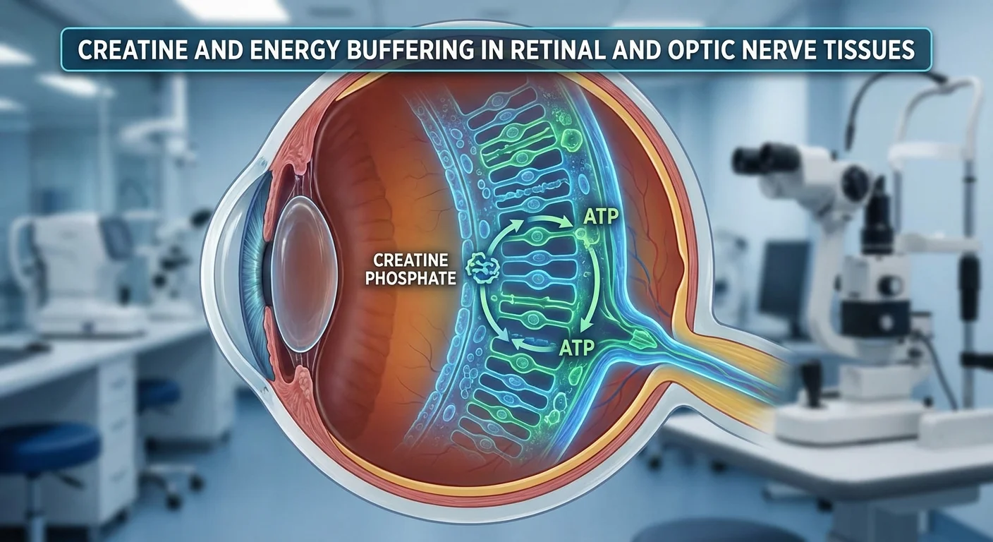
The Creatine–Phosphocreatine Energy Buffer
Creatine is a natural molecule made in the liver, kidney and pancreas (from arginine, glycine, methionine) and stored mostly in muscle (≈95%) and also in brain and other tissues (pmc.ncbi.nlm.nih.gov) (pmc.ncbi.nlm.nih.gov). Inside cells, creatine is converted back and forth to phosphocreatine (PCr) by the enzyme creatine kinase (CK). This PCr–CREATINE system serves as an energy buffer: when ATP is used up quickly (for example during muscle contraction or neuron signaling), PCr donates its phosphate to adenosine diphosphate (ADP) to reform ATP. Simply put, PCr can regenerate ATP far more quickly than mitochondria alone (pmc.ncbi.nlm.nih.gov).
In practical terms, within a few seconds of intense activity, a resting cell’s ATP is depleted, but the CK system steps in by converting PCr back to ATP to keep energy levels stable (pmc.ncbi.nlm.nih.gov). After the burst of activity, excess ATP can again recharge creatine back into PCr for the next cycle. This reversible cycle makes creatine/PCr a “ready reserve” of energy, especially important in cells with high and rapid energy needs (pmc.ncbi.nlm.nih.gov) (pmc.ncbi.nlm.nih.gov).
Importantly, this system exists not only in muscle but in nerve cells. Neurons (including RGCs) express CK isoforms that enable them to use creatine. In fact, retinal neurons express predominantly mitochondrial CK, while retinal glial cells use cytosolic CKs (docslib.org). By storing a pool of PCr in cells, tissues like the retina can get an instant ATP supply when needed.
Creatine in the Retina and Optic Nerve
Role of Creatine in RGC Metabolism
In the retina, RGCs have very high energy demands. Even brief impulses require substantial ATP for ion pumps and signaling. When IOP rises or blood flow drops, RGCs can become ischemic, meaning oxygen and nutrients can’t meet demand. In such situations, the PCr reserve is crucial. Research notes that when optic nerve blood flow is poor (as may happen in glaucoma), tissues rely on PCr to keep ATP levels from crashing (pmc.ncbi.nlm.nih.gov). In other words, phosphocreatine acts as a local energy “battery” that RGCs can draw on during stress (pmc.ncbi.nlm.nih.gov).
Experimental work in other nerves supports this: adding creatine before an induced ischemia protected brain axons and prevented ATP depletion (pmc.ncbi.nlm.nih.gov). These findings suggest RGCs could similarly benefit from extra creatine under IOP-induced stress. The idea is that if RGCs are better able to maintain ATP via the CK–PCr system, they might resist damage and death.
Laboratory Studies of Creatine and Retinal Neurons
Several studies have tested creatine’s effect on retinal neurons. In rat retinal cell cultures, adding creatine to the medium protected neurons (including RGCs) from death due to metabolic toxins or glutamate excitotoxicity (docslib.org). In those in vitro experiments, creatine dramatically reduced cell loss caused by energy poisons (like sodium azide) or by NMDA (a glutamate agonist) (docslib.org). Blocking CK eliminated the protection, confirming the effect was through the creatine energy buffer (docslib.org). These results show creatine can directly support retinal neurons when their energy production is deliberately impaired.
However, translating this to intact eyes has been challenging. In live rat models of retinal injury (either NMDA excitotoxicity or brief high IOP ischemia), giving animals oral creatine did raise retinal creatine levels but did not significantly improve RGC survival (docslib.org). In other words, despite creatine entering the retina in vivo, it failed to save RGCs from acute injury in those studies (docslib.org). The reasons for this discrepancy are not fully clear; it may involve differences in delivery, timing, or the severity of injury.
Overall, the lab data suggest that while creatine can protect retinal neurons under controlled conditions, its benefit in whole-animal glaucoma models is unproven. This gap highlights the need for more research on creatine dosing, formulation (to cross barriers or stay longer), and timing in eye tissues.
Other Neurodegenerative Model Insights
Creatine’s potential reaches beyond the eye. It has been widely studied in other neurological conditions characterized by energy failure. For example, creatine shows broad neuroprotective actions in models of stroke and brain hypoxia (pmc.ncbi.nlm.nih.gov). Clinical interest has spanned Parkinson’s disease, Huntington’s disease, amyotrophic lateral sclerosis (ALS), Alzheimer’s disease, and even psychiatric disorders (pmc.ncbi.nlm.nih.gov) (pmc.ncbi.nlm.nih.gov). In animal models of Parkinson’s (with toxin-induced mitochondrial dysfunction), dietary creatine improved neuronal survival in early studies. In humans, creatine has been tested in clinical trials for PD and memory impairment, given its antioxidant and ATP-buffering properties (pmc.ncbi.nlm.nih.gov).
While these fields are separate from ophthalmology, they share a key concept: neurons that lose energy balance tend to die. If creatine can slow neurodegeneration in one system, it may help in another. Thus, lessons from brain and spinal cord studies support exploring creatine for the retina. In fact, Nicotinamide (Vitamin B3), which indirectly boosts cellular energy, has been shown to protect RGCs in glaucoma models (pmc.ncbi.nlm.nih.gov) – hinting that metabolic support can help RGCs. Creatine is a logical candidate in this category.
Systemic Aging and Functional Benefits
Beyond the eyes, creatine has known benefits for aging muscle and brain function. In older adults, creatine supplementation (often combined with exercise) improves muscle mass, strength, and bone health (pmc.ncbi.nlm.nih.gov). Meta-analyses of older populations show creatine + resistance training significantly increases lean body and muscle mass compared to training alone (pmc.ncbi.nlm.nih.gov). This can translate to better physical function and independence in the elderly.
Cognitively, there are promising signs that creatine may help. Aging is associated with a natural decline in brain creatine levels, and trials have found that older people taking creatine sometimes perform better on memory or intelligence tests. One review noted that creatine “could enhance cognition in elderly subjects,” though mechanisms are not fully understood (pmc.ncbi.nlm.nih.gov). Safety and efficacy data suggest creatine crosses the blood–brain barrier, so it raises brain PCr as well as muscle PCr (pmc.ncbi.nlm.nih.gov) (pmc.ncbi.nlm.nih.gov). This has led researchers to propose creatine as an adjunct in mild cognitive impairment or early dementia, though large trials are still needed.
In summary, creatine is not just for athletes – it is increasingly viewed as a general energy booster for aging tissues. Its track record in preserving muscle and possibly brain function supports the idea that “if it works there, maybe it will help the stressed optic nerve too”.
Safety Considerations: Renal and Fluid Effects
Creatine is widely used and generally safe at recommended doses (typically a loading of ~20 g/day for a week followed by 3–5 g/day maintenance). Its safety profile has been carefully studied. The main observed effect in many studies is a small weight gain, usually just a couple of kilograms, due to water retention in muscles (pmc.ncbi.nlm.nih.gov). No serious harmful side effects consistently appear in healthy people.
A large meta-analysis of studies (over 400 subjects) reported that apart from weight gain, there were no differences in hydration or kidney volume between creatine users and controls (pmc.ncbi.nlm.nih.gov). In fact, increased intracellular water seems to stay within muscle cells, without changing blood pressure or blood plasma volume significantly (pmc.ncbi.nlm.nih.gov) (pmc.ncbi.nlm.nih.gov). Thus, while athletes speculated about cramps or dehydration, controlled data show creatine simply pulls more water into cells – something that normal hydration and monitoring can manage.
The most common concern is on kidney function. Creatine breakdown produces creatinine, a normal waste. Blood creatinine levels do rise slightly after creatine use, which can mimic kidney impairment in standard lab tests. However, up-to-date evidence shows this is a benign lab change, not actual damage. A 2025 systematic review found that creatine supplementation caused a very small, transient rise in serum creatinine but caused no change in glomerular filtration rate (GFR) (bmcnephrol.biomedcentral.com) (bmcnephrol.biomedcentral.com). In plain terms, creatine users had a higher creatinine number on lab tests (because of more turnover), but their kidneys were filtering just as well as non-users. The conclusion: when used responsibly in healthy adults, creatine does not harm kidney function (bmcnephrol.biomedcentral.com) (bmcnephrol.biomedcentral.com). Of course, people with pre-existing kidney disease should consult a doctor before using any supplement.
Fluid balance is another consideration. As noted, creatine tends to increase total body water – mostly inside cells. Early studies showed that a week of creatine loading increased total body water significantly (pmc.ncbi.nlm.nih.gov). This is usually not dangerous; it just makes muscle feel fuller. A recent large population study (NHANES dietary data) examined how different dietary creatine intakes affected hydration markers across thousands of people. It found that very high creatine intakes (above typical dietary levels) were actually associated with slightly lower total body water and fluid volumes and subtle shifts in blood osmolality (pmc.ncbi.nlm.nih.gov). This was unexpected, and suggests the relationship between creatine and hydration is complex. The take-home for patients is minimal: modest creatine use might cause a bit of water retention, but should not dehydrate you. Drinking normal amounts of water remains advisable when taking creatine, especially during exercise.
In terms of overall safety, a broad review of older adults taking creatine found no increase in any side effect versus placebo (pmc.ncbi.nlm.nih.gov). Creatine has been evaluated by regulatory bodies (e.g. the FDA) and is confirmed as safe for healthy use. The most frequently reported issues are mild gastrointestinal upset (rare) or muscle cramping (disputed), but these occur no more often than in controls. Given this safety record, adding creatine in older patients to improve energy balance is a reasonable proposition, if done under medical guidance.
Relevance to Glaucoma and Research Directions
Putting this together for glaucoma: glaucoma is now understood not just as high pressure, but as a chronic RGC energy crisis. Studies in mouse glaucoma models (e.g. the DBA/2J mouse) show that high IOP and aging drain ATP in the optic nerve well before cells die (pmc.ncbi.nlm.nih.gov). The logic is that bolstering the RGC energy supply might slow or prevent degeneration. Creatine, by replenishing ATP via PCr, is a plausible neuroprotective agent in this context (pmc.ncbi.nlm.nih.gov) (docslib.org).
To translate this idea, new research is needed with specific eye-focused endpoints and biomarkers. Key recommendations include:
-
Ocular imaging endpoints: Future trials should include structural imaging of the optic nerve and retina. Optical coherence tomography (OCT) can measure the thickness of the retinal nerve fiber layer (RNFL) and ganglion cell layer. These quantitative measures are sensitive to early RGC loss. For example, RNFL/OCT thinning is strongly associated with glaucoma severity (pmc.ncbi.nlm.nih.gov). Any neuroprotective treatment should aim to slow thinning. Another imaging modality is optical coherence angiography (OCTA), which visualizes retinal blood flow; since energy delivery involves circulation, OCTA could monitor vascular changes.
-
Functional tests: Visual function testing is crucial. Standard visual fields detect vision loss from glaucoma, but more specific tests like the pattern electroretinogram (PERG) or multifocal VEP can measure RGC function directly. Including PERG amplitude or latency as an endpoint could reveal early functional benefits of creatine that precede field changes.
-
Metabolic imaging: Creatine’s effect on energy metabolism might be tracked by advanced imaging. Magnetic resonance spectroscopy (^31P-MRS) can noninvasively measure PCr and ATP levels in neural tissue (demonstrated in brain). It has also been applied in optic pathways (pubmed.ncbi.nlm.nih.gov). ^31P-MRS of the optic nerve or visual cortex after supplementation could directly show if PCr levels rise in the visual system. Likewise, near-infrared spectroscopy (NIRS) or retinal oximetry could monitor changes in oxygen/glucose usage in the retina.
-
Clinical trial design: Randomized trials in glaucoma patients or high-risk individuals would be needed. Important factors are dosing (likely similar to sports usage, ~3-5 g/day), duration (months to years), and control of other risk factors (IOP, blood pressure). Endpoints should combine ocular imaging and function (as above) with neurodegenerative biomarkers (e.g. neurofilament light chain) if available. Given creatine’s profile, trials could start with normal-tension glaucoma patients, who already show RGC vulnerability, to see if vision decline slows without pressure changes.
-
Safety monitoring: Even though creatine is safe generally, ocular studies should monitor kidney markers and fluid status as a precaution. In older glaucoma patients, renal function and hydration should be checked, especially if they have comorbidities or take other meds.
Overall, current evidence is not sufficient to recommend creatine for glaucoma yet. But its known systemic benefits on muscle and possibly brain in aging, coupled with specific data that it can support RGCs in culture (docslib.org) and energy metabolism in nerves (pmc.ncbi.nlm.nih.gov), make it a promising avenue. Well-designed trials with ocular endpoints (OCT/PERG) and perhaps metabolic imaging (MRS) would clarify whether creatine supplementation can indeed energize the optic nerve and protect vision.
Conclusion
Glaucoma may be seen as an energy-starvation disease of RGCs. Creatine, by bolstering the phosphocreatine energy buffer, offers a rational way to sustain neuronal ATP under stress. In vitro studies show clear benefits for retinal neurons (docslib.org), and neurodegenerative research suggests broader potential (pmc.ncbi.nlm.nih.gov) (pmc.ncbi.nlm.nih.gov). Creatine’s safety and aging-related benefits (muscle, possibly brain) further support its exploration in eye health. The next step is targeted research: trials and animal studies designed with optic nerve imaging and RGC function tests, to see if this weight-training supplement can also weight-bear the energy needs of the retina.