Introduction
Yes – glaucoma can indeed strike one eye while sparing the other, or at least cause one eye to be much worse than its fellow. In fact, it is surprisingly common for glaucoma to present asymmetrically: a large fraction of newly diagnosed patients have one eye with higher pressure, more optic nerve damage, or worse visual field loss than the other. Indeed, even classic forms of glaucoma that are generally bilateral often begin or are detected in one eye first. Clinicians report that roughly one- to two-thirds of patients show significant inter-eye differences in at least one measure at diagnosis. For example, one study found that interocular pressure differences of 3 mmHg or more were highly predictive of glaucoma [64] and other research shows that meaningful visual field or nerve-fiber loss asymmetry can occur in on the order of tens of percent of patients. This asymmetry is clinically important – it both preserves useful vision (since the “good” eye compensates) and masks the disease (because the patient doesn’t notice a problem until very late).
In this article we will explore unilateral and asymmetric glaucoma in depth. We begin with why one eye can be affected more than the other, then discuss how the healthy eye hides the loss in the bad eye. We review all known causes of truly one-sided glaucoma (from eye trauma to inflammatory disease to eye structure differences). We explain why one optic nerve might be more vulnerable than the other even under similar eye pressure, and why we sometimes see glaucoma only in one eye in angle-closure disease. We cover how doctors use the two eyes as a pair of internal controls to diagnose and track glaucoma, and what else must be ruled out when damage is very uneven. Finally, we talk about what life is like for a patient with glaucoma in “only one eye,” including the coping, driving, and treatment issues unique to this situation. In each section we will point out relevant studies or clinical observations to back up the discussion (pubmed.ncbi.nlm.nih.gov) (pubmed.ncbi.nlm.nih.gov) (pmc.ncbi.nlm.nih.gov). With any luck, you’ll come away with a clear understanding that asymmetric glaucoma is not mild – it’s sneaky, and requires special vigilance in both diagnosis and treatment.
How and Why Glaucoma Can Affect One Eye More Than the Other
Under ideal conditions, glaucoma tends to be a bilateral disease (affecting both eyes) because many risk factors – like blood pressure, genetics, and age – are systemic. However, eyes are not identical twins, and even in the same person there can be meaningful differences between the two eyes. These differences can occur at every level: eye pressure, drainage anatomy, history of injury or disease, and even the susceptibility of the optic nerve. Below we list the main factors that can lead to one eye developing glaucoma (or worse glaucoma) when the other eye is relatively healthy.
-
Inter-eye pressure asymmetry. In most people, the pressures in the right and left eye are similar, but often one eye runs a bit higher. Even a seemingly small difference can matter. Studies show a clear dose–response effect: as the gap in pressure grows, the risk of glaucoma soars. For example, one multicenter study found that when the IOP (intraocular pressure) difference between eyes was 3 mmHg, the chance of having glaucoma rose to about 6%; if one eye was >6 mmHg higher, that risk jumped to ~57% (pubmed.ncbi.nlm.nih.gov). In plain terms, if one eye gradually operates a few millimeters of mercury higher than its fellow over many years, the higher-pressure eye will tend to accumulate more damage (deepening the optic nerve “cup” and losing more visual field) in a dose-dependent way. Small baseline anatomical differences – such as slightly tighter drainage channels (trabecular meshwork) in one eye – can underlie these pressure asymmetries. Over decades, even a 3–5 mmHg difference can compound into dramatically worse disease in the higher-pressure eye.
-
Traumatic angle-recession for one eye. A blunt injury to one eye (like a punch or car accident) can tear or damage the drainage angle. This “angle recession” often does not cause immediate problems – the IOP may remain normal for years – but eventually the injured eye can develop glaucoma decades later as the damaged tissue scars and stops draining fluid properly. Notably, trauma usually hits just one eye. A patient may have forgotten a 20-year-old injury, only to have that same eye later develop pressure spikes and optic nerve loss, whereas the untouched fellow eye is fine.
-
Inflammatory causes in one eye. Infections or inflammation often affect just one eye. For example, herpes simplex or herpes zoster (shingles) can cause severe uveitis (inflammation inside the eye) in one eye. The inflammation itself can raise pressure, and the steroids used to treat it can further raise pressure. Unilateral uveitic glaucoma from herpes is a well-known scenario. Similarly, Posner-Schlossman syndrome (a recurrent swollen-iris condition often linked to tiny herpes particles) typically causes repeated pressure spikes in one eye, leading to damage over time.
-
Preglaucoma deposits in one eye. Some eye conditions deposit material in the drainage angle unilaterally. The classic example is pseudoexfoliation syndrome (PXF). Although PXF is fundamentally a body-wide disorder of elastin mishandling, it often appears to involve one eye first. Clinically, many patients are noted to have PXF material on the lens and iris of only one eye while the other looks clear (pubmed.ncbi.nlm.nih.gov). Research suggests that even when only one eye shows fibrils by exam, both eyes may eventually have microscopic deposits. But clinically, one eye can sit years or decades behind the other. PXF elevates IOP by clogging the outflow pathways, so it can cause severe glaucoma in just one eye while the other remains PXF-free or only mildly affected.
-
Fuchs’ heterochromic iridocyclitis. This is a rare chronic uveitis that always hits one eye (the name even implies different-colored irises). Fuchs’ can quietly lead to glaucoma in that one eye over time. Patients typically have a slightly whiter eye and tiny blood vessel changes on the iris of only one side. Because the other eye is completely normal, this glaucoma is unilateral by definition.
-
Pigment dispersion syndrome. In younger, myopic patients, a bowed iris can rub against the lens and fling pigment into the drainage angle. Sometimes one eye’s iris anatomy is enough different (more concave or floppy) that it sheds pigment and raises pressure more than the other eye. Although true pigmentary glaucoma is often bilateral, subtle anatomical asymmetry can cause one eye to exhibit a clearly progressive pigment-related pressure rise while the other remains stable.
-
Blood vessel obstructions. A retinal vein occlusion (blockage of a large retinal vein) in one eye can lead to neovascular glaucoma in that eye. For instance, a central retinal vein occlusion causes retinal ischemia, which triggers growth of abnormal new vessels over the iris and angle. That “neovascular glaucoma” usually occurs in just the badly ischemic eye, leaving the fellow eye untouched.
-
Unilateral steroid response. Perhaps surprisingly, one eye can be a “steroid responder” even if the other isn’t. If a patient uses steroid eye drops (for allergies or post-surgery inflammation), sometimes only one eye’s pressure will soar. The precise reason isn’t fully understood, but could relate to small genetic/pharmacologic differences in the trabecular meshwork cells between eyes. In practice, any incompletely explained rise in pressure in one eye after topical steroids while the other eye’s pressure stays normal suggests a unilateral steroid response.
In short, true unilateral glaucoma is often secondary (trauma, uveitis, pigment, pseudoexfoliation, vein occlusion, etc.) because those conditions are inherently one-sided. But even in presumed primary open-angle glaucoma, one eye may have higher baseline IOP due to tiny anatomical differences in outflow, causing better eye forgiveness. Any process that is asymmetric – outsiders it.
Asymmetric Vulnerability of the Optic Nerve
Even if two eyes had identical eye pressure and drainage anatomy, one optic nerve can still be more prone to glaucomatous damage than the other. Nature did not guarantee absolute symmetry. Several optic nerve head factors can differ from one eye to the other:
-
Disc size and shape. Larger optic disks contain more nerve fibers, but they also have a larger lamina cribrosa (the sieve-like structure the nerves pass through) which may be mechanically weaker. In essence, a big optic nerve head might lose more fibers visibly with a given pressure stress. Conversely, a very small disk can appear to have a deep cup even before any disease (giving a misleading impression). Clinically, a doctor must always factor in disc size when judging damage (pmc.ncbi.nlm.nih.gov). A large disc in one eye could mean more potential damage than a smaller disc under the same pressure.
-
Optic nerve anatomy quirks. One eye may have a tilted insertion of the optic nerve or more peripapillary atrophy (surface thinning around the nerve). Tilt or coiled entry can stretch or stress nerves, making them more susceptible to pressure. Differences in lamina cribrosa thickness between eyes have been measured with advanced OCT scans: one eye might have a thinner, more concave lamina, so pressure pulls harder on it. In effect, even identical pressures can cause more strain in one eye if its lamina pores are wider or its supporting connective tissue is weaker.
-
Ocular blood supply differences. The optic nerve head is nourished by tiny branches of the posterior ciliary arteries. In some people, vascular anatomy is lopsided: one eye might naturally receive slightly less choroidal or ciliary blood flow. A weaker perfusion pressure in one optic nerve (especially during nocturnal blood pressure dips) could aggravate damage from the same IOP. While direct measurement in humans is tricky, ophthalmologists recognize that asymmetries in ocular circulation can exist. For example, one internal carotid or vertebral artery might branch differently, or cilioretinal vessels may supply only one eye.
-
Sleeping position (“the pillow hypothesis”). Many glaucoma patients report sleeping habitually on one side. Recent studies noticed a correlation: patients tend to have worse glaucoma on the side they sleep on【40†】. The theory is that lying face-down or on one pillow repeatedly raises the pressure in that eye each night. Over years, that subtle nocturnal pressure elevation on the “pillow side” could lead to significant damage on that side. While research is ongoing, the idea is gaining traction: the side you sleep on may become the side with the worse visual field loss. (One study directly interviewed patients about sleep position and found a significant link between preferred side-sleeping and the worse-eye field loss.)
In summary, even absent any other differences, two eyes in the same person are biologically not interchangeable. Subtle anatomical and vascular differences can make one optic nerve head a “weak link,” so that that eye suffers more damage under the same disease process.
Angle-Closure Glaucoma: Why One Eye Can Attack First
Angle-closure glaucoma has its own story of asymmetry. By definition, angle-closure diseases stem from narrow or closed drainage angles, which often affects both eyes anatomically. However, in practice the acute attack pattern and chronic progression can seem unilateral:
-
Acute attack in one eye. Many patients wake up with an abrupt, painful red eye from angle-closure in, say, the left eye. The right eye at that moment, though similarly narrow in angle, has not yet equaled that pressure. It is essentially a ticking time bomb. In acute angle-closure, the first eye to go “pop” usually does so while the other eye is still above the threshold. Ophthalmologists always treat the fellow eye prophylactically because its risk is so high (often nearly identical angle anatomy). A laser peripheral iridotomy (LPI) is typically done in both eyes: one eye to stop the attack and prevent further pressure spikes, and the other to prevent its own emergency attack (pubmed.ncbi.nlm.nih.gov). Even if the second eye appears normal at a given exam, it usually has equally crowded angles behind the scenes.
-
Chronic asymmetric progression. In chronic angle-closure, one eye may develop more synechiae or lens thickening than the other, meaning one eye’s angle gradually seals off more. Slight differences in lens tilt, iris insertion, or axial length can cause one eye to build pressure gradually while the other lags behind. Over years, one eye can end up with glaucomatous damage while the fellow eye still only has “narrow angles” but no field loss.
-
Truly unilateral anatomical causes. Rarely, an acute angle-closure can be truly one-sided due to a unique cause. Examples include an iris cyst or tumor on one side, or a small lens subluxation (partial dislocation) in one eye pushing the iris forward. In those cases, only the affected eye has mechanical blockage. Another dramatic example is a posterior segment mass (like a ciliary body melanoma) pushing the iris of one eye forward. In all of these cases, the unaffected eye’s angle is completely normal, so the glaucoma is one-eye-only. These situations are usually obvious once identified (for example, an ultrasound or scan finds the cyst or tumor). They require different management (often tumor removal or cyst drainage, not just standard glaucoma care) on that one eye.
Diagnostic Significance of Asymmetry
When an eye doctor sees marked side-to-side differences in glaucoma measures, it immediately raises a diagnostic flag:
-
Red flag for “secondary” cause. Ophthalmologists are taught: if one eye is clearly worse than the other, think secondary glaucoma or other pathology before complacently calling it routine primary open-angle glaucoma. For instance, very asymmetric optic nerve cupping might prompt a doctor to check for an optic nerve sheath meningioma, pituitary lesion, or orbital mass affecting the worse eye. Similarly, if a patient has one eye with cupping and the other eye is completely healthy, the doctor will carefully ask about past trauma, steroid use, or uveitis that could explain a secondary cause on that side.
-
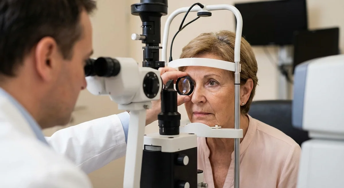
Each eye as a control. A powerful diagnostic tool is simply comparing the two eyes’ anatomy and function. The “good” eye often defines that patient’s personal normal. For example, on OCT scans the fellow eye’s nerve fiber layers, retinal ganglion cell maps, and optic disc size are the patient’s baseline. By overlaying maps of the two eyes (side-by-side OCT ganglion cell or nerve-fiber analyses), an ophthalmologist can spot thinning in the worse eye even if it is borderline. Small abnormalities that might not reach “outside normal limits” on one eye alone can become glaring when contrasted with the normal sister eye. In fact, software in modern OCT machines often allows “mirror” or asymmetry analysis to highlight where one eye is thinner than its counterpart, helping catch early unilateral glaucoma even before absolute thresholds are met.
-
Excluding other causes of one-eyed damage. True asymmetric nerve damage mandates consideration of non-glaucomatous causes. For example:
- A compressive optic neuropathy (like an optic nerve tumor or brain lesion) can produce glaucoma-like cupping, but it is usually one-sided (or severely asymmetric). Studies show that when optic nerve cupping is pronounced but only affects one eye, neuroimaging is recommended (pmc.ncbi.nlm.nih.gov).
- Anterior ischemic optic neuropathy (AION) – a stroke of the optic nerve – often affects only one eye (the “first eye” usually), causing optic disc pallor and some cupping later on. Without attention, one might mistake this for glaucoma in that eye, but the clinical clues (optic nerve swelling during the event, altitudinal field loss, risk factors like giant cell arteritis) lead away from glaucoma.
- Optic neuritis (demyelinating optic neuropathy) typically hits one eye in multiple sclerosis and causes vision and color loss, and can leave a pale optic nerve. Again, this must not be mistaken for glaucoma.
- Congenital optic disc anomalies – some people are born with one nerve that looks abnormally cupped (e.g. coloboma, tilted disc, or congenital asymmetrical cupping). These are non-glaucomatous cupping that also must be distinguished.
Because these other conditions require urgent treatment (e.g. neurosurgery or stroke workup), eye doctors take one-sided optic nerve damage very seriously. The rule of thumb is: with pronounced asymmetry, do a careful workup that may include visual-evoked potentials or brain/orbit MRI to exclude compressive or neurologic causes, before concluding “just glaucoma.”
Why the Patient Often Doesn’t Notice Until Late
Paradoxically, having one eye that’s still fine delays recognition of how bad the diseased eye is. This is because of binocular vision:
-
Binocular compensation. With both eyes open, the brain mostly relies on the better eye for everyday tasks and “fills in” where the worse eye has deficits. If one eye has a patchy field defect or blurred vision from glaucoma, the other eye can cover in just about all routine activities – driving, reading, walking – without the person realizing anything’s wrong. The patient might still pass distance vision tests (using the good eye) and only fail the visual field test in the poorer eye. In other words, vision is functionally binocular, and the healthy eye masks the blind spots of the bad eye.
-
Delayed symptoms and adherence issues. Because patients feel “normal,” they are often surprised to learn about significant damage. This creates a tricky situation: the patient might ask, “If I can see just fine with both eyes, why do I need treatment?” They have no pain and no obvious vision trouble. This can lead to poor adherence – they might skip drops or appointments because “I feel okay.” Unfortunately, glaucoma damage cannot be reversed, so by the time one good eye left sees trouble or double vision, it is too late for the bad eye.
-
Driving and daily life. Legally, driving vision is often assessed by the better eye. If one eye still has good central acuity and a mostly full field, many patients continue to drive legally even when the other eye has advanced loss on its side. However, peripheral blind areas on the damaged side do reduce hazard detection. For example, a patient with right-sided glaucoma field loss might not notice pedestrians or cars approaching from the right as quickly, even though central vision is fine. This poses safety risks that are not obvious to the patient. Likewise, jobs requiring wide field vision (like piloting or some heavy machinery) can be compromised by one-sided field defects, even if the patient “sees well” in a static eye chart sense.
-
Anxiety and psychological burden. Patients eventually do recognize that one eye has sunk into a deep problem while the other is a lifeline. This creates a unique anxiety: “My good eye is all I have.” They often become very motivated to protect the healthy eye, but that carries its own stress. People may become hyper-vigilant, fearing that even a minor eye injury or illness could suddenly leave them blind. Living with asymmetric glaucoma imposes a psychological burden above and beyond symmetric disease, because the patient knows the gulf between the eyes is huge. Counseling and support can be important, since anxiety itself can lead patients to abandon life-enriching activities out of undue fear of their condition.
Management Considerations for One-Eyed Glaucoma
Treating glaucoma is always about risk vs reward, and this becomes more complex when only one eye is at immediate risk:
-
To treat or not to treat the fellow eye? If one eye is significantly damaged and the fellow eye looks totally normal (maybe only mild ocular hypertension or suspicious anatomy), do we act on it? Opinions vary.
-
Pros of treating the “good” eye: Some doctors advocate low-level prophylactic therapy for the less-affected eye (especially if pressures are high-normal or optic nerve is borderline) because the second eye has a higher-than-average risk of conversion (pmc.ncbi.nlm.nih.gov). The rationale is preventive – lower its pressure and possibly delay or prevent any damage. Since every patient’s future risk of bilateral disease is essentially 100%, some feel both eyes should be protected.
-
Cons of unnecessary treatment: Others caution that dropping medication into a healthy eye has downsides: cost, inconvenience, side effects (red eye, allergy, systemic absorption), and the psychological burden of “taking medicine for a disease I don’t have.” If the “normal” eye has truly no sign of disease and normal pressure, immediate treatment might not change the outcome and could undermine quality of life or adherence. Many doctors will watch the fellow eye very closely instead, adding therapy only if it shows any sign of progression.
The decision is individualized. Factors include how high the better-eye pressure is, whether any optic nerve thinning is already present, and patient preference. Shared decision-making is key.
-
-
Setting separate target pressures. In asymmetric cases, each eye often has its own “target” IOP goal. The damaged eye’s target must be very low so as to slow its progression: it already has damage. The good eye’s target might be more moderate, aiming just to prevent any future damage. For example, an eye with moderate glaucoma may have a goal IOP of <15 mmHg, whereas the unaffected eye might have a goal of <18 mmHg. This means the doctor will fine-tune each eye’s drops or schedule differently.
-
Using one eye as a barometer for progression. The relatively healthy eye serves as a control. If at follow-up one notices new thinning or field loss in the once-normal eye, it suggests the underlying disease (or systemic factors) have worsened. For instance, if years later the previously good eye begins to show nerve fiber loss despite treatment, that signals it is probably beginning its glaucomatous journey. This would prompt the physician to tighten therapy in both eyes or seek new causes. In practice, an asymmetric pair is monitored more critically: any change in the better eye is taken very seriously as an early warning sign.
-
Surgery timing and risks. Considering surgery (like trabeculectomy or tube shunt) in unilateral glaucoma involves extra caution. Operating on the diseased eye may be necessary if medical therapy fails, but complications (like infection, bleeding, or hypotony) could be devastating since that eye is already compromised. Surgeons must balance the risk of intervention against the already-high stakes. In some cases, if the worse eye’s damage is near-blind vision threshold, the surgeon might operate earlier to preserve what’s left; in others, they might delay in hopes that the eye never worsens so severely. It is never a casual decision. Likewise, one would never operate on the only good eye (to lower pressure prophylactically) because a complication there would eliminate the patient’s functional vision entirely.
Conclusion
In summary, glaucoma in “one eye” is real – and serious. It is far from the mild end of the spectrum; if anything, it can be more treacherous than symmetrical disease. The presence of a good eye masks the problem in the bad eye, leading to later detection and a false sense of security. At the same time, the healthy fellow eye is a blessing – giving patient’s functional binocular vision – and a diagnostic aid, setting a personalized baseline.
Patients and doctors alike should remember that an eye that “sees okay” cannot be taken for granted in unilateral cases. Rigorous monitoring of both eyes is essential, and therapy must be tailored eye-by-eye. If only one eye has detectable glaucoma today, it means the other eye is on notice. The core message is: always protect the healthy eye with as much vigor as the bad eye. Only by vigilant care of both eyes can we prevent the ultimate disaster of sequential bilateral blindness.