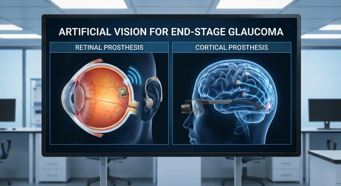
Artificial Vision for End-Stage Glaucoma: Retinal vs. Cortical Prostheses
Advanced glaucoma kills the optic nerve and retinal ganglion cells (RGCs), leaving patients blind. Artificial vision (visual prosthesis) aims to bypass such damage. Most existing prostheses target the retina or optic nerve, but in end-stage glaucoma those routes are gone. Instead, researchers are exploring implants that directly stimulate the visual cortex (brain). This article compares inner-retinal (eye-based) versus cortical (brain-based) prostheses for glaucoma blindness.
In conditions like retinitis pigmentosa or macular degeneration, the photoreceptors die but RGCs and optic nerve remain intact (pmc.ncbi.nlm.nih.gov) (pmc.ncbi.nlm.nih.gov). Retinal implants work here. For example, Argus II (an epiretinal implant) received FDA approval for retinitis pigmentosa in 2013 (pmc.ncbi.nlm.nih.gov). These devices can restore basic light perception and motion detection (pmc.ncbi.nlm.nih.gov) (pmc.ncbi.nlm.nih.gov). However, glaucoma is different: the RGCs and optic nerve are destroyed, so a signal from a retinal implant has nowhere to go (pubmed.ncbi.nlm.nih.gov) (pmc.ncbi.nlm.nih.gov). In novel tests like ORION’s early trial, the goal is to “bypass diseased eye anatomy” altogether by sending electrical signals directly to the brain’s visual cortex (pmc.ncbi.nlm.nih.gov).
In short, retinal implants require surviving retinal neurons and an intact optic nerve (pmc.ncbi.nlm.nih.gov) (pmc.ncbi.nlm.nih.gov). They are designed for outer-retinal disease (photoreceptor loss) where RGCs still exist (pmc.ncbi.nlm.nih.gov) (pmc.ncbi.nlm.nih.gov). Conversely, cortical implants target patients with severe inner-retinal degeneration or optic nerve damage (pmc.ncbi.nlm.nih.gov) (pmc.ncbi.nlm.nih.gov). For advanced glaucoma (no RGCs), cortical approaches are the only realistic prosthetic option (pmc.ncbi.nlm.nih.gov) (pmc.ncbi.nlm.nih.gov).
Inner-Retinal Implants
Inner-retinal prostheses (often called “bionic eyes”) use an external camera (usually on goggles) to capture images and convert them into electrical pulses. These pulses are delivered via a microelectrode array placed on or under the retina (pmc.ncbi.nlm.nih.gov). Epiretinal implants (like Argus II) sit on the retinal surface adjacent to RGCs, while subretinal implants lie beneath the retina among the photoreceptors. There are also suprachoroidal designs (electrodes between the retina and sclera). In all cases, the goal is to electrically stimulate the remaining retinal neurons.
Requirements and Candidates
Candidates for retinal implants must have lost photoreceptor vision but still retain an intact inner retina (ganglion and bipolar cells) (pmc.ncbi.nlm.nih.gov) (pmc.ncbi.nlm.nih.gov). Typical candidates are those with end-stage retinitis pigmentosa or geographic atrophy (advanced macular degeneration), not glaucoma patients. Glaucoma patients lack viable RGCs, so retinal prostheses generally cannot work for them (pmc.ncbi.nlm.nih.gov) (pmc.ncbi.nlm.nih.gov).
Surgical Procedure and Risks
Implanting a retinal device requires a vitreoretinal surgeon. Procedures like a pars plana vitrectomy (removing the vitreous gel) and attaching the electrode array are needed (www.sciencedirect.com). Compared to brain surgery, retinal surgery is less complex. Epiretinal implants are “less complicated and [carry] lower levels of risk during implantation” than cortical devices (pmc.ncbi.nlm.nih.gov). Nevertheless, serious eye-related complications can occur. In a 30-patient Argus II trial, for example, half of the participants experienced device- or surgery-related adverse events over 5 years (pmc.ncbi.nlm.nih.gov). The most common problems were conjunctival erosion (pink eye) and ocular hypotony (abnormally low eye pressure) (pmc.ncbi.nlm.nih.gov). Overall, about 40% of subjects had a serious adverse event (half of which were reversible) (pmc.ncbi.nlm.nih.gov). Systematic reviews confirm that epiretinal implants tend to have more complications than sub- or suprachoroidal designs (pmc.ncbi.nlm.nih.gov).
Spatial Resolution and Visual Performance
Current retinal implants produce very low-resolution vision. For instance, the Argus II’s 6×10 array (60 electrodes) is analogous to a 6×10 pixel camera. In practice, most subjects can perceive only very basic patterns of light and dark. Studies report that epiretinal implant users can localize high-contrast shapes and detect motion better with the device on (pmc.ncbi.nlm.nih.gov) (pmc.ncbi.nlm.nih.gov). In one systematic review, performance on a high-contrast “square localization” task improved by up to 89% with the implant active (pmc.ncbi.nlm.nih.gov). Still, visual acuity with these devices remains extremely poor – even the best cases recorded were only around 20/460 to 20/550 in Snellen terms (pmc.ncbi.nlm.nih.gov) (far below the 20/200 threshold for legal blindness).
Subretinal implants (like the Alpha IMS/AMS system) have achieved slightly higher density (hundreds of photodiode pixels instead of tens of electrodes). A recent summary reported grating acuities up to 3.33 cycles per degree (equivalent to roughly 20/460 vision) and some ability to recognize simple motions and large letters (pmc.ncbi.nlm.nih.gov). However, even these represent only rudimentary vision.
Training and Adaptation
Users of retinal implants require extensive training and rehabilitation to interpret the unusual visual signals. Clinical trial patients typically undergo months of training with vision therapists. For example, Argus II trial participants were “meticulously selected” and given extensive training on device use (pmc.ncbi.nlm.nih.gov). They learned tasks like locating a bright square, detecting motion, and recognizing basic shapes. This nurturing lab environment may not reflect real-world use, though; one study found that commercial users often rely less on the implant day-to-day, due to usability issues (pmc.ncbi.nlm.nih.gov).
Functional Outcomes and Quality of Life
Even with thousands of dollars of technology, the restored vision is very limited. Realistic goals for retinal implants include navigation and orientation and coarse object localization, rather than reading or face recognition. In the Argus II trials, patients performed notably better on orientation and mobility tasks with the implant activated (pmc.ncbi.nlm.nih.gov). Many could detect doorways or windows, follow bright lines on the ground, and locate large objects. In daily life, users report improvements in mobility and independence. A systematic review found that recipients of retinal implants reported better orientation and mobility, helping them with daily tasks (pmc.ncbi.nlm.nih.gov). However, vision remains far from normal: study authors caution that even with the device, acuity stayed below 20/200 (legally blind) (pmc.ncbi.nlm.nih.gov).
Cortical Implants
Cortical visual prostheses take a very different approach: they bypass the eye and stimulate the brain directly. Electrodes are placed on or in the visual cortex (the brain region at the back of the head that processes sight). An external camera and processor convert images into electrical stimulation patterns sent wirelessly to these cortical electrodes. Since the signal skips the retina and optic nerve, this method works even when those structures are destroyed.
How They Work
For example, Second Sight’s ORION system uses a grid of electrodes placed on the occipital cortex (creating phosphenes—dots of light—in the patient’s visual field) (pmc.ncbi.nlm.nih.gov). Another design (the Illinois ICVP) uses arrays of penetrating electrodes in the cortex (pmc.ncbi.nlm.nih.gov) (pmc.ncbi.nlm.nih.gov). In one trial, a 96-electrode Utah array was implanted into the visual cortex of a blind volunteer. Electrical stimulation there produced simple blue-white flashing dots that followed the visual field. Impressively, the patient could distinguish the borders of objects and even identify different letters by their outlines (pmc.ncbi.nlm.nih.gov). These initial results show that a cortical implant can indeed create patterned vision in a human brain.
Candidate Criteria
Cortical implants are intended for people with complete, untreatable blindness and no useful vision. In practice, trial protocols require no light perception in either eye (www.ninds.nih.gov) (pmc.ncbi.nlm.nih.gov). For example, an Orion clinical trial accepts patients who are bilaterally blind (bare light perception or worse) due to damage anywhere along the visual pathway (retinal disease, optic nerve injury, glaucoma, etc.) (www.ninds.nih.gov). Any remaining sight would make someone ineligible, since invasive brain surgery could risk that residual vision (pmc.ncbi.nlm.nih.gov). In fact, studies emphasize that people eligible for retinal implants (those with intact inner retina) generally are poor candidates for cortical implants (pmc.ncbi.nlm.nih.gov). Thus, cortical devices target the final option for vision restoration in totally blind individuals.
Surgical Risks
Cortical prostheses require neurosurgery (craniotomy) to implant electrodes on the brain. This is obviously more invasive than ocular surgery. Potential risks include stroke, infection, or neurological damage. In practice, small feasibility trials have shown surgical safety so far, but sample sizes are tiny. For example, an initial ORION trial implanted its 60-electrode array in six blind subjects without any implant-related serious adverse events (pmc.ncbi.nlm.nih.gov). (All six reported seeing phosphenes with the device on.) However, experts caution that brain surgery must be approached very carefully, which is why only completely blind candidates are considered (pmc.ncbi.nlm.nih.gov).
Spatial Resolution and Visual Performance
Spatial resolution for cortical implants is also very low at present. Existing devices have at most a few dozen electrodes. ORION, for instance, used 60 electrodes on the cortex (pmc.ncbi.nlm.nih.gov). Even with dozens of electrodes, the “image” a patient perceives is extremely grainy. In the aforementioned trial with 96 electrodes, patients could only perceive very simple patterns (lines, simple letters). Achieving finer detail would require hundreds or thousands of electrodes spread across multiple patches of cortex — technology that does not exist yet for humans.
Training is similarly intensive. Like retinal systems, cortical patients must learn to interpret their abnormal visual input. Early reports suggest patients can learn to recognize large shapes and letters over time, but this warrants long rehabilitation. There is not yet published data on formal training regimens for cortical implants, but by analogy to retinal trials, we expect years of specialized training would be needed to reach any practical utility.
Functional Outcomes
No cortical prosthesis has yet produced a level of vision close to normal. So far, the goals are conservative: navigation and object localization. Trials focus on tasks like identifying large bright objects, detecting motion, and avoiding obstacles. As one review noted, visual prostheses may best help with orientation and navigation, tasks where current blind technologies (like smartphone apps) still fall short (pmc.ncbi.nlm.nih.gov). In experimental settings, blind animals with brain stimulation prostheses have successfully navigated mazes using artificial cues. In human trials, the one patient who identified letters via ORION only recognized very large, isolated characters (pmc.ncbi.nlm.nih.gov).
In summary, early cortical implant studies show that patients can perceive simple patterns of light and use them to guess shapes or letters. This hints at possible benefits for tasks like locating doors or finding edges. However, fine object recognition (e.g. faces or text) remains well beyond current capabilities. Quality-of-life measurements for cortical implants have not yet been reported; ongoing trials plan to include patient-reported outcomes and functional tests (www.ninds.nih.gov) to see if any daily-living improvements occur.
Comparative Summary
-
Surgical complexity: Retinal implants involve eye surgery (vitrectomy and retinal electrode placement). Cortical implants require brain surgery (craniotomy and cortical electrode array). Epiretinal surgery is fairly routine for retinal specialists, whereas cortical implantation carries inherently higher neurological risk. A recent review notes that even a mild remaining vision would be lost if operating on the brain (pmc.ncbi.nlm.nih.gov), underscoring the risk of cortical surgery.
-
Spatial resolution: Both approaches currently deliver very low-resolution vision. Retinal devices use on the order of 60–150 electrodes or pixels, yielding only crude light patterns (pmc.ncbi.nlm.nih.gov) (pmc.ncbi.nlm.nih.gov). Cortical arrays also number in the tens to low hundreds. Neither approach can produce more than very rough shapes. Reported visual acuity in retinal devices rarely exceeds ~20/500 (pmc.ncbi.nlm.nih.gov). Cortical acuity is even harder to define but is expected to be similarly low given the small electrode count.
-
Training/adaptation: All implanted patients need extensive rehabilitation. Clinical trial participants receive intensive, guided training to learn how phosphenes correspond to their environment (pmc.ncbi.nlm.nih.gov). This is true for both retinal and cortical implants. In practice, many users find the constant training burdensome, which may limit how often they actually use the device (pmc.ncbi.nlm.nih.gov) (pmc.ncbi.nlm.nih.gov).
-
Quality of life: Retinal implants have demonstrated some modest quality-of-life benefits. Users often report improved orientation, easier navigation of familiar spaces, and help with day-to-day tasks that rely on detecting light versus dark or large shapes (pmc.ncbi.nlm.nih.gov) (pmc.ncbi.nlm.nih.gov). No cortical implant has yet proven a clear QOL improvement, but researchers are incorporating QOL and daily function measures into ongoing trials (pmc.ncbi.nlm.nih.gov) (www.ninds.nih.gov).
-
Realistic outcomes: Neither approach will restore normal vision. Patients should expect only very basic vision-like sensations. Practical goals include detecting doorways or windows, differentiating light from dark rooms, localizing large obstacles, or recognizing very large letters or shapes. Navigation (walking a room without bumping into objects) is a realistic near-term use-case (pmc.ncbi.nlm.nih.gov) (pmc.ncbi.nlm.nih.gov). Reading or recognizing faces remains beyond current capability.
-
Candidate selection: In practice, patients who are still potential retinal-implant candidates (having some intact inner retina) will not be routed to cortical trials (pmc.ncbi.nlm.nih.gov). Conversely, cortical trials require patients with total blindness (no residual vision) from any cause (including glaucoma) (www.ninds.nih.gov) (pmc.ncbi.nlm.nih.gov). For now, a typical cortical implant candidate is someone who cannot benefit from retinal devices at all (e.g. RGC layer destroyed or optic nerve severed).
Timeline and Regulatory Status
Retinal prosthesis development began over 20 years ago. The Argus II (Second Sight) is a milestone: FDA approval came in 2013 (pmc.ncbi.nlm.nih.gov) (after earlier CE mark in Europe). Argus II remained the only commercially approved system worldwide until the company halted production in 2019 (pmc.ncbi.nlm.nih.gov). Around the same time, some subretinal devices achieved CE approval in Europe – for example the Alpha IMS (2013) and AMS (2016) systems (pmc.ncbi.nlm.nih.gov) – but none are FDA-approved. Many retinal devices are now discontinued or in transition, with only a few (like Pixium’s PRIMA and Bionic Vision’s suprachoroidal implant) in current trials (pmc.ncbi.nlm.nih.gov) (pmc.ncbi.nlm.nih.gov).
Cortical prostheses are much earlier in the pipeline. The first human cortical implant (Brindley-Dobelle in 1970s) was purely experimental. Today, Second Sight (now called Cortigent/Vivani) has led a new effort: their ORION system began an early feasibility trial in 2017 (www.cortigent.com), and as of 2022 they’ve merged into Vivani Medical working on an ORION II upgrade (pmc.ncbi.nlm.nih.gov). No cortical device has yet obtained regulatory approval. Current studies (like Orion’s and the Illinois ICVP study (pmc.ncbi.nlm.nih.gov)) are listed as early feasibility trials (e.g. NCT03344848), with the goal of assessing safety and basic function.
Key milestones to date: Argus II (retina) trials 2007–2009, FDA approval 2013, discontinuation 2019 (pmc.ncbi.nlm.nih.gov) (pmc.ncbi.nlm.nih.gov). Alpha IMS subretinal CE 2013, upgraded AMS CE 2016 (pmc.ncbi.nlm.nih.gov). Orion (cortical) early trials began ~2017, with follow-up studies ongoing (no approvals yet) (pmc.ncbi.nlm.nih.gov). Upcoming: Phase I trials of other cortical systems (CORTIVIS in Spain, ICVP in USA) are recruiting (pmc.ncbi.nlm.nih.gov) (www.ninds.nih.gov). Over the next decade, developers hope to increase electrode counts and brain interfacing to improve outcomes, but for now both retinal and cortical prostheses remain highly experimental.
Conclusion
For most glaucoma patients with end-stage optic nerve damage, retinal implants are not an option because the neural pathway is severed. Cortical visual prostheses thus represent the only viable artificial-vision strategy. Early studies show cortical devices can elicit simple light patterns even when the eye is blind (pmc.ncbi.nlm.nih.gov) (pmc.ncbi.nlm.nih.gov). However, both retinal and cortical approaches currently offer only rudimentary vision. Retinal implants have slightly more clinical history (some FDA/CE approvals in other diseases) and demonstrable, if limited, improvements in mobility and orientation (pmc.ncbi.nlm.nih.gov) (pmc.ncbi.nlm.nih.gov). Cortical implants are at the feasibility stage: they avoid the optic nerve but must overcome the challenges of brain surgery, signal mapping, and patient training. Realistic goals for either approach are basic navigation and object localization, not high-detail vision. Timeframes: retinal devices have been in trials for 15–20 years (with a few commercial products emerging then fading) (pmc.ncbi.nlm.nih.gov), whereas cortical systems are only now beginning human testing. Regulatory clearance in the US/EU for cortical implants is still years away.
In summary, for severe glaucoma with no optic nerve, cortical implants offer hope. But patients and doctors should understand current limitations: even the most advanced “bionic vision” devices restore only on/off light perception and simple shapes. Ongoing research aims to increase electrode counts, improve biocompatibility, and refine signal processing, but at present the functional vision from any prosthesis will be extremely basic. Patients must have realistic expectations (e.g. distinguishing a door from a wall, not reading text) and be prepared for extensive rehabilitation. Future milestones to watch include completion of ongoing cortical implant trials and any approvals of next-generation devices.