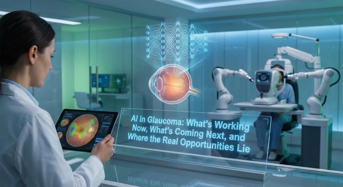
Introduction
Glaucoma is a group of eye conditions that damage the optic nerve and can lead to irreversible blindness. Often called “the silent thief of sight,” glaucoma affects millions worldwide. In fact, an estimated 111.8 million people will have glaucoma by 2040 (medicalxpress.com). Early detection and treatment are critical because vision loss cannot be fully recovered. This is where artificial intelligence (AI) is making inroads: by analyzing eye images and test data, AI can help screen, diagnose, and monitor glaucoma more efficiently. In this article we explore how AI is being applied today in glaucoma care – citing real-world tools and studies – and examine emerging opportunities, especially in vision restoration research. We focus on proven results (e.g. sensitivity and specificity of AI tools) and on concrete future applications, providing practical guidance for patients and researchers alike.
AI in Current Glaucoma Screening and Diagnosis
Smartphone and Fundus Image Analysis
One major use of AI today is automated analysis of fundus photographs (images of the retina) to screen for glaucoma. Research teams have paired portable fundus cameras or smartphone attachments with AI classifiers to flag glaucomatous optic discs. For example, a recent prospective study in India tested an offline AI model embedded on a smartphone fundus camera (Medios AI-Glaucoma on Remidio’s FOP NM-10 device). This system detected patients needing referral (“referable glaucoma”) with about 94% sensitivity and 86% specificity compared to a full clinical workup (pmc.ncbi.nlm.nih.gov). In numbers, the AI correctly identified 93.7% of true glaucoma cases and correctly excluded 85.6% of non-glaucoma cases (pmc.ncbi.nlm.nih.gov). Such high accuracy shows that smartphone-based AI screening can reliably find patients with glaucoma changes in their optic discs.
Another study used a similar AI-camera setup across all severities of glaucoma. It found the AI achieved 91.4% sensitivity and 94.1% specificity for detecting glaucoma or suspect cases (pmc.ncbi.nlm.nih.gov). Performance was slightly lower for very early disease (about 87% sensitivity) and highest for advanced cases (96% sensitivity) (pmc.ncbi.nlm.nih.gov). These results come from outpatient clinics and show that AI tools can match specialist exams in flagging suspicious eyes. They also highlight that AI often errs on the side of caution by flagging mild or suspect cases; in one study most false-positives were eyes labeled “disc suspect” by specialists (pmc.ncbi.nlm.nih.gov). This conservative approach helps avoid missing true disease at the cost of some extra referrals.
Commercial and research groups are already developing such systems. For instance, the Medios AI-Glaucoma system (Remidio, India/ Singapore) integrates on a smartphone fundus camera and has shown the results above (pmc.ncbi.nlm.nih.gov) (pmc.ncbi.nlm.nih.gov). Other AI platforms (e.g. BegIA) use smartphone images to estimate cup-to-disc ratios or even analyze facial images for eye abnormalities (glaucoma.org). In one clinical evaluation, a smartphone app reported an area under curve (AUC) of 0.966 for glaucoma detection, with 95.4% sensitivity and 87.3% specificity (glaucoma.org).
Telemedicine and Remote Screening
AI-enabled apps are also used in telemedicine for glaucoma. For example, the iPredict cloud platform runs AI on uploaded fundus images. In a real-world trial, this telemedicine tool achieved ~89.7% accuracy (83.3% sensitivity, 93.9% specificity) in identifying glaucoma suspects from retinal photos (pmc.ncbi.nlm.nih.gov). The AI classified optic discs into “glaucoma suspect” vs. normal by measuring vertical cup/disc ratio, matching expert graders 93.9% of the time (pmc.ncbi.nlm.nih.gov). The system showed 100% agreement between in-person and AI-processed remote grading for a test set (pmc.ncbi.nlm.nih.gov). This means a patient in a rural clinic could get a real-time screening result via AI, with immediate referral advice if needed. Such platforms make screening more accessible and consistent, especially in underserved areas (glaucoma.org) (pmc.ncbi.nlm.nih.gov).
OCT, Visual Fields and Data Integration
Beyond photos, AI is applied to other glaucoma tests. Deep learning models can segment optical coherence tomography (OCT) scans to measure retinal nerve fiber layer (RNFL) thickness or optic nerve head features. They can also analyze visual field (VF) tests for subtle progression. For instance, convolutional neural networks have been trained to distinguish glaucoma patterns on VF maps. Other AI tools combine multiple data sources – pressure readings, OCT, VFs, patient history – to calculate glaucoma risk scores. While many of these are in development or early trials, they promise to assist clinicians by highlighting patients whose disease may worsen and need closer care. One review reports DL systems that successfully predict future VF loss up to several years ahead by learning from past VF series (pmc.ncbi.nlm.nih.gov). These cutting-edge methods have so far been tested on retrospective data, indicating the feasibility of AI forecasting disease progression, but they have not yet become routine in practice.
Measurable Impact and Performance in Practice
Several studies demonstrate tangible performance of AI tools in clinic-like settings. As noted, smartphone-fundus-AI achieved ~91-94% sensitivity and ~86-94% specificity in large patient cohorts (pmc.ncbi.nlm.nih.gov) (pmc.ncbi.nlm.nih.gov). The telemedical AI project reported ~89.7% overall accuracy (pmc.ncbi.nlm.nih.gov). These are impressive figures – in research settings AI is already on par with trained ophthalmologists for screening picks. Importantly, some false negatives were only mild early glaucoma, while false positives tended to be “disc suspects” not clear-cut normals (pmc.ncbi.nlm.nih.gov).
Equally important is adoption. Systems like Medios and the iPredict model are being rolled out in parts of India and elsewhere for population screening. Though detailed adoption data is emerging, initial reports (for example, Remidio’s outreach programs) suggest hundreds of clinics using AI-driven camera units. AI is also making its way into hospital workstations: several OCT device manufacturers are integrating AI segmentation and analysis features to flag RNFL thinning or predict RNFL loss. In academia, many clinics now trial AI models on existing data to refine diagnostics.
That said, adoption in Western clinical practice is still limited by regulatory approval and workflow integration. No FDA-approved AI is yet standard for glaucoma screening (unlike diabetic retinopathy where AI systems like IDx exist). However, promising field trials and peer-reviewed validations suggest rapid progress. As AI in glaucoma screening has a clear public health benefit (catching disease before vision loss), we can expect some of these tools to seek regulatory clearance in the next few years.
Emerging AI Applications: What’s Next
Predictive Analytics and Personalized Care
The next wave of AI in glaucoma will focus on prediction and personalization. Machine learning models can combine clinical, imaging, and genetic data to forecast an individual’s risk of vision loss or of converting from ocular hypertension to glaucoma (pmc.ncbi.nlm.nih.gov). For example, neural nets trained on patient records purportedly identify who is most likely to progress. In coming years, such systems could help doctors tailor treatment aggressiveness. Imagine an AI score that weighs IOP, corneal thickness, ethnicity, family history, and more to compute a “time until blindness” estimate – helping prioritize therapy. Large datasets now exist (from biobanks and eye hospitals), so AI can learn intricate patterns beyond simple risk factors.
Glaucoma Monitoring and Home Testing
AI could also revolutionize monitoring. Wearable intraocular pressure (IOP) sensors or smart contact lenses are under development, and AI could analyze their continuous data to alert patients of dangerous spikes. Similarly, smartphone-based visual field apps are improving (for example apps that project perimetry charts on the phone). When coupled with AI, these could become home glaucoma tests. Patients might one day self-administer quick eye exams at home, with an app using AI to detect new changes and inform their doctor, rather than visiting the clinic. Early prototypes of home tonometry and vision testing exist, but AI-driven analysis will make them clinically useful by ensuring reliability and flagging real deterioration.
Surgical Planning and Outcome Prediction
Surgical interventions (trabeculectomy, shunts, MIGS) are another frontier. AI could help predict which patients will best respond to which surgery, by analyzing thousands of past cases. For example, a machine-learning tool might learn that patients with X pattern on imaging and Y genetics do well with a drainage implant, while others do better with laser trabeculoplasty. Such decision-support tools are under research in many fields; glaucoma surgery could benefit similarly. Additionally, AI could guide robotic eye surgery in future, though that is longer-term.
Vision Restoration and Regeneration – Untapped Opportunities
One of the most exciting frontiers is vision restoration after glaucoma damage. Currently there is no therapy to regrow optic nerves or replace lost retinal ganglion cells (RGCs). However, researchers are intensely working on neuroprotection, gene therapies, stem cell transplants, and prosthetics. AI has only begun to influence these areas, but the opportunities are real:
-
AI-Assisted Drug Discovery: A striking example is a 2024 study where AI screeners identified small molecules that protect RGCs under glaucoma-like stress. Using large language models and graph neural nets, the researchers predicted candidate inhibitors of RIPK3 (a cell-death kinase). After lab tests, one compound (HG9-91-01) was found to preserve RGC structure in an acute glaucoma model (pmc.ncbi.nlm.nih.gov). In fact, all five AI-recommended molecules in that study helped RGC survival under low-oxygen stress, with HG9-91-01 giving the best protection. This AI-enabled neuroprotective drug discovery shows how computational methods can accelerate preclinical glaucoma therapy development (pmc.ncbi.nlm.nih.gov). (A popular science report described this as AI “aids discovery of potential glaucoma drug candidates” (medicalxpress.com).)
-
Neural Prosthesis Design: For patients who have already lost vision, technologies like retinal or optic nerve implants might offer a way to regain some sight. Designing such devices is extremely complex. Here too AI and modeling play a role. For example, a 2024 paper developed a detailed computational model of the optic nerve and visual brain to evaluate “optic nerve stimulation” prostheses. The team used machine-learning–simulated images to test how electrode arrays on the optic nerve might restore wide-field vision (pmc.ncbi.nlm.nih.gov) (pmc.ncbi.nlm.nih.gov). Their findings suggest optic nerve implants could potentially produce broader visual fields than current retinal prostheses, and importantly they provided a modeling framework to optimize electrode placement and stimulation strategies (pmc.ncbi.nlm.nih.gov) (pmc.ncbi.nlm.nih.gov). This kind of work shows how in silico tools and AI-driven image processing can guide the next generation of vision-restoring implants.
-
Future Gene/Cell Therapies: Regenerative approaches – such as reprogramming Müller cells into RGCs, transplanting RGCs, or using gene editing to reactivate growth – are under intense basic research. AI could eventually accelerate these by analyzing large genetic and molecular datasets. For example, a 2024 Development paper performed a huge CRISPR screen to uncover genes controlling RGC regeneration【65†】. Machine learning methods could help mine these complex results to prioritize targets. Moreover, AI-driven protein design (e.g. AlphaFold or generative models) might create novel therapeutic proteins or gene constructs for regeneration. While such AI applications have not yet been reported in glaucoma, the field of genomics and stem cell therapy is ripe for AI. Computational tools might predict which gene combinations encourage axon regrowth, or optimize viral vectors for safer gene delivery.
Currently, integrating AI into RGC regeneration research is limited, but it represents a high-value opportunity. As regenerative therapies (nanoparticles, stem cells, optogenetics) advance, AI could help optimize their design and delivery. For instance, computer simulations might model how new RGCs connect to the brain, or how drug-release contact lenses respond to IOP. In short, AI could inform the very strategies to repair the optic nerve – a goal not yet achieved clinically. Researchers interested in “vision restoration” should consider collaborations between AI experts and neurobiologists to explore these untapped possibilities.
Realistic Timelines
It’s important to be realistic. AI tools for screening and diagnosis are already here – several high-performing models exist and are moving toward clinical use. We might see FDA approval of an AI glaucoma screening tool within the next few years, given the successful trials. Telemedicine apps are also close to practice. However, vision-restoring cures (true regeneration of nerves) are likely years or decades away from clinical reality. AI will speed the science, but therapies like RGC regeneration face biological hurdles. In the meantime, AI’s practical gains will mainly come in earlier detection and smarter management.
Conclusion
AI is already improving glaucoma care today by enabling faster, cheaper screening and more accurate diagnosis. Numerous studies confirm high accuracy: for example, a smartphone fundus-AI achieved ~94% sensitivity/86% specificity (pmc.ncbi.nlm.nih.gov), and a telemedicine platform reached ~89.7% overall accuracy (pmc.ncbi.nlm.nih.gov). These tools can triage patients and reduce missed cases. For patients, this means that soon they may have access to glaucoma checks outside specialist clinics – even on mobile phones. Early detection powered by AI can save vision through timely treatment.
Looking ahead, AI’s greatest impact may be in where it’s not yet used. The frontier lies in protecting and restoring vision after damage. AI-driven drug discovery (as with the RIPK3 inhibitor or others) and computational modeling of implants show the way. High-value research directions include combining AI with genomics, imaging, and tissue engineering to crack the problem of nerve regeneration.
In summary, AI promises significant practical benefits in glaucoma screening and management within the coming years. For scientists, the big opportunities are at the intersection of AI with biology: using computational models and large-scale data to drive breakthroughs in neuroprotection and regeneration. As technology and medicine converge, both patients and researchers should stay informed. Evidence-based AI tools are coming, and they will complement – but not completely replace – traditional glaucoma care. Diligent validation and thoughtful integration into clinical practice will ensure that AI’s promises translate into better outcomes and restored sight.