What Does Glaucoma Look Like
Glaucoma is often called the “silent thief of sight” because it slowly steals vision with few obvious symptoms (www.medicalnewstoday.com) (www.zeiss.com). In the United States it is the second leading cause of permanent blindness (www.medicalnewstoday.com). But what does glaucoma look like – to the patient, and to the eye doctor? In early stages, most people notice nothing at all. Over time, glaucoma chips away at peripheral vision (the edges of the field of view) one tiny blind spot at a time (www.medicalnewstoday.com). Because these “dots” in the side vision come on slowly and the brain fills in gaps, many patients don’t realize anything is wrong until more serious loss occurs (www.medicalnewstoday.com). By the time glaucoma is advanced, however, the remaining sight can narrow to a small central tunnel or even total blackness.
Patient’s Experience: How Vision Changes
Early stage. In early glaucoma, most vision—especially central vision—is normal, so patients usually feel fine. The earliest signs are subtle blind spots at the edges of vision, often near the nose (nasal visual field). These spots are so small that people rarely notice them. For example, a patient information article explains that early glaucoma “creates blind spots in the outer edges of the visual field,” which usually go unnoticed (www.medicalnewstoday.com). People may only start to notice trouble when the blind spots grow or come closer to central vision.
Mid stage (tunnel vision). As glaucoma progresses, the vision gradually constricts inward. Side vision gets cut off first, producing what patients call “tunnel vision.” Imagine looking through a narrow tunnel: objects at the very edges begin to disappear. Many patients describe this stage as if they can only see through a pipe or a small keyhole. One study of glaucoma patients found that as visual field loss worsened, people reported that objects to one or both sides became hard to see, “as if looking through dirty glasses,” and that they had trouble distinguishing edges and colors (pmc.ncbi.nlm.nih.gov). At this point you may bump into things at your side or have trouble watching traffic from the periphery.
Advanced stage. In very advanced glaucoma, little – or no – vision may remain. The visual field can shrink to tiny islands of sight or go completely dark. For example, one overview notes that if left untreated, glaucoma can “eventually cause blindness” by wiping out nearly all side and central vision (www.medicalnewstoday.com). People who are blind from glaucoma usually have almost zero field of vision. They might only perceive light versus dark, but no clear images.
Brain compensation (filling-in). One reason glaucoma often goes undetected is that the brain “fills in” missing visual information. Even if the eye has an actual blind spot, the brain may use surrounding patterns and context to cover it up. This same filling-in happens for everyone’s natural blind spot (at the optic nerve) and for small scotomas (vision gaps) in any eye condition. So when glaucoma causes a blank in the side vision, the brain usually ignores it. The result is that a person with mild or moderate glaucoma often sees a surprisingly normal world, because subtle deficits are automatically hidden. Only when the blind areas are large or start encroach on central sight do most people finally notice. This is why routine screening exams are crucial – patients often remain unaware of significant vision loss until it is irreversible (www.medicalnewstoday.com).
What Doctors See: Ophthalmic Examination Findings
Eye doctors have tools to look for signs of glaucoma even when the patient feels fine. A comprehensive glaucoma exam includes observing the optic nerve, measuring eye pressure, checking the drainage angle, and testing the visual field.
Optic Nerve Appearance
On an eye exam (using an ophthalmoscope or slit lamp), the doctor looks at the optic nerve head at the back of the eye. In glaucoma, the optic disc (the visible nerve “head”) takes on a cupped or hollowed-out appearance. Normally, the optic disc has a pink rim of nerve tissue called the neuroretinal rim, with a small central “cup” that is pale. In glaucoma, that rim thins out, especially at the top and bottom (vertical) parts of the disc, making the cup look larger and vertically stretched (www.msdmanuals.com). For example, the MSD Manual notes that moderate glaucoma often shows “thinning of the neuroretinal rim with an increased cup:disk ratio, vertical elongation of the cup (cupping)… and wedge-shaped dark areas” where nerve fibers are missing (www.msdmanuals.com).
Doctors often describe this by the cup-to-disc ratio (C/D ratio) – the size of the cup divided by the overall optic disc size. A higher C/D ratio means more cupping. Normally, the vertical C/D ratio is about 0.3 (30%) (entokey.com). In glaucoma the cup grows, so the ratio may rise above 0.6 or more. (One telltale sign is if the vertical C/D ratio becomes larger than the horizontal ratio, or if one eye’s C/D is much higher than the other eye’s (entokey.com).) The doctor also watches for notches or wedge-shaped losses in the rim, vessel streeting (the retinal blood vessels bend at the rim), splinter hemorrhages, and loss of healthy stripes of nerve fibers. All of these indicate glaucomatous optic nerve damage.
Retinal Nerve Fiber Layer (OCT)
Modern clinics use optical coherence tomography (OCT) to scan the retina and optic nerve. OCT gives a cross-sectional image of the retinal nerve fiber layer (RNFL) around the optic disc. In glaucoma, OCT typically shows thinning of the RNFL compared to a normal eye. Areas where nerve fibers have died appear as dark wedges on the OCT thickness map. In practice, OCT aids doctors by quantifying how much nerve layer has been lost, especially in early glaucoma where clinicians suspect damage but it may be subtle by eye exam alone. Studies confirm that eyes with glaucoma have significantly thinner RNFL on OCT than healthy eyes (www.ncbi.nlm.nih.gov).
Eye Pressure (Tonometry)
Most glaucoma cases involve intraocular pressure (IOP) higher than normal. IOP is measured in millimeters of mercury (mm Hg) using tonometry (Camorair to measure). The normal IOP range is about 11–21 mm Hg (entokey.com). When IOP rises above this range, it is a major risk factor for glaucoma. Many patients with glaucoma will have pressure readings above 21 mm Hg. This elevated pressure eventually damages the optic nerve. (However, some people can develop glaucoma even with normal pressures – so-called normal-tension glaucoma.) In any case, tonometry is an easy initial test: chronic open-angle glaucoma patients often have high or fluctuating pressure readings.
Drainage Angle (Gonioscopy)
Gonioscopy is the examination of the anterior chamber drainage angle (between the iris and cornea) using a special contact lens. It tells whether the angle is open or narrow/closed. In primary open-angle glaucoma, the angle looks wide open and normal – the problem is that the tiny drainage channels (trabecular meshwork) are clogged even though they appear unblocked. In angle-closure glaucoma, gonioscopy reveals a very narrow or completely shut angle. For example, in acute angle-closure glaucoma (an emergency), the drainage angle is anatomically shallow or the iris is pushed forward to block outflow (www.ncbi.nlm.nih.gov). In such cases, doctors often see the iris overlapping the drainage area (no gap is visible between iris and cornea) and may see new blood vessels in secondary cases. If the iris is flush against the cornea all around (a 360° “closed angle”), that is classic angle-closure. In contrast, open-angle glaucoma shows normal angle width.
Visual Field Testing (Perimetry)
Visual field testing maps out exactly which parts of vision are lost. Standard automated perimetry is used. In glaucoma, fields often show characteristic patterns:
- Nasal step: A common early defect is a small step-like deficit near the nose side of vision. This happens because the nerve fibers respect the horizontal midline and leave a small gap or “step” between damaged and intact areas.
- Arcuate (arc) scotoma: Another hallmark is an arcuate (arc-shaped) scotoma that curves from near the blind spot into the nose, following the nerve fiber layer. This is sometimes called a Bjerrum scotoma.
- Paracentral scotoma: Defects just adjacent to central vision, within a few degrees of fixation, can appear.
- Enlarged blind spot: The normal blind spot (where the optic nerve is) often gets larger in glaucoma.
Studies of typical patterns find that nasal steps and arcuate/ paracentral defects are very common in glaucoma. For example, one analysis reported that over half of early glaucoma fields had a nasal step, and many had arcuate or paracentral blind spots (entokey.com). These visual field defects often respect the horizontal midline (because of nerve fiber anatomy) and form dense arcs or half-moon shapes. The exact pattern depends on where on the optic disc the rim was lost. By carefully analyzing the visual field map, doctors can both confirm glaucoma and monitor it over time.
Glaucoma Types and Their Signs
Glaucoma comes in different forms, and the visible signs vary among them. Whether open-angle or angle-closure, primary or secondary, each type has typical clues.
Primary Open-Angle Glaucoma (POAG)
Primary open-angle glaucoma is the most common form. It is “open-angle” because the drainage angle looks normal on gonioscopy, and “primary” because it occurs without another eye disease causing it. POAG usually progresses painlessly and symptomlessly. There is no red eye or acute pain. Vision loss begins in the periphery and moves inward, often unnoticed as described above (www.medicalnewstoday.com). An eye exam will show an open angle and usually raised IOP, optic disc cupping, and matching field defects, but the patient typically reports no acute symptoms. Because it develops slowly, most people only discover it on routine eye tests. An article explains that except for an acute attack (see below), glaucoma is usually only noticed once considerable optic nerve damage has already occurred (www.zeiss.com).
Acute (Primary) Angle-Closure Glaucoma
Open-angle glaucoma is often symptom-free until late. In contrast, an acute angle-closure attack is a dramatic, painful emergency. In acute angle-closure glaucoma, the eye’s drainage angle suddenly closes, abruptly stopping fluid outflow. This causes a very rapid rise in IOP and severe symptoms. Patients describe sudden onset of excruciating eye pain or headache**, often on one side, along with blurry vision** (www.ncbi.nlm.nih.gov). Common symptoms include seeing rainbow-colored rings or halos around lights and nausea or vomiting (www.ncbi.nlm.nih.gov). The affected eye is red, feels hard and tight, and the pupil may be mid-dilated and not react to light. Patients usually notice their vision and color change (the roof of their field looks dark), unlike the sneakiness of open-angle. On examination, doctors see a cloudy cornea (from edema) and a very high IOP on tonometry. Gonioscopy will reveal a closed angle (ligament strongly apposed to iris). In summary, acute angle-closure glaucoma looks like a sudden red, painful eye with halos, in contrast with the silent course of open-angle glaucoma (www.ncbi.nlm.nih.gov) (www.zeiss.com).
Chronic Angle-Closure and Secondary Angle-Closure
There is also chronic angle-closure glaucoma, where the angle narrows slowly and permanently but without acute pain. These cases can look like open-angle vision loss until a pressure spike. Eye doctors may find pigment or inflammatory materials clogging the angle, or peripheral anterior synechiae (iris glued to cornea). But unless an acute attack has occurred, the patient often feels nothing until vision is lost.
Congenital (Infantile) Glaucoma (Buphthalmos)
Glaucoma is rare in infants, but when it happens it is usually obvious. Congenital glaucoma causes the developing eye to grow abnormally. A classic sign is buphthalmos (Greek for “ox-eye”): the entire eyeball becomes enlarged and the cornea looks too big. Parents may notice an unusually large, cloudy eye (often bluish tint) in a baby. The corneal diameter exceeds normal: usually >12 mm in newborns and >13 mm in older children (www.ncbi.nlm.nih.gov). Infants often have tearing, light sensitivity, and corneal haziness (swelling). On exam, the cornea is enlarged with broken Descemet’s membrane lines (“Haab’s striae”) and edema (www.ncbi.nlm.nih.gov). The optic nerves show severe cupping from high pressure. In short, congenital glaucoma looks like a big, cloudy, protruding eye (www.ncbi.nlm.nih.gov), unlike the eye of an adult.
Secondary Glaucomas: Pigmentary, Pseudoexfoliative, Neovascular
Some glaucomas arise from other eye problems:
-
Pigmentary Glaucoma (Pigment Dispersion): In this type, pigmented granules from the iris flake off and clog the drainage. On slit-lamp exam, the doctor may see a Krukenberg spindle (vertical spindle-shaped deposit of brown pigment on the cornea) and heavy brown pigment coating the trabecular meshwork (www.ncbi.nlm.nih.gov). The iris often shows radial dark lines on transillumination. Patients are often younger (30s–40s) and may have myopia. Vision loss pattern is like open-angle, with gradual peripheral loss, but the distinctive pigment sign in the anterior chamber distinguishes it (www.ncbi.nlm.nih.gov).
-
Pseudoexfoliation (PEX) Glaucoma: This is an age-related condition where flaky, dandruff-like white material accumulates on the lens capsule and at the pupil margin (www.ncbi.nlm.nih.gov). On exam, the physician will spot fine white flakes on the front of the lens, iris, or in the angle (www.ncbi.nlm.nih.gov). (It almost looks like someone poured glue that dried on the eye.) These deposits can clog the drainage angle and cause pressure spikes. The lens head may also have a uneven pupil margin. Because PEX material is eye-easy to see, any glaucoma work-up that finds it raises red flags for higher pressure. Vision usually fades gradually like in primary open-angle, but the presence of pseudoexfoliative material is the giveaway (www.ncbi.nlm.nih.gov).
-
Neovascular Glaucoma: This type is caused by abnormal new blood vessels growing over the iris and angle (often from diabetes or retina disease). On exam the iris will be covered with fine new blood vessels (rubeosis iridis). The angle likewise develops new vessels and scar tissue, which closes it. The eye looks red and irritable, the pupil may not react, and vision usually deteriorates quickly. StatPearls notes that neovascular glaucoma is simply “characterized by new vessels on the iris and the angle” (www.ncbi.nlm.nih.gov). If the conditions causing it (like diabetic retinopathy) are known, the doctor will look especially for these vessels. Seeing these tiny blood vessels on the iris is a clear sign of neovascular glaucoma (www.ncbi.nlm.nih.gov).
Each secondary glaucoma has its own telltale sign during slit-lamp exam or gonioscopy: brown pigment for pigmentary, white flakes for PEX, new vessels for neovascular. Recognizing these can alert the clinician to the underlying cause and the type of glaucoma.
How Glaucoma Differs from Other Eye Diseases
Patients often confuse glaucoma with other common eye problems. Below are key differences so you can spot the warning signs and know when to get checked.
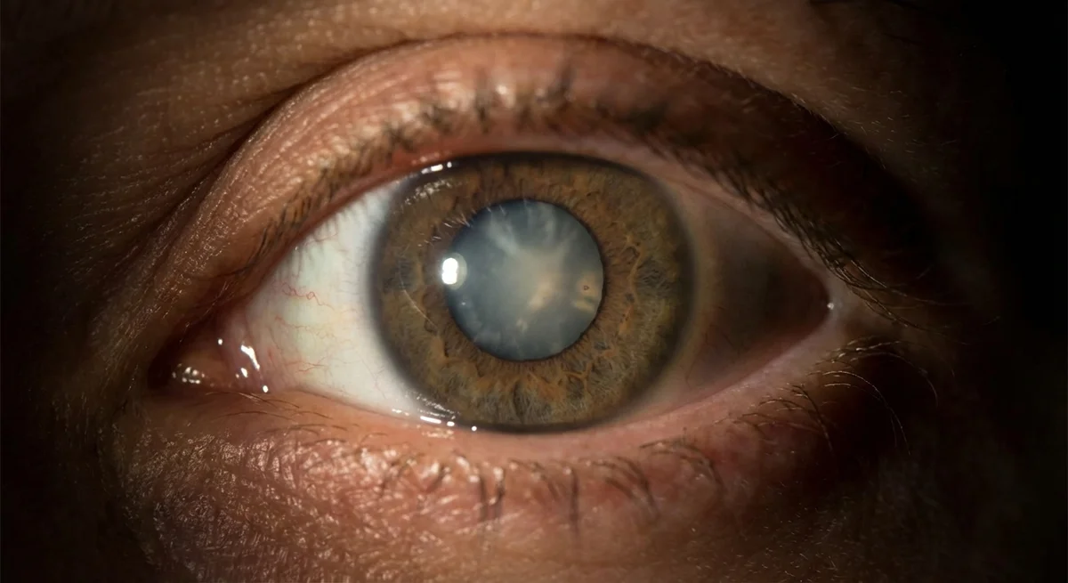
-
Glaucoma vs Cataract. Cataracts cloud the lens inside the eye, producing overall blurry or misty vision and glare from lights, especially halos and fading of colors in low light (dusk) (www.zeiss.com). Glaucoma, by contrast, initially knocks out peripheral vision but leaves central clarity intact. In cataract, you might notice gray haze, trouble with night driving, or bright glare; in glaucoma you won’t have those symptoms until very late. For example, one source notes cataract patients experience "fading colors and contrasts, trouble seeing at dusk... greater glare" (www.zeiss.com). Those are color and light issues, not field loss. True glaucoma vision loss is patchy and on the sides, not just blur from lens opacity.
-
Glaucoma vs Macular Degeneration (AMD). Macular degeneration affects the central retina (macula), causing loss of central sharp vision. Patients with AMD see distortion or a dark/blurred spot right in the center of their vision – e.g. straight lines look wavy, print is missing, faces become hard to recognize. A summary explains dry AMD causes reduced clarity in the center – “letters at the edge are clear, but those in the middle [are] slightly blurred” and gradually a blind spot forms there (www.zeiss.com). In glaucoma, patients generally retain central vision until very late, but lose side vision. They do not see a blind spot in the very middle early on. Thus, if someone sees a dark or wavy area in front of their gaze, think macular degeneration or another central retinal issue, not glaucoma.
-
Glaucoma vs Diabetic Retinopathy. Diabetic eye disease darkens vision in a different way. Diabetic retinopathy can cause floaters or cloudiness from bleeding or leaking vessels in the retina, and blurred patches especially if the macula takes fluid. Patients often describe seeing specks, cobwebs, or shadows drifting across the vision (diabetes.org). The American Diabetes Association notes that floaters or “little spots or shapes that float in your vision” may indicate diabetes-related retinopathy (diabetes.org) – these floaters are actually shadows from broken blood vessels. In contrast, glaucoma does not cause floaters or cobwebs; it causes actual gaps (scotomas) in peripheral vision. Also, a retina specialist examining a diabetic eye will see patched hemorrhages or new funny vessels on the retina, which are not features of glaucoma. Diabetic vision problems tend to fluctuate with blood sugar and are usually more central; glaucoma field loss is permanent and peripheral. Therefore, if you notice floaters, flashes, or blotchy blurred spots, get checked for diabetic retinopathy or retinal tear rather than glaucoma.
In short, glaucoma’s hallmark is peripheral field loss with normal-looking lens and retina. Fading colors, night glare, or floaters usually suggest something else. If you notice your side vision narrowing (for example, bumping into things at the edges), or colored halos around lights plus eye pain, or an unusually large eye in a baby, those are classic glaucoma warning signs. Any one of these should prompt an urgent evaluation by an eye doctor.
Conclusion
Glaucoma itself doesn’t cause pain or obvious symptoms until late, making it tricky to notice. From the outside, early glaucoma “looks” normal – patients have clear eyes and feel fine. But inside, the optic nerve is slowly being damaged. The main clues are what doctors see: increasing optic nerve cupping, thinning nerve fiber layers on OCT, high pressures, and characteristic visual field losses (nasal steps, arcuate blind spots, etc.) (www.msdmanuals.com) (entokey.com).
By understanding the visual effects of glaucoma – from the patient’s tunnel vision to the doctor’s view of the optic disc – you can recognize when something is wrong. Remember that glaucoma’s changes (blind spots in the side vision) are very different from cataract (overall blur), macular degeneration (central distortion), or diabetic retinopathy (floaters and blotches) (www.zeiss.com) (www.zeiss.com) (diabetes.org). Regular eye exams, especially for adults over 40 or anyone with risk factors, are crucial because glaucoma can take your vision without warning. If you ever experience any of the classic symptoms described – such as side vision loss, an episode of red painful eye with halos, or seeing spots and shadows – seek immediate evaluation. Early detection and treatment are the best ways to preserve vision once glaucoma begins to strike.