Cortical Plasticity and Perceptual Learning: Can the Brain Compensate for Optic Nerve Damage?
Interestingly, many glaucoma patients have little awareness of their blind spots. This perceptual filling-in – where the brain “fills” missing...
Deep research and expert guides on maintaining your visual health.
Interestingly, many glaucoma patients have little awareness of their blind spots. This perceptual filling-in – where the brain “fills” missing...
Track peripheral vision changes between eye doctor visits. Start your free trial and get results in under 5 minutes.
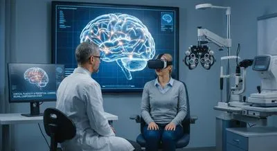
Functional magnetic resonance imaging, commonly called fMRI, is a brain imaging method that measures changes in blood flow to infer which parts of the brain are active. It uses a strong magnet and radio waves to create detailed pictures of the brain and detects a signal called the BOLD response, which stands for blood-oxygen-level-dependent contrast. When a brain region becomes more active, it consumes more oxygen and nearby blood flow increases, and fMRI picks up that change. The technique is noninvasive, meaning it doesn’t require injections or surgery, and it can map activity across the whole brain at once. Researchers and clinicians use fMRI to study how different brain areas work together, to track changes over time, and to see how the brain adapts after injury or disease. It matters because it gives a window into brain function that helps explain behavior, perception, and recovery processes. However, fMRI measures blood flow indirectly rather than the electrical firing of neurons, so its signals are slower and less precise in time than the brain’s actual activity. It also can be expensive, sensitive to motion, and not suitable for people with certain metal implants. Despite these limitations, fMRI has become a powerful tool for understanding brain organization, guiding research, and sometimes aiding clinical decisions about disorders that affect thinking and sensation.