Introduction
Glaucoma and other optic nerve diseases gradually destroy the eye’s nerve cells, causing visual field loss. Although patients often don’t notice slowly expanding blind spots, researchers wonder if the brain can adapt and use remaining vision. In other words, can cortical plasticity (the brain’s ability to reorganize itself) and perceptual learning help compensate after optic nerve damage? This question is under active study. Brain imaging shows that glaucoma not only kills retinal ganglion cells but also leads to changes along the visual pathway (pmc.ncbi.nlm.nih.gov) (pmc.ncbi.nlm.nih.gov). Researchers have found that as glaucomatous damage worsens, activity in the visual cortex (the brain area for sight) declines in the matching visual field regions (pmc.ncbi.nlm.nih.gov). Yet the overall map of vision in the brain often remains intact (pmc.ncbi.nlm.nih.gov).
Interestingly, many glaucoma patients have little awareness of their blind spots. This perceptual filling-in – where the brain “fills” missing peripheral information – is thought to reflect neural compensation. For example, a brain imaging study noted that glaucoma patients (even with severe field loss) did not feel their vision loss soon because their brains effectively masked or “filled in” the defective areas (pmc.ncbi.nlm.nih.gov). These findings suggest the adult visual cortex retains some plasticity, even after long-term eye disease (pmc.ncbi.nlm.nih.gov) (pmc.ncbi.nlm.nih.gov).
Cortical Reorganization in Glaucoma
Glaucoma destroys retinal ganglion cells and their axons in the optic nerve. Autopsy and animal studies show that glaucoma also causes “upstream” damage: thinning of the lateral geniculate nucleus (a relay in the brain) and even neuronal loss in the primary visual cortex (V1) (pmc.ncbi.nlm.nih.gov) (pmc.ncbi.nlm.nih.gov). In vivo fMRI studies of human glaucoma support this: the strength of V1 activity correlates with visual field sensitivity loss (pmc.ncbi.nlm.nih.gov). A leading study demonstrated that areas of V1 corresponding to blind portions of the field had lower blood-oxygen signals, closely matching the eye’s loss of sensitivity (pmc.ncbi.nlm.nih.gov). In short, the eyes’ damage is reflected in weaker cortical responses where nerve input is gone.
On the other hand, the layout of visual cortex in glaucoma often looks broadly normal. One recent fMRI study found that large-scale retinotopic organization (which part of the brain corresponds to which part of vision) was largely preserved in glaucoma patients (pmc.ncbi.nlm.nih.gov). Even with peripheral field loss, the coarse map from central to far-off vision stayed in the correct order (pmc.ncbi.nlm.nih.gov). What did change were small local properties: receptive fields in early visual areas tended to shift and sometimes enlarge toward the intact regions (pmc.ncbi.nlm.nih.gov) (pmc.ncbi.nlm.nih.gov). In other words, neurons adjacent to a scotoma (blind spot) sometimes started responding to nearby seeing regions. These subtle shifts suggest there is a localized plasticity in the adult visual cortex (pmc.ncbi.nlm.nih.gov). Importantly, the degree of these pRF (population receptive field) changes correlated with disease severity (pmc.ncbi.nlm.nih.gov), implying that more advanced glaucoma triggers more cortical adaptation.
In summary, imaging studies of glaucoma show that the visual brain does change when the eyes are damaged: cortical activity drops in lost field regions, and minor remapping occurs near scotomas (pmc.ncbi.nlm.nih.gov) (pmc.ncbi.nlm.nih.gov). This reorganization may help explain why many patients are unaware of early field loss – the brain “fills in” information and masks the defect (pmc.ncbi.nlm.nih.gov). However, the changes are limited. Most studies find that adult V1 does not dramatically rewrite its map: gross organization remains, and neurons do not suddenly recover lost input.
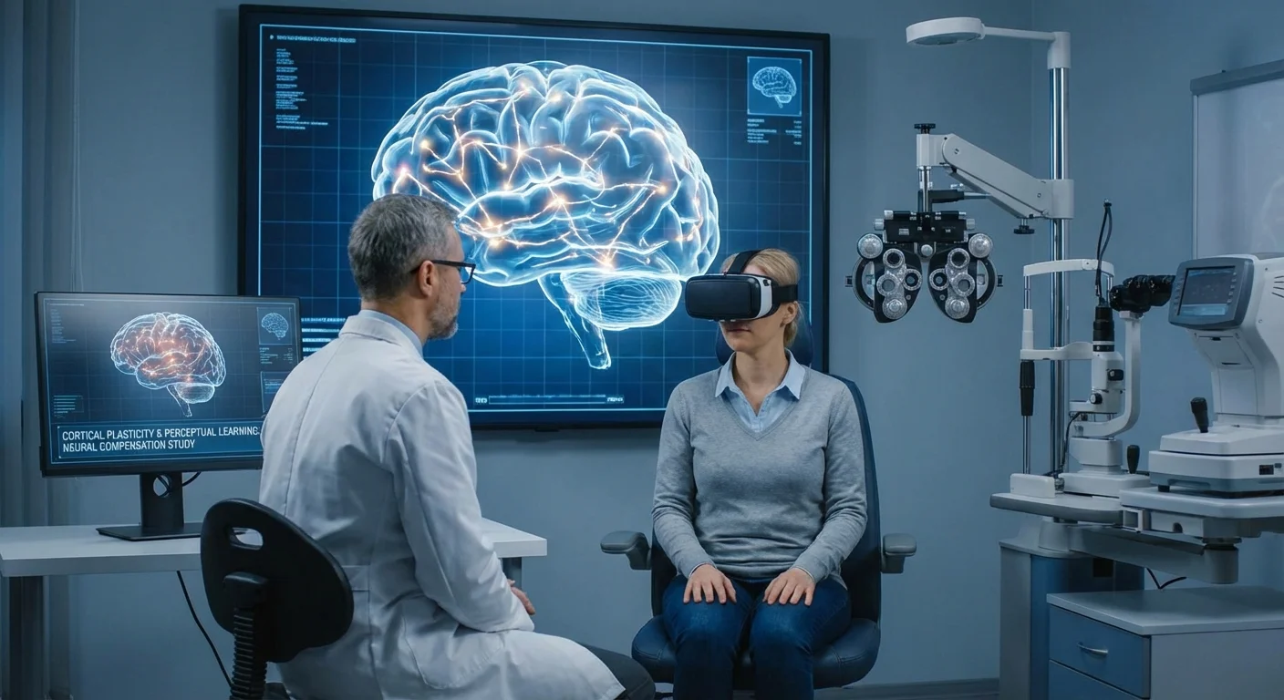
Perceptual Learning and Vision Training
Perceptual learning refers to systematic practice on visual tasks that can improve perceptual abilities. In medicine, specialized vision training programs are being developed to help patients with visual field defects (from glaucoma, stroke, or macular disease) make the most of their remaining sight. These programs often use computer or virtual reality exercises where patients repeatedly discriminate patterns in or near their blind regions. The idea is to reinforce any weak signals and retrain the brain to better detect them.
Several training platforms have been tested. For example, one commercial system (NovaVision’s “Vision Restoration Therapy”) has users do hours of visual exercises per day targeting the edges of their blind fields. Other approaches use contrast patterns, Gabor patches, or motion stimuli in virtual reality headsets. There are even biofeedback devices that convert brain signals (like VEPs) into sounds, so patients can “tune” their brain responses in real time (pmc.ncbi.nlm.nih.gov) (pmc.ncbi.nlm.nih.gov).
Clinical Trial Evidence
Despite the excitement, rigorous trials have yielded mixed results. Early enthusiastic reports of large field gains drew criticism. A prominent review noted that pioneers of computerized training reported dramatic improvements (some patients gaining tens of degrees of field). However, when independent, controlled testing was done, those gains vanished (pmc.ncbi.nlm.nih.gov) (pmc.ncbi.nlm.nih.gov). In one analysis, after-training perimetry with careful fixation showed no significant field improvement despite patients’ subjective sense of better vision (pmc.ncbi.nlm.nih.gov). In essence, initial studies often used the same software for training and outcome testing, which can overestimate benefits (pmc.ncbi.nlm.nih.gov). Critics pointed out that subtle eye movements during training could mimic field expansion: patients learned to make tiny saccades into the blind side, so visual stimuli were seen even though the scotoma hadn’t really shrunk (pmc.ncbi.nlm.nih.gov).
More recent randomized trials have adopted stricter controls. A 2021 multicenter trial in stroke-induced hemianopia used 6 months of home training. Patients performed motion discrimination tasks in their field. The treated group saw very small improvements (~0.6–0.8 dB in visual field sensitivity), which were not significantly greater than the control group’s changes (www.sciencedirect.com) (www.sciencedirect.com). This suggests routine training in the blind field did no better than control (training in the seeing field) at enlarging the defect.
However, not all studies have been negative. A new trial (May 2025) using a personalized virtual-reality visual discrimination program showed clear benefits. Stroke patients using the VR headset for 12 weeks had significantly more regions of improved sensitivity (by ≥6 dB) compared to no-training controls (pmc.ncbi.nlm.nih.gov). By standard perimetry, trained patients improved by ~0.7–1.2 dB in their affected field, whereas controls had essentially zero change (pmc.ncbi.nlm.nih.gov). These gains translated into statistically and clinically better field scores. This suggests that intense, tailored training can indeed strengthen visual sensitivity in chronic field loss.
Other work using audio-VEP biofeedback (mentioned above) also found encouraging but preliminary results. In an uncontrolled pilot, a brief course of VEP-guided auditory feedback improved visual acuity and roughly tripled the amplitude of the VEP signal (pmc.ncbi.nlm.nih.gov). While evidence is still scarce, these studies hint that carefully designed training may drive measurable cortical improvements.
Effect Sizes and Controversies
It’s important to set expectations. Even when training shows statistically significant effects, the size of improvement is usually modest. Changes of less than 1 dB in visual threshold (in decibels of contrast) are typical (www.sciencedirect.com) (pmc.ncbi.nlm.nih.gov). For context, a 1 dB gain in a Humphrey visual field is hardly noticeable, and test-retest variability can be similar. Also, many trials only report short-term gains immediately after training. Very few have long-term follow-up, so we don’t know how durable these effects are. Patients are often left to continue exercises indefinitely to maintain any benefit.
The controversies center on whether measured improvements reflect true neural recovery or other factors. Critics warn that some gains may be due to better fixation stability or practice effects on the tests. As noted, careful studies found that brain-based training often fails to produce field recovery when eye position is strictly controlled (pmc.ncbi.nlm.nih.gov). In short, while perceptual learning holds promise, the evidence is mixed. Some high-quality trials show small but real benefits (pmc.ncbi.nlm.nih.gov), but others find no reply to sham training (www.sciencedirect.com).
Cortical Compensation vs Retinal Recovery
A key distinction is whether training leads to cortical compensation or actual recovery of the eye’s nerve cells. True recovery would imply that damaged retinal ganglion cells or optic nerve fibers regenerate or reconnect, which is biologically improbable. The adult human optic nerve has virtually no capacity to regrow lost neurons. Therefore, most experts assume that any visual gains from training are due to brain-level changes.
For example, optical coherence tomography (OCT) can measure the thickness of retinal nerve fiber and ganglion cell layers. Almost all studies of vision training show no meaningful increase in these thicknesses (and no new axons), underscoring that the nerve injury remains. Interestingly, one small study reported slight thickening in parts of the macula after virtual-reality training (pubmed.ncbi.nlm.nih.gov), but this is exceptional and might be due to measurement variability or transient changes in tissue. In general, it is safer to assume the visual system is making better use of residual signals rather than truly regenerating tissue.
In contrast, cortical compensation means the brain reweights and reorganizes its existing inputs. Training might recruit spared neural circuits or heighten sensitivity in higher processing areas. For instance, as one study observed, regions of visual cortex that still responded weakly despite blindness – so-called “neural reserve” – were exactly where field improvements occurred after training (pmc.ncbi.nlm.nih.gov). In other words, if the brain already had some disabled-but-restorable activity in a blind spot, training mostly amplified that latent response. Any modest enlargement of perceived fields is therefore often due to these intracortical adjustments, not retinal healing.
Monitoring Brain Changes: fMRI and VEP Biomarkers
Because distinguishing brain-level changes from retinal changes is crucial, researchers use objective biomarkers. Two main tools are functional MRI (fMRI) and visual evoked potentials (VEP).
-
Functional MRI: This non-invasive brain scan measures blood flow changes when the visual cortex is active. In glaucoma and other conditions, fMRI can map “retinotopy,” revealing which parts of cortex respond to which part of the visual field. Studies have used fMRI to confirm that V1 signals drop in scotomas and to detect subtle remapping (pmc.ncbi.nlm.nih.gov) (pmc.ncbi.nlm.nih.gov). In a rehabilitation context, fMRI can show whether training stimulates greater cortical activity. For example, one study found that patients who had so-called “neural reserve” (cortical responses without conscious vision) in their blind field showed the greatest post-training gains (pmc.ncbi.nlm.nih.gov). This implies fMRI could eventually predict who will benefit from therapy: areas that light up on fMRI even when the patient isn’t aware of seeing might be ripe for training enhancement.
-
Visual Evoked Potentials: VEPs are scalp EEG recordings of the brain’s electrical response to flashes or patterns. They directly measure cortical response strength and timing. In practice, a checkerboard or flash is presented, and electrodes pick up the characteristic P100 wave ~100 ms after the stimulus. Larger amplitude or shorter latency generally means stronger cortical processing. Training studies have shown that these measures can improve. For example, a recent pilot using VEP-directed feedback reported that the P100 amplitude roughly tripled after training, paralleling gains in visual acuity (pmc.ncbi.nlm.nih.gov). This kind of change strongly suggests cortical learning. Because VEPs are objective and quantitative, they serve as a useful biomarker: if a vision training boosts VEP amplitude, it indicates real neural plasticity in the visual pathways.
By combining these methods with eye imaging (OCT) and standard visual field tests, clinicians can separate cortical adaptation from any retinal anomaly. For instance, if after months of training a patient’s OCT layers are unchanged but their VEP and fMRI responses are stronger, that points to brain-level plasticity.
Conclusion
In summary, cortical plasticity exists even in adults with optic nerve damage, but its effects are limited. Brain imaging shows that glaucoma patients retain a largely stable visual map, with only local receptive field shifts and amplitude changes (pmc.ncbi.nlm.nih.gov). Perceptual training can tap into this plasticity: in some cases, carefully designed exercises have boosted visual sensitivity and acuity, likely by enhancing cortical processing. However, clinical trial results are mixed. Many trials show only tiny improvements (often within test noise), and some of the early excitement has been dampened by rigorous controls (pmc.ncbi.nlm.nih.gov) (www.sciencedirect.com).
Crucially, any improvement seen with training should not be mistaken for true optic nerve repair. Current evidence suggests that vision gains come from the brain learning to use remaining signals, not from the retinal cells growing back. To monitor such changes, researchers use neuroimaging and electrophysiology (fMRI, VEP) alongside eye exams. These biomarkers can document cortical reorganization that underlies any functional gains.
For patients, the message is cautious optimism. The brain can adapt to some extent, and systematic vision exercises may yield small benefits for residual vision. Yet these are enhancements of the existing input, not cures. Understanding and leveraging cortical plasticity is an active research area. Future therapies may integrate imaging-guided training or closed-loop biofeedback to maximize the brain’s natural adaptability, but for now, any such approach should be seen as an adjunct to standard eye care, not a replacement.