Introduction
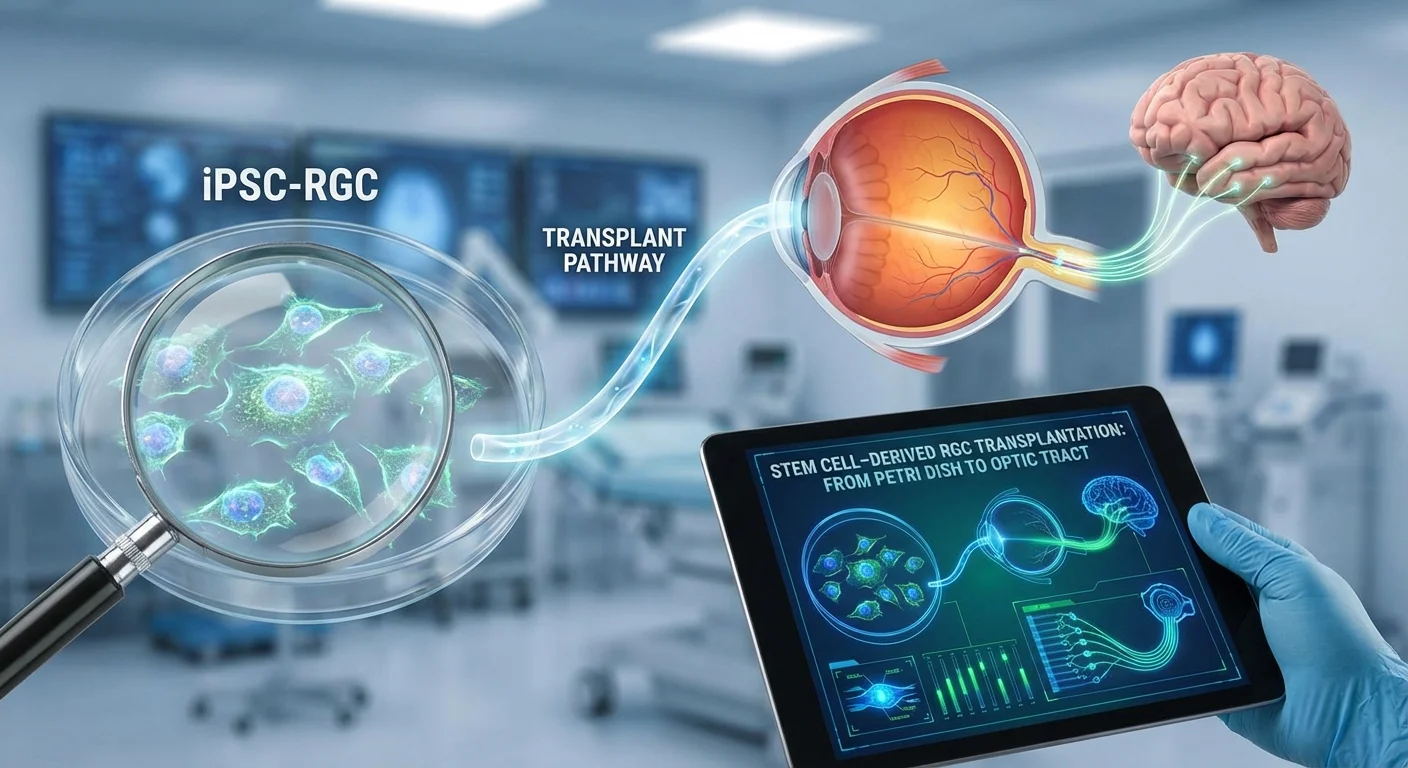
Glaucoma is a leading cause of irreversible blindness worldwide because the retinal ganglion cells (RGCs) that connect the eye to the brain die and cannot regenerate (pmc.ncbi.nlm.nih.gov). Without RGCs, visual signals from the retina cannot reach brain centers (like the lateral geniculate nucleus and superior colliculus), so vision is lost. Current glaucoma treatments (e.g. lowering intraocular pressure) can protect surviving RGCs but cannot restore those already lost (pmc.ncbi.nlm.nih.gov) (pmc.ncbi.nlm.nih.gov). Stem-cell therapy aims to replace lost RGCs by differentiating human pluripotent stem cells (either embryonic stem cells, ESCs, or induced pluripotent stem cells, iPSCs) into RGCs and transplanting them into the eye (pmc.ncbi.nlm.nih.gov) (pmc.ncbi.nlm.nih.gov). In principle this could supply an unlimited source of retinal neurons (pmc.ncbi.nlm.nih.gov). But realizing this vision requires surmounting enormous challenges: the new RGCs must survive, grow axons through the eye’s exit (the lamina cribrosa) into the optic nerve, navigate long distances to precise brain targets, form functional synapses, and become myelinated – all in the inhibitory environment of the adult central nervous system.
This article reviews the state of the art in deriving RGCs from human stem cells and transplanting them in animal models. We then discuss critical barriers to success – axon extension through the lamina cribrosa, guidance to thalamic and collicular targets, synapse formation, and myelination – as well as safety issues (immune rejection, tumor risk) and delivery methods (intravitreal vs. subretinal injection). Finally, we give a realistic outlook for when “first-in-human” trials in glaucoma might be feasible and what outcome measures they would require. Throughout, we strive for clarity: key terms are kept bold and any technical concepts are explained for a lay audience.
Differentiating RGCs from Human Pluripotent Stem Cells
Scientists have developed many protocols to turn human ESCs or iPSCs into RGC-like neurons. Typically, stem cells are first guided into a retinal progenitor state using combinations of growth factors and small molecules that mimic eye development (for example, FGF, IGF, BMP, Wnt and Notch pathway modulators) (pmc.ncbi.nlm.nih.gov). Under the right conditions these cells will further differentiate into RGCs, which can be confirmed by RGC markers. Key markers include the transcription factors BRN3B (POU4F2) and ISL1, the RNA-binding protein RBPMS, the neuronal cytoskeletal protein β-III tubulin (TUJ1), and synuclein-γ (SNCG). Indeed, one study showed PSC-derived cultures expressing multiple RGC markers: “transcription factors such as BRN3, ISL1, and SNCG” appeared alongside long neurites, confirming an RGC identity (pmc.ncbi.nlm.nih.gov). These stem-cell RGCs resemble their natural counterparts in gene expression and morphology, extending long processes and firing action potentials.
RGCs are not a uniform cell type. Dozens of RGC subtypes exist (e.g. motion-sensitive direction-selective cells, on/off center cells, intrinsically photosensitive melanopsin cells, alpha-RGCs, etc.), each with distinct functions (pmc.ncbi.nlm.nih.gov) (pmc.ncbi.nlm.nih.gov). Animal studies have cataloged 30+ RGC subtypes by anatomy and molecular markers (pmc.ncbi.nlm.nih.gov), and evidence suggests humans have on the order of 20 or more subtypes with unique connectivities (pmc.ncbi.nlm.nih.gov). In theory, stem-cell protocols could be tuned to produce specific subtypes by adjusting developmental cues. In practice most current methods aim for a mixed RGC population. Researchers then verify subtype diversity by co-staining for marker combinations: for example, one human RGC differentiation study identified candidate on–off direction-selective RGCs (expressing CART) and alpha-RGCs (expressing SPP1/osteopontin) within their BRN3+ cells (pmc.ncbi.nlm.nih.gov). Optimizing subtype specification is an active research area, since each RGC subtype (with its own pre- and *post-*synaptic partners) will need appropriate integration in vivo (pmc.ncbi.nlm.nih.gov).
The efficiency and speed of RGC generation have improved. Early protocols took several weeks or months, but newer methods accelerate the process. For instance, Luo et al. engineered overexpression of the transcription factor NGN2 plus a neurotrophic medium to produce RGC-like neurons in only two weeks, compared to 1–2 months in earlier 2D or 3D cultures (pmc.ncbi.nlm.nih.gov). These cells expressed RGC markers and, when transplanted into adult rat eyes, “successfully migrated into the ganglion cell layer in 1 week” (pmc.ncbi.nlm.nih.gov). Similarly, pluripotent stem cells grown as 3D retinal organoids (which recapitulate eye development) naturally yield RGCs along with other retinal neurons. Organoid-derived RGCs tend to have gene expression profiles closer to fetal RGCs than do 2D cultures, and many groups now harvest RGC-enriched cells from organoids for transplantation experiments (pmc.ncbi.nlm.nih.gov).
Despite this progress, yields remain modest and cultures are heterogeneous. Protocols often produce a mixed retinal cell population with a minority of RGCs, and survival in culture can be limited (pmc.ncbi.nlm.nih.gov) (pmc.ncbi.nlm.nih.gov). Researchers typically use cell sorting (e.g. Thy1 or BRN3 reporters) to purify RGCs before transplant. A major goal is to achieve very high purity, because any undifferentiated or off-target cells risk forming tumors. A recent study cautioned that “for translational studies it will be critical to determine the purity of donor RGCs to reduce the risk of teratoma formation” (pmc.ncbi.nlm.nih.gov).
Transplantation in Animal Models: Survival and Integration
A number of preclinical studies have now tested human stem-cell–derived RGCs in animal models. Goals include demonstrating that transplanted RGCs can survive, integrate into the host retina, send out axons, and (ultimately) transmit signals. Experiments have been done mostly in rodents (mice, rats), but also in larger animals (cats) and non-human primates.
After differentiating or isolating RGCs in vitro, investigators deliver them into the eye of the host. Two main strategies are intravitreal injection (injecting cells into the vitreous, the eye’s inner cavity) or subretinal delivery (placing cells beneath the retina). Results vary:
-
Intravitreal injection is technically straightforward for targeting RGCs (which reside on the inner retinal surface). Several groups have injected suspension of human RGCs or retinal organoid-derived RGCs into rodent vitreous. For example, Vrathasha et al. injected about 50,000 human iPSC-RGCs intravitreally into WS mice and found the transplanted cells localized within the ganglion cell layer and survived at least five months post-transplant (pmc.ncbi.nlm.nih.gov). These cells elaborated normal dendritic arbors and elicited light-driven action potentials nearly identical to native mouse RGCs (pmc.ncbi.nlm.nih.gov), proving they could integrate functionally at least in the retina. Luo et al. (2020) similarly showed hESC-derived RGC-like cells (overexpressing NGN2) migrated into the ganglion layer of adult rats within a week (pmc.ncbi.nlm.nih.gov). These outcomes are encouraging, but the number of cells that truly integrate is generally small. Vrathasha reported an average of ~672 surviving donor cells per mouse retina (pmc.ncbi.nlm.nih.gov) – a tiny fraction of normal RGC numbers – highlighting the challenging environment.
One problem with simple intravitreal suspensions is that cells often clump or fail to adhere. In a cat model of RGC injury, Becker et al. found that intravitreal injection of a cell suspension yielded cell aggregation and little true integration (pmc.ncbi.nlm.nih.gov). They noted that using a scaffold could improve survival and retinal migration. Indeed, some studies now embed RGCs on biomaterial scaffolds or organoid tissue to support them. For example, human retinal organoids (harvesting RGCs at developmental day 60–70) were transplanted subretinally into feline eyes. With systemic immunosuppression, these organoid grafts survived at least 1 month and appeared to form synaptic contacts with host neurons (pmc.ncbi.nlm.nih.gov). The subretinal approach ensured firm contact between donor tissue and the retina, whereas intravitreal cell suspensions tended to float or clump. On the other hand, subretinal delivery is a more complex surgery and may be limited by available space (the subretinal space is thin in quadrupeds and primates).
In small rodents, intravitreal delivery remains the most common approach. After injection, successful donor cells have been identified migrating to the host retinal ganglion cell layer and expressing RGC markers (BRN3, RBPMS) for weeks to months (pmc.ncbi.nlm.nih.gov) (pmc.ncbi.nlm.nih.gov). Some studies report donor cells extending new dendrites and even initial axon sprouts toward the optic nerve head. For example, transplanted hiPSC-RGCs in mice showed elaborate dendritic trees and (when stimulated by light) generated postsynaptic potentials, indicating they had formed synapses with bipolar/amacrine interneurons (pmc.ncbi.nlm.nih.gov) (pmc.ncbi.nlm.nih.gov). However, it is important to be cautious: experiences with photoreceptor transplants show that transferred fluorescent markers can sometimes make it appear that transplant cells have integrated when in fact they only passed dye to host cells (pmc.ncbi.nlm.nih.gov). Rigorous labeling and functional testing are needed to confirm true integration. In all cases so far, only a subset of injected RGCs survive and integrate. For instance, Vrathasha et al. injected 500,000 cells but later counted only ~0.13% (about 650 cells) as surviving at 5 months (pmc.ncbi.nlm.nih.gov). Clearly, host retinal environment imposes strong selective pressures, and survival remains a limiting factor.
Delivery Routes: Intravitreal versus Subretinal
The choice of how to deliver RGCs into the eye has practical and biological implications. Intravitreal injections place cells in the eye’s gel (vitreous) next to the retina. This route directly bathes the inner retina but can also expose cells to diffusive challenges (they must adhere to the retinal surface to integrate). As noted above, cell suspensions without support can clump; survival may be poor unless the cells quickly migrate to the host tissue. Several studies have found that scaffolded or organoid-based grafts (rather than single-cell suspensions) improve outcomes (pmc.ncbi.nlm.nih.gov) (pmc.ncbi.nlm.nih.gov). Intravitreal delivery has the advantage of relatively simple technique (it’s already used for drug injections and gene therapy vectors) and direct targeting of RGCs.
By contrast, subretinal delivery (placing cells between the retina and the retinal pigment epithelium) is traditionally used for photoreceptor or RPE transplants. For RGC transplants it is less intuitive but can provide advantageous contact. In the feline study by Singh et al., human retinal organoids were implanted subretinally with close apposition to the host retina. Despite the need for immunosuppression, these grafts survived for weeks and showed signs of synapse formation with retinal ganglion cells (pmc.ncbi.nlm.nih.gov). The narrow subretinal space kept the donor cells in place. However, in cats and primates this space is extremely thin, making targeting challenging. Subretinal surgery also carries higher risk to the host retina. Thus, intravitreal injection remains the standard approach in rodents, while subretinal or epiretinal (onto the retinal surface) strategies may be explored in larger eyes.
In summary, intravitreal injection is easiest but often requires scaffolds or high cell numbers for any survival (pmc.ncbi.nlm.nih.gov). Subretinal grafts/clusters can achieve firm contact (as in the Singh cat study (pmc.ncbi.nlm.nih.gov)), but pose surgical challenges. Both routes are being investigated, and it is possible that future protocols will combine cell embedding in biocompatible scaffolds or gels to maximize donor-host interfacing.
Barriers to Axon Regeneration and Connectivity
Even if transplanted RGCs survive and position themselves in the eye, major obstacles block their ability to transmit vision to the brain. In a normal (adult) central nervous system, injured optic nerve fibers do not regrow well. Transplanted RGCs face the same hostile environment. Key barriers include:
Axon Growth through the Lamina Cribrosa
The lamina cribrosa is a sieve-like structure at the optic nerve head where RGC axons exit the eye. It is a major choke point for regrowth. In animal experiments, researchers find that few transplanted RGC axons cross this barrier. One careful study reported that “when RGCs were injected into the vitreous, few integrated into the retina. Of the RGCs that successfully integrated into the GCL, many sprouted axons that grew toward the optic nerve head but few grew past the lamina cribrosa (~10%)” (pmc.ncbi.nlm.nih.gov). In other words, 90% of new axons stalled at the lamina. The lamina contains dense glial and extracellular matrix that likely produces inhibitory signals and physical barriers. Overcoming this roadblock might require either engineering the donor axons (for instance, by upregulating pro-growth pathways like mTOR or Wnt) or modifying the lamina environment (for example, applying enzymes or neutralizing inhibitory molecules). This problem is analogous to any spinal cord injury: the CNS property of axon regeneration failure. It suggests that even if we place RGCs in the eye, getting their axons into the optic nerve will require very strong pro-regenerative stimuli.
Guidance to Brain Targets
Assuming RGC axons can exit the eye, the next challenge is axon guidance over long distances to the correct targets (primarily the lateral geniculate nucleus (LGN) in the thalamus and the superior colliculus in the midbrain). During development, RGC axons are guided by molecular gradients (e.g. ephrin-A/EphA proteins) and spontaneous retinal activity. Adult brains generally lack these cues. Some rodent studies have shown it is possible to direct regenerating RGC axons to reconnect with the superior colliculus: for example, one optic tract lesion model upregulated pro-growth genes (mTOR, JAK/STAT) and observed new synapses in the colliculus (pmc.ncbi.nlm.nih.gov). However, these regenerated axons did not restore vision unless they were artificially supported (see myelination below). In short, finding the right guidance signals (or providing them) is an open research question. The transplanted RGC axons would ideally recapitulate embryonic guidance cues to form the correct retinotopic map in the brain, but it remains unclear how to achieve that in adults.
Synapse Formation
New axons must ultimately form synapses with the correct target neurons. Encouragingly, evidence suggests transplanted RGCs can form synaptic connections at least within the retina. In the study by Johnson et al., hiPSC-derived RGCs that migrated into the host GCL developed normal dendritic arbors. Using synaptic-marker staining and light stimulation, the authors “demonstrated the formation of novel and functional synapses between donor RGCs and host retina” (pmc.ncbi.nlm.nih.gov). In other words, transplanted RGCs were able to connect with bipolar/amacrine interneurons and transmit signals to downstream host cells, although the responses were somewhat weaker than native cells. This finding indicates that, at least at the level of the inner retina, appropriate wiring can occur.
Synapse formation in the brain is even more difficult to achieve and measure. Some regeneration studies (not transplant studies per se) have induced RGC axons to regrow towards the colliculus and form synapses (pmc.ncbi.nlm.nih.gov). In the optic tract lesion model mentioned above, new axons in the suprachiasmatic/collicular region did make synapses, but the mice still had no measurable visual behavior. This was later attributed to a lack of myelin (see next section) rather than faulty synapses (pmc.ncbi.nlm.nih.gov). Bottom line: Synaptogenesis is possible in principle, but ensuring robust, precisely targeted synapses that restore vision is a major hurdle. It will likely require “development-like” cues, such as patterned light stimulation (retinal waves) or co-transplantation of supporting glia, to guide and strengthen new connections.
Myelination of Regenerated Axons
Finally, RGC axons normally become myelinated only after they pass through the lamina cribrosa – an interesting design feature of the eye. Oligodendrocytes (the CNS myelinating cells) are kept out of the retina by the lamina (pubmed.ncbi.nlm.nih.gov). If a transplanted RGC’s axon exits the eye, it enters the CNS, which has myelinating glia. However, in many experimental cases new axons remain unmyelinated. This matters because unmyelinated long CNS axons conduct impulses very poorly. In the optic tract regeneration study (described above), the authors found that the newly formed axons were unmyelinated, and the mice showed no visual improvement unless given 4-aminopyridine (4-AP) – a drug that blocks potassium channels and boosts conduction in demyelinated fibers (pmc.ncbi.nlm.nih.gov). In effect, 4-AP partially restored vision by compensating for the lack of myelin. This result underscores the point: even if an RGC axon reaches its target, without myelin it won’t conduct signals fast enough for vision. Ensuring proper myelination – perhaps by co-transplanting oligodendrocyte precursors or stimulating host glia – will be crucial.
In summary, transplanted RGCs face a gauntlet: only a few grow past the lamina cribrosa (pmc.ncbi.nlm.nih.gov), they must find the correct corridor to brain targets, make appropriate synapses, and then be ensheathed in myelin. Each step currently has only partial success in animal models. Overcoming these barriers is an active area of research in neuro-regeneration.
Immune and Safety Challenges
The eye is relatively immune-privileged, but transplantation of cells still risks immune attack. If donor cells are autologous (from a patient’s own iPSCs), rejection is minimal but technical complexity is high. Allogeneic cells (from another donor or a stem cell line) are easier to produce but may be attacked by the host immune system. In animal studies, researchers often use immunosuppressive drugs to promote graft survival. For example, in the cat organoid transplant study, systemic immunosuppression was required for the graft to survive and form connections (pmc.ncbi.nlm.nih.gov). Without immunosuppression, xenogeneic cells are rapidly cleared. Interestingly, most preclinical studies of retinal transplants report only low-grade inflammation rather than full rejection – a benefit of the eye’s barriers (pmc.ncbi.nlm.nih.gov). However, long-term success will likely require either transient immunosuppression or advanced techniques (such as “cloaking” cells with immune-evasive coatings) (pmc.ncbi.nlm.nih.gov). Any future human trial would need to address this so that donor RGCs are not killed by host T-cells.
A related concern is tumorigenicity. Pluripotent stem cells can form teratomas if undifferentiated cells are transplanted. Even a small number of contaminant PSCs in the RGC preparation could be disastrous. Thus, researchers emphasize high purity of the grafted population. Vrathasha et al. note it is “critical to determine the purity of donor RGCs to reduce the risk of teratoma formation” (pmc.ncbi.nlm.nih.gov). This requires thorough quality control – for instance, sorting cells via RGC-specific reporters or using flow cytometry, and testing by genome methylation or gene expression assays to ensure no pluripotent cells remain (pmc.ncbi.nlm.nih.gov). So far, no tumors have been reported in the small animal RGC transplant experiments, but clinical translation will mandate extremely stringent purification and release testing of any stem-cell product.
Outlook: Toward Human Trials for Glaucoma
Given the formidable challenges above, when might one reasonably expect a first clinical trial of RGC replacement in glaucoma patients? Unfortunately, the answer is likely “not soon.” The field is still in early preclinical stages (pmc.ncbi.nlm.nih.gov). To date, no human trial is registered specifically for RGC transplantation in glaucoma. Existing “stem cell clinics” (for example, misleading trials of autologous adipose or bone-marrow cells) have focused on ad hoc approaches and, glaringly, have caused harm (pmc.ncbi.nlm.nih.gov). Patients should be wary of unproven therapies that circumvent FDA oversight. Legitimate first-in-human trials would require solid proof-of-concept in animals addressing each barrier, and robust safety data. This could take many years.
A pragmatic outlook is that small safety trials might start in the late 2020s or 2030s, if progress continues. Candidates would likely be patients with very advanced disease (where the retina and optic nerve may be largely disconnected), or conversely those in mid-stage disease (to maximize chance of any benefit). The primary endpoints would initially be safety: absence of adverse inflammatory reactions or tumor formation in the eye. Secondary endpoints would aim to detect any anatomical or functional signs of graft “take.” For example, imaging of the retina (optical coherence tomography) could look for a thickening of the retinal nerve fiber layer or ganglion cell layer where cells were injected. Electrophysiological tests, like pattern electroretinogram (PERG) or visual evoked potentials (VEP), might reveal electrical responses originating from the grafted cells. Ultimately, functional vision tests (like visual fields or contrast sensitivity) would be important, but even demonstrating restoration of a tiny arc of vision would be groundbreaking. By analogy, recent gene therapy trials for inherited retinal disease measure outcomes in structural vs. functional categories (pmc.ncbi.nlm.nih.gov); similar categories (OCT anatomy, electrophysiology, visual function, patient-reported vision) would apply.
In summary, while there is cautious optimism, any practical timeline is long. Each of the steps outlined above needs refinement. A realistic first trial might be designed in the mid to late 2030s, contingent on breakthroughs in axon regeneration and safety profiles. Candidates and endpoints would be chosen carefully: probably safety-first endpoints, followed by surrogates of integration (imaging, electrophysiology) before expecting measurable vision gains. In other words, the field must balance hope with realism – pursuing RGC replacement will be a marathon of research rather than a quick sprint.
Conclusion
Replacing lost RGCs in glaucoma with lab-grown counterparts is an exciting but nascent idea. In vitro, human pluripotent stem cells can be coaxed into RGC-like cells expressing key markers and even some subtype characteristics (pmc.ncbi.nlm.nih.gov) (pmc.ncbi.nlm.nih.gov). Transplant studies in animals have shown that a fraction of these cells can survive for months, integrate into the retinal circuitry, and potentially form synapses (pmc.ncbi.nlm.nih.gov) (pmc.ncbi.nlm.nih.gov). However, enormous barriers remain. Axon growth beyond the lamina cribrosa is poor (pmc.ncbi.nlm.nih.gov), guidance to central targets is insufficiently controlled, synapses are weak or absent, and axons lack myelin (pmc.ncbi.nlm.nih.gov) (pubmed.ncbi.nlm.nih.gov). On top of that, immune rejection and tumor risks must be managed. For now, researchers continue to tackle each challenge in turn. Until we can reliably grow, deliver, and connect stem-cell RGCs, vision-restoring transplants will stay in the lab. But the steady progress gives a measure of hope: with continued innovation and caution, the dream of “petri-dish to optic tract” RGC replacement may one day move from experiment to cure.