Red Cell Distribution Width (RDW): A Window into Eye Health
Glaucoma is an eye disease where tiny blood vessels and nerves in the eye gradually become damaged, leading to vision loss. Researchers are exploring whether simple blood tests can hint at this damage. One measure is Red Cell Distribution Width (RDW) – a number on a standard complete blood count (CBC) that reflects how varied your red blood cells (RBCs) are in size. Normally RDW is about 12–15% (pmc.ncbi.nlm.nih.gov). A higher RDW means there is a wide mix of small and large red blood cells. Doctors know that RDW can rise when the bone marrow makes blood cells unevenly, often due to stress, poor nutrition, or illness. Some studies suggest that a higher RDW might signal oxidative stress and problems in the tiny blood vessels (microcirculation) that supply the eye (pmc.ncbi.nlm.nih.gov). In glaucoma research, scientists are asking: Could RDW serve as a simple marker of the microvascular stress that contributes to glaucoma?
What Is RDW and Why Does It Matter?
RDW is one number in a CBC blood test, which most people can get through their doctor or even direct-to-consumer lab services. The CBC reports your hemoglobin, hematocrit, and other values – including RDW. If the blood lab shows an RDW above the normal ~11–15%, it means your red blood cells vary a lot in size (pmc.ncbi.nlm.nih.gov). This can happen for many reasons. For example, deficiencies in iron or vitamins (like B12 or folate) cause some cells to be too small or too big. Chronic kidney disease or inflammation can also disturb RBC production. In simple terms, a higher RDW suggests impaired erythropoiesis (uneven red blood cell production) and shorter RBC survival, which often happens when the body is under stress (pmc.ncbi.nlm.nih.gov) (pmc.ncbi.nlm.nih.gov).
Patients can access these tests themselves: you can order a CBC from many laboratories or ask your doctor for one. The report will list RDW-CV (RDW coefficient of variation) in percent. If it’s high, one should check related tests. For example, low hemoglobin or iron levels (from the same blood draw) might indicate iron-deficiency anemia, which raises RDW. Low B12 or folate can be detected by their specific blood tests. A simple blood panel can also include kidney function (creatinine) to see if kidney issues are present. Interpreting these results means comparing them to normal ranges: high RDW plus low iron or B12 suggests a deficiency; high RDW with high creatinine points to kidney stress; high RDW alone might suggest inflammation or oxidative stress even if anemia isn’t obvious.
Importantly, RDW has been linked to various diseases. In heart and vascular research, higher RDW is often found in people with cardiovascular diseases (CVD) – like heart failure, heart attack, or stroke (pmc.ncbi.nlm.nih.gov). These links are well documented in medical journals. However, genetics studies show RDW is usually a marker of these diseases, not the cause (pmc.ncbi.nlm.nih.gov). In one genetic study, researchers found that the genes raising RDW levels did not independently increase heart disease risk (pmc.ncbi.nlm.nih.gov). Instead, factors like body weight (BMI) influenced RDW. In simple terms, RDW often signals that something else (like inflammation or poor nutrition) is going on, rather than being directly harmful itself (pmc.ncbi.nlm.nih.gov) (pmc.ncbi.nlm.nih.gov).
RDW, Oxidative Stress, and Erythropoiesis
Why does RDW track with stress? One idea is oxidative stress – an imbalance of damaging molecules (free radicals) in the body. RBCs carry oxygen and are exposed to oxidative forces. Laboratory experiments show that exposing RBCs to oxidative stress can increase RDW by shrinking some cells or damaging them (pmc.ncbi.nlm.nih.gov). A large research team analyzed over a million blood tests and found that oxidative stress led to higher RDW and smaller red cells, even without anemia (pmc.ncbi.nlm.nih.gov). They concluded that when the body faces oxidative damage, it often makes uneven red cells, raising RDW. This helps explain why RDW is higher in conditions like chronic illness or aging, where oxidative stress is common.
Impaired erythropoiesis also raises RDW. This means the bone marrow isn’t producing blood cells in a balanced way. For instance, if someone lacks B12 or folate (needed for making DNA), their cells grow too large (macrocytosis), mixing with normal cells and increasing RDW. Conversely, iron deficiency causes many small cells (microcytosis) and some normal cells, again raising RDW. In either case, the CBC will show a wide range of cell sizes. While experts often think of RDW in anemia, the oxidative stress research suggests an elevated RDW can occur even before full-blown anemia appears (pmc.ncbi.nlm.nih.gov) — essentially a subtle warning sign.
RDW and Vascular (Blood Vessel) Disease
Researchers have long noticed that people with blood vessel diseases tend to have higher RDW. For example, one review noted that RDW is consistently associated with the presence and severity of heart disease and stroke (pmc.ncbi.nlm.nih.gov). Preventing and treating heart disease often involves traditional risk factors like cholesterol and blood pressure, but RDW adds extra information about the body’s state. However, careful studies using genetics indicate that RDW itself probably does not cause these vascular diseases (pmc.ncbi.nlm.nih.gov). It likely reflects underlying issues [Source 4] (a key evidence).
Studies show associations like this: One large analysis found that very high RDW made it more likely someone had coronary artery disease or heart failure (pmc.ncbi.nlm.nih.gov). Another found high RDW predicts worse outcomes after a heart attack. It is thought to capture systemic problems (inflammation, poor nutrition, blood flow disturbances) that also harm vessels. A specialist explained that RDW combines both harmless factors (like aging) and harmful ones (like chronic illness) that affect blood cell production (pmc.ncbi.nlm.nih.gov). In practice, doctors are learning that an unexplained high RDW should prompt looking at the whole picture (nutrition, inflammation, organ health) (pmc.ncbi.nlm.nih.gov).
RDW and Glaucoma: What We Know
Glaucoma’s exact causes are complex. High eye pressure is one factor, but blood flow and vascular health in the eye also matter. If the small blood vessels around the optic nerve head or retina are unhealthy, the nerve cells can die faster. Scientists have begun checking if blood markers like RDW relate to glaucoma appearance or progression.
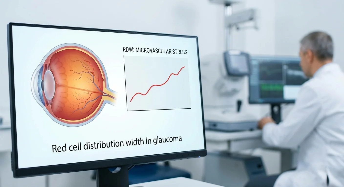
A recent Chinese study examined 1,191 patients with primary angle-closure glaucoma (PACG) and 982 healthy controls. They found that those with glaucoma had a higher average RDW (about 13.01%) than the controls (12.65%) (pmc.ncbi.nlm.nih.gov). Moreover, glaucoma that was more severe (worse vision defects) had even higher RDW (pmc.ncbi.nlm.nih.gov). In fact, patients with mild glaucoma had lower RDW than those with severe glaucoma. After adjusting for things like age and gender, a higher RDW more than doubled the odds of having glaucoma in this study (pmc.ncbi.nlm.nih.gov). In other words, elevated RDW tracked with both the presence and severity of PACG (pmc.ncbi.nlm.nih.gov) (pmc.ncbi.nlm.nih.gov). The authors suggested RDW could help predict who will have worse glaucoma, although they noted more research is needed.
Another study from India looked at pseudoexfoliation glaucoma (PEXG), an eye condition related to glaucoma. They found the same pattern: patients with the eye disease had higher RDW than healthy people (pmc.ncbi.nlm.nih.gov). Those with just the early syndrome (PEX) had intermediate RDW, and it rose further in full glaucoma (PEXG). In fact, as RDW climbed from control to PEX to PEXG, the odds of eye disease increased. Even after accounting for other factors, each 1-point jump in RDW (percentage) raised glaucoma risk by about 76% (pmc.ncbi.nlm.nih.gov). The authors concluded RDW might be a useful blood marker for early detection of eye disease (pmc.ncbi.nlm.nih.gov). These studies match a growing idea: RDW appears up in glaucoma patients, possibly reflecting the underlying stress or damage to their small blood vessels (pmc.ncbi.nlm.nih.gov) (pmc.ncbi.nlm.nih.gov).
It’s important to note that these are associations, not proof of cause. They show that many people with glaucoma also have high RDW. And some hints of biological mechanisms are present: glaucoma involves inflammation and oxidative stress in the eye, which could also raise RDW in the body (pmc.ncbi.nlm.nih.gov). But to be sure RDW can predict glaucoma progression, scientists need long-term data.
Retinal Blood Flow: OCTA Capillary Density and Vision Field
Doctors now have advanced imaging like Optical Coherence Tomography Angiography (OCTA) to look at tiny blood vessels in the eye without dye. OCTA can measure the capillary density—basically how many tiny vessels are present—in the retina or optic nerve head. Studies show that glaucoma eyes tend to have lower capillary density. For example, a study of glaucoma patients found that eyes with faster early loss of optic nerve head capillary density on OCTA had about double the risk of later visual field worsening, compared to eyes with slower vessel loss (pmc.ncbi.nlm.nih.gov) (pmc.ncbi.nlm.nih.gov). In simple terms, losing blood vessels measured by OCTA predicts that the patient’s field of vision will get worse faster (pmc.ncbi.nlm.nih.gov) (pmc.ncbi.nlm.nih.gov).
Another big UK Biobank study (over 42,000 people) used regular retina photos to measure blood vessel density. It found that lower vessel density and complexity in the retina were strongly linked to a higher risk of developing glaucoma over years (pmc.ncbi.nlm.nih.gov). These and other studies confirm the role of tiny blood vessels in glaucoma: eyes with weaker microcirculation suffer more damage (pmc.ncbi.nlm.nih.gov) (pmc.ncbi.nlm.nih.gov).
Putting these pieces together: if RDW is truly a microvascular stress marker, then we would expect high RDW patients to have thinner (sparser) OCTA vessels and faster vision loss. This is the hypothesis researchers want to test in cohort studies.
Linking RDW with OCTA and Glaucoma: A Research Approach
Imagine a large group of glaucoma patients followed over time – a cohort study. Each patient has regular OCTA scans of the optic nerve and retina (measuring capillary density) and visual field tests (measuring vision loss). They also have blood tests including CBC (with RDW) and measures of hemoglobin, iron, B12, folate, and kidney function (creatinine). Researchers would analyze whether a patient’s baseline RDW (or changes in RDW over time) relate to their OCTA and vision outcomes.
-
Adjusting for confounders: Analysts must account for other causes of high RDW. For example, if someone has iron-deficiency anemia, their RDW will be high due to small RBCs. So the study would adjust for hemoglobin level and iron/ferritin tests (to see if RDW predicts outcomes beyond anemia). Similarly, doctors would include vitamin B12 and folate levels (since deficiency can raise RDW) and kidney function (since chronic kidney disease can slightly increase RDW). Including these in statistical models helps isolate the independent effect of RDW on eye health.
-
Handling nonlinear effects: The relationship might not be simple “twice as bad.” It could be that RDW only matters above a certain threshold, or that its effect plateaus. Researchers can use flexible models or even machine-learning approaches to check for nonlinear associations (pmc.ncbi.nlm.nih.gov). This means not assuming every extra point of RDW has the same effect. For example, going from RDW 12% to 13% might not impact much, but going from 15% to 16% could jump risk significantly. Checking nonlinearity ensures no hidden pattern is missed.
-
Within-person change: Another strategy is to look at each patient’s own changes. If a patient’s RDW rises over a year, does their capillary density fall more than someone whose RDW stayed stable? This within-person analysis (often using mixed models) reduces the noise of comparing different people. It asks: “When RDW goes up in this person, do their eye vessels worsen, regardless of their age, genetics, etc.?” This approach can strengthen evidence that rising RDW tracks with disease progression for that individual.
No published study has yet combined all these elements (RDW, OCTA, adjustment factors) in glaucoma, but retrospective cohorts or biobank data could be used. For instance, a hospital might have records of glaucoma patients with blood test results and OCTA scans. Or future eye biobanks could collect this data. By carefully designing the analysis as above, researchers can test if high RDW truly signals worse microvascular health in the eye.
Mendelian Randomization: Genetic Clues to Causality
Beyond observational studies, genetics can help answer cause-vs-effect. Mendelian randomization (MR) uses genetic variants as natural experiments. Scientists have identified many DNA variants that influence RDW from large genome-wide studies. If RDW causally leads to glaucoma, then people with the “high-RDW” genetic variants would be more likely to develop glaucoma (or faster vessel loss) regardless of lifestyle. By contrast, if RDW is just a side-effect, those variants would not raise glaucoma risk.
A similar approach was already done for heart disease. One study created a genetic “risk score” for RDW and checked if it associated with cardiovascular events. They found no evidence that genetically higher RDW caused heart disease (pmc.ncbi.nlm.nih.gov) (pmc.ncbi.nlm.nih.gov). The only trait that did link to RDW genetically was body mass index (pmc.ncbi.nlm.nih.gov), suggesting RDW rises with obesity but doesn’t itself trigger vessel blockages. For glaucoma, no such MR study exists yet, to our knowledge. But researchers could use MR in the future: combine genome data (like from the International Glaucoma Genetics Consortium) with RDW genetics. If high-RDW variants also predict glaucoma, that would hint at a causal role. If not, it suggests RDW remains a useful marker of underlying stress, rather than a cause.
Overall, these genetic tools add rigor. So far, the heart research suggests caution: RDW often reflects health state more than drives disease (pmc.ncbi.nlm.nih.gov). That may turn out true for glaucoma too, but only study can tell.
Conclusion
In summary, RDW is an inexpensive, widely available blood test measure that captures how varied red blood cells are in size. It rises with nutrient deficiencies, inflammation, kidney disease, and oxidative stress (pmc.ncbi.nlm.nih.gov) (pmc.ncbi.nlm.nih.gov). Emerging eye research shows glaucoma patients often have higher RDW in their blood (pmc.ncbi.nlm.nih.gov) (pmc.ncbi.nlm.nih.gov). This fits the idea that glaucoma involves microvascular damage and oxidative stress in the eye, which also affects blood cell production.
New imaging studies (using OCTA) confirm that loss of tiny retinal capillaries predicts vision loss in glaucoma (pmc.ncbi.nlm.nih.gov) (pmc.ncbi.nlm.nih.gov). A recommended next step is a well-controlled cohort analysis: track glaucoma patients’ OCTA capillary density and visual fields along with their RDW over time, adjusting for anemia and nutrient levels. If RDW still predicts vessel loss and vision decline, it would strengthen its case as a microvascular stress marker in glaucoma. Genetic studies (Mendelian randomization) could add evidence on whether high RDW truly causes eye changes or just flags them.
For patients, the takeaway is that a routine blood test (CBC) can provide hints about eye health. If you have glaucoma or are at risk, you can ask your doctor for a CBC and check your RDW. You might also get iron, B12, folate, and kidney tests checked to understand the context. Keep in mind: no single blood number tells the whole story. But combined with eye exams (like OCTA) and vision tests, RDW could become part of a more complete “stress profile” of your eye health. Ongoing research may soon clarify how much weight to give this number. Until then, an elevated RDW on your report is a signal to investigate further – medically, and possibly with your eye doctor – to ensure all factors in your health are optimized for your vision.