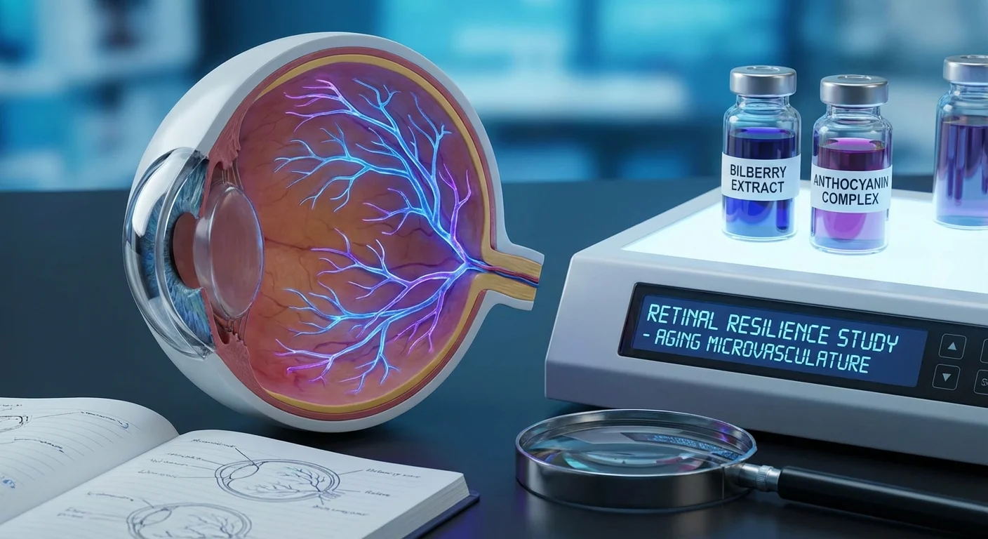
Anthocyanins and Bilberry Extracts: Retinal Resilience and Aging Microvasculature
The flavonoids anthocyanins (pigments in berries) have long been claimed to benefit eye health, and modern studies suggest they do concentrate in ocular and vascular tissues (pmc.ncbi.nlm.nih.gov). These compounds are powerful antioxidants and anti‐inflammatory agents: they scavenge free radicals, stabilize blood vessel walls, and even inhibit platelet aggregation and inflammatory mediators (pmc.ncbi.nlm.nih.gov). In the retina – a high‐metabolism organ especially vulnerable to oxidative stress – anthocyanins from bilberry (Vaccinium myrtillus) may bolster the defense against aging and disease.
Antioxidant and Anti‐Inflammatory Effects in the Retina
Animal research confirms that bilberry anthocyanins protect retinal cells by enhancing antioxidant systems and damping inflammation. In a rabbit model of light‐induced retinal damage, oral bilberry extract (high in anthocyanins) preserved retinal function and structure. Treated rabbits showed higher levels of antioxidant enzymes (superoxide dismutase, glutathione peroxidase, catalase) and total antioxidant capacity than controls, along with lower malondialdehyde (a marker of lipid oxidation) (pmc.ncbi.nlm.nih.gov). At the same time, pro‐inflammatory and angiogenic signals such as interleukin‐1β and VEGF were suppressed (pmc.ncbi.nlm.nih.gov). These changes indicate that bilberry anthocyanins can neutralize excess reactive oxygen species (ROS) in the retina and prevent the downstream inflammation that would otherwise damage retinal cells.
In a mouse model of retinal inflammation (endotoxin‐induced uveitis), anthocyanin‐rich bilberry extract preserved photoreceptor health. Treated mice had better electroretinogram (ERG) responses (reflecting photoreceptor function) and intact photoreceptor outer segments compared to untreated mice. This protective effect was linked to blockade of inflammatory signaling (specifically, bilberry suppressed IL-6/STAT3 activation) and reduction of ROS‐driven NF-κB activation (pubmed.ncbi.nlm.nih.gov). In short, bilberry anthocyanins curtailed the molecular cascade of inflammation and oxidative stress that would otherwise impair vision.
Retinal ganglion cells (RGCs) – the neurons whose axons form the optic nerve – also appear to benefit from anthocyanins. In a mouse optic nerve‐crush model (mimicking glaucoma‐like injury), oral bilberry extract dramatically increased RGC survival. This neuroprotective effect was accompanied by upregulation of endoplasmic‐reticulum chaperones (Grp78 and Grp94) around the RGC layer and a decrease in stress/apoptosis genes (Chop, Bax, Atf4) (pmc.ncbi.nlm.nih.gov). In other words, anthocyanins helped activated cellular “stress machines” that prevent cell death under injury. These experimental results suggest bilberry anthocyanins can support RGC resilience in situations of oxidative or ER stress (as in glaucoma), likely through antioxidant and anti‐apoptotic pathways (pmc.ncbi.nlm.nih.gov).
Vascular Effects on the Optic Nerve Head and Peripapillary Retina
Beyond direct neural protection, anthocyanins may improve ocular microcirculation, especially around the optic nerve (peripapillary region). In patients with normal‐tension glaucoma (NTG), daily supplementation with a standardized bilberry anthocyanin extract (50 mg of total anthocyanins per day) for six months significantly increased blood flow in the optic nerve head and peripapillary retina, measured by laser Doppler flowmetry (www.mdpi.com). In that study, intraocular pressure remained unchanged, suggesting the blood flow improvement was due to vascular effects of the supplement (www.mdpi.com).
A longer randomized controlled trial (24 months) in open‐angle glaucoma (OAG) patients also supports a vascular role. In the treated group (using 50 mg/day anthocyanins), progression of visual field loss was slower and ocular blood flow (around the nerve head) improved compared to placebo (www.mdpi.com). Remarkably, glaucoma patients often have low levels of endothelin‐1 (ET-1), a vascular regulator. Anthocyanin treatment normalized ET-1 levels to those of healthy controls (www.mdpi.com). Since ET-1 helps regulate blood vessel tone, restoring it may underlie the better optic nerve perfusion observed. In summary, clinical data suggest that bilberry anthocyanins can boost peripapillary perfusion and correlate with stabilized vision in glaucoma patients (www.mdpi.com) (www.mdpi.com).
Visual Performance Outcomes
Studies of visual function have produced mixed results. For healthy individuals, rigorous trials generally fail to show meaningful night‐vision improvements with bilberry. A systematic review found that most well‐designed placebo‐controlled trials showed no benefit of bilberry anthocyanosides on dark adaptation or low‐light visual acuity in normal subjects (pubmed.ncbi.nlm.nih.gov). (Notably, smaller and weaker studies did sometimes report positive effects, often using higher or unstandardized doses (pubmed.ncbi.nlm.nih.gov).) In short, any putative night-vision benefit of bilberry remains unproven in healthy eyes.
By contrast, improvements are sometimes seen in people with ocular disease. For example, an uncontrolled clinical report in NTG patients found that long-term anthocyanin supplementation (often with Ginkgo biloba) coincided with better visual acuity and visual field indices (pmc.ncbi.nlm.nih.gov). In that chart review, mean best‐corrected visual acuity (logMAR) improved from 0.16 to 0.11 (p=0.008) and Humphrey visual field mean deviation improved from –6.44 to –5.34 (p=0.001) after about two years of anthocyanin treatment (pmc.ncbi.nlm.nih.gov). While open‐label and retrospective by design, such observations hint that, in already compromised optic nerves, anthocyanin therapy might slow functional loss.
Other small trials suggest modest benefits for visual performance in specific contexts. For instance, standardized bilberry extracts have reduced ciliary muscle spasm and eye fatigue in near‐work or video display tasks (improving accommodation) (www.mdpi.com). These effects could potentially contribute to subjective vision ease, although robust replication is still limited. Overall, clinical outcomes on visual performance vary: normal eyes seem largely unaffected by anthocyanins (pubmed.ncbi.nlm.nih.gov), whereas eyes under stress (glaucoma, inflammation, intense work) show occasional gains, at least in small studies (www.mdpi.com) (pmc.ncbi.nlm.nih.gov).
Dosages, Standardization and Consistency
A major challenge is that bilberry products vary widely in how much anthocyanin they actually deliver. Some extracts (e.g. Mirtoselect®) are standardized to high anthocyanin content (>36%), while others are only ~25%. Human trials have used a broad range of doses. For example, one controlled study gave subjects 43.2 mg of bilberry anthocyanins per day (contained in 120 mg of extract) (www.researchgate.net). Other glaucoma trials used about 50 mg/day (www.mdpi.com). Animal studies typically use much higher mg/kg doses to see effects (pmc.ncbi.nlm.nih.gov).
Importantly, outcomes have not been consistent across dosage differences. The night-vision review noted that positive results generally came from higher-dose, less rigorous trials, whereas the better-designed studies (often with lower anthocyanin content) were uniformly negative (pubmed.ncbi.nlm.nih.gov). Similarly, the plateau in visual gains in some glaucoma cases suggests a ceiling effect beyond which more anthocyanin may not add benefit. No official “optimum” dose is established, but most ocular trials fall roughly in the 50–100 mg of anthocyanins per day range.
Beyond dose, extract composition matters. Bilberry contains multiple anthocyanin subtypes (cyanidins, delphinidins, etc.), and non-anthocyanin polyphenols may also play roles. Some preliminary studies hint the non-anthocyanin fraction might influence efficacy, but data are scarce (pubmed.ncbi.nlm.nih.gov). In practice, product to product differences are huge: one analysis of commercial supplements found that the anthocyanin content per dose ranged from as little as 0.04 mg up to 14.37 mg (pmc.ncbi.nlm.nih.gov), a 100‐fold difference between products. This variability likely contributes to inconsistent trial outcomes. In short, researchers have used different extracts and doses, and while many report some improvement in blood flow or visual indices, the lack of standardized anthocyanin delivery makes results hard to compare (pubmed.ncbi.nlm.nih.gov) (www.mdpi.com).
Systemic Vascular Aging and Cognitive Implications
Intriguingly, the vascular benefits of anthocyanins in the eye mirror findings elsewhere in the body. Good blood vessel health is vital for both vision and brain function, and anthocyanin‐rich diets have been linked to better cognitive aging. Recent reviews of human trials show that berry anthocyanins improve vascular function and memory. One 2024 review of 20 clinical trials reported consistent positive effects of anthocyanin intake on verbal and working memory, often backed by imaging evidence of increased cerebral blood flow in memory-related brain regions (pubmed.ncbi.nlm.nih.gov). Endothelial function (e.g. flow‐mediated dilation) tended to improve as well, though effects on blood pressure were mixed (pubmed.ncbi.nlm.nih.gov). Another comprehensive review (49 intervention studies) found that berry anthocyanins boosted memory and attention, enhanced flow-mediated dilation, and in many cases modestly lowered blood pressure (pmc.ncbi.nlm.nih.gov). These systemic findings suggest that anthocyanins help aging blood vessels generally – including those in the eye – and thus may secondarily support tissue health and cognition.
The parallel is appealing: improved microvascular perfusion in the retina may accompany wider benefits in brain microcirculation and function. Thus, the visual gains seen with bilberry could be one part of a pan-neurovascular effect. In other words, anthocyanins might help the aging retina and the aging brain together by improving endothelial health (pubmed.ncbi.nlm.nih.gov) (pmc.ncbi.nlm.nih.gov).
Safety, Quality Issues, and Future Research Needs
A key benefit of bilberry anthocyanins is safety. Large analyses have found no serious toxicity. Bilberry extracts up to 1000 mg/day (containing high anthocyanins) have been tested, and side effects are rarely more than mild gastrointestinal upset (www.ncbi.nlm.nih.gov). There are no confirmed cases of liver injury from bilberry, and overall liver safety risk is rated “unlikely” (www.ncbi.nlm.nih.gov). (One pharmacologic caution: anthocyanins can inhibit platelet aggregation. Patients on anticoagulants should use bilberry carefully to avoid bleeding effects (www.ncbi.nlm.nih.gov).)
In contrast to its apparent safety, product quality is a major concern. Analyses of herb supplements routinely find adulteration or mislabeling. For example, one survey of bilberry products found that ~50% of so-called bilberry extracts (and 33% of finished supplements) did not match authentic bilberry anthocyanin profiles (pubmed.ncbi.nlm.nih.gov). Many products had far less anthocyanin content than claimed. Similarly, a broader study of Vaccinium-based supplements found over 30% contained no true listed fruit, and anthocyanin levels varied wildly among those that did (pmc.ncbi.nlm.nih.gov). Consumers may be ingesting cheap fillers (like chokeberry or food dyes) without knowing it, or wasting money on minimal active fruit content. This inconsistency greatly confounds research; promising effects of one high-quality extract may not translate to another supplement that is largely inert.
Given the mixed clinical data and quality issues, rigorous trials are a priority. Future studies should use well-characterized, high-quality bilberry extracts with confirmed anthocyanin content. They should be double-masked, placebo-controlled, and sufficiently large, with clearly defined endpoints. For ocular effects, ideal measures include objective blood flow (e.g. OCT angiography of the optic nerve head and retina) and functional tests (visual fields, contrast sensitivity, low-light acuity). Longitudinal cognitive and vascular biomarkers (blood pressure, flow‐mediated dilation, or brain perfusion imaging) would help link eye outcomes to systemic aging. Dose-ranging studies could establish the minimal effective anthocyanin intake. Ultimately, carefully designed trials will clarify whether bilberry can truly slow ocular aging and neurodegeneration, and inform how eye findings relate to generalized vascular and cognitive health.
Conclusion: Anthocyanin-rich bilberry extracts show promise as retinal antioxidants and microcirculation modulators. Bench studies reveal neuronal and vascular protective mechanisms, and small clinical reports hint at improved optic nerve blood flow and visual function in some patients. However, results are inconsistent, partly due to variable dosing and product quality. Crucially, bilberry is safe and well tolerated. The field now needs large-scale, standardized trials to determine if these natural pigments can offer meaningful prevention or therapy for retinal aging and whether such eye benefits reflect broader gains in vascular and cognitive aging.