Creatine and Energy Buffering in Retinal and Optic Nerve Tissues
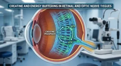
Importantly, this system exists not only in muscle but in nerve cells. Neurons (including RGCs) express CK isoforms that enable them to use creatine....
Deep research and expert guides on maintaining your visual health.
Importantly, this system exists not only in muscle but in nerve cells. Neurons (including RGCs) express CK isoforms that enable them to use creatine....
Screen your peripheral vision from home — no downloads, no waiting rooms. Sign up for a free trial and test in under 5 minutes.
OCT imaging stands for optical coherence tomography and is a noninvasive way to take very detailed cross-sectional pictures of tissues using light. It works a bit like ultrasound but uses light waves instead of sound, measuring how light reflects from different layers to build a near-microscopic map. In eye care, OCT lets doctors see the layers of the retina and the optic nerve head so they can spot swelling, thinning, or fluid that you cannot detect with regular exams. The images show fine structural detail, so clinicians can follow disease progression over time and judge whether a treatment is working. OCT is quick, painless, and does not use radiation, so patients can have repeated scans during follow-up visits. It matters because many sight-threatening conditions, like glaucoma, macular degeneration, and diabetic eye disease, develop subtle changes that OCT can detect early. The technology continues to improve, giving higher resolution and faster scans that make it easier to image moving or uncooperative patients. However, OCT has limits: it gives structural information rather than direct measures of cell function and can be affected by poor image quality from cataracts or eye movement. For a full diagnosis, OCT results are usually combined with vision tests and other clinical information, making it a powerful and practical tool for preserving vision.