Trabeculectomy vs Tube Shunts in the Modern Era: Long-Term Safety and Durability
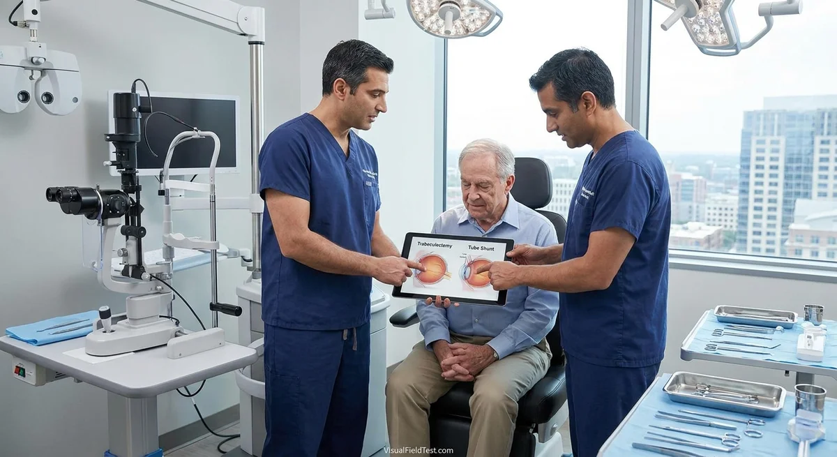
Glaucoma is often treated surgically by creating a new pathway for fluid to drain out of the eye. Two main approaches exist: trabeculectomy (making a new small flap/“bleb” in the eye’s wall) and tube shunt implants (silicone tubes that divert fluid to a distant reservoir). Over the past decades, doctors have shifted increasingly toward tube shunts, especially in complex cases (pmc.ncbi.nlm.nih.gov). However, patients and surgeons still debate which is safer and more durable in the long run. Large clinical trials and patient series have compared these surgeries. In general, tubes tend to be more reliable at keeping pressure from jumping up, whereas trabeculectomies often achieve lower pressure with less medication. Each method has different risks: for instance, trabeculectomy blebs can leak or get infected, while tubes can cause double vision or corneal problems. Importantly, how the surgery is done – the dose of antifibrotic medication, suture techniques, and careful follow-up – can greatly affect outcomes. This article will summarize the key long-term findings from major studies, detail typical complications, and explain how technique and postoperative care influence safety. We will also offer guidance on which procedure may be best suited for eyes needing very low target pressure or eyes with “refractory” glaucoma (e.g. after failed prior surgery).
Comparing Long-Term Results of Trabeculectomy and Tube Shunts
Tube versus Trabeculectomy (TVT) Study – Eyes with Prior Surgery
An important trial known as the Tube Versus Trabeculectomy (TVT) Study looked at patients who had already had cataract or glaucoma surgery that failed (pmc.ncbi.nlm.nih.gov) (pmc.ncbi.nlm.nih.gov). Here, one group received a large Baerveldt tube implant (350 mm² endplate), and the other had a trabeculectomy with mitomycin C (MMC). In the first year, both surgeries lowered eye pressure (intraocular pressure, IOP) similarly. However, tubes were more likely to maintain good pressure control long-term and needed fewer repeat surgeries. For example, at 1 year the failure rate (by strict criteria including high IOP, very low IOP, or need for more surgery) was significantly lower with tubes (3.9%) than with trabeculectomy (13.5%) (pmc.ncbi.nlm.nih.gov). In practical terms, tube patients were less likely to need another glaucoma surgery or to have dangerously low pressure. Both groups lost vision at similar rates (about 32–33% lost ≥2 lines of vision, usually due to non-surgical causes) (pmc.ncbi.nlm.nih.gov).
Over longer follow-up, the advantage for tubes continued. At 3 years, IOPs were effectively the same (around 13 mmHg on average) between the groups, and use of glaucoma medicines was similar (pubmed.ncbi.nlm.nih.gov). But tubes failed less often: the 3-year chance of failure was 15% with tube versus 31% with trabeculectomy (a statistically significant difference) (pubmed.ncbi.nlm.nih.gov). Postoperative complications (mostly mild and transient) were also more common after trabeculectomy. In the first year 60% of trabeculectomy patients had some complication versus 39% with a tube (pubmed.ncbi.nlm.nih.gov). Notably though, severe complications harming vision occurred at about the same rate (~20–27%) in both groups (pubmed.ncbi.nlm.nih.gov). Key findings of the TVT Studies can thus be summarized as:
- Both surgeries significantly lowered IOP long-term, but tubes required slightly more medical therapy initially (pmc.ncbi.nlm.nih.gov).
- Tube shunts had higher success rates (fewer failures and reoperations) in eyes with prior surgery (pubmed.ncbi.nlm.nih.gov).
- Trabeculectomy achieved lower IOP without meds, but had more postoperative problems like bleb leaks (pmc.ncbi.nlm.nih.gov).
- Over 5 years, there was no clear winner for vision loss or glaucoma control – other factors like patient/doctor preference and follow-up patterns matter (pmc.ncbi.nlm.nih.gov).
(For completeness, a more recent “Primary Tube vs Trabeculectomy (PTVT)” trial in eyes without prior surgery found somewhat different results. At 1 year in that trial, trabeculectomy with MMC actually had a higher success rate and lower IOP (mean 12.4 mmHg vs 13.8 mmHg) than tubes (pubmed.ncbi.nlm.nih.gov). However, most serious complications occurred in the trabeculectomy group (7% vs 1% for tubes) (pubmed.ncbi.nlm.nih.gov). This suggests that in eyes where healing is normal, trabeculectomy can give a lower pressure but may carry more risk. By contrast, in complex eyes (like in TVT), tubes had the edge.)
Ahmed vs Baerveldt (Tube versus Tube)
There have also been head-to-head trials comparing different types of tube shunts. The two most common are the Ahmed valve (flow-restricted device) and the Baerveldt plate (non-valved, larger plate). The Ahmed Versus Baerveldt (AVB) Study randomized hundreds of refractory glaucoma patients to one of these devices. At 3 years, both implants had similar pressure control (mean IOP ~15 mmHg) (pubmed.ncbi.nlm.nih.gov), but Baerveldt eyes needed fewer medicines (1.1 vs 1.8 meds on average) (pubmed.ncbi.nlm.nih.gov). More importantly, failure (defined as inadequate IOP or vision loss) was lower with the Baerveldt (34% failure) than Ahmed (51% failure) at 3 years (pubmed.ncbi.nlm.nih.gov). The main difference was pressure: the Baerveldt produced lower IOP (mean ~14.4 mmHg) than the Ahmed (~15.7 mmHg), though this just missed statistical significance (P=0.09) (pubmed.ncbi.nlm.nih.gov). However, hypotony (too-low pressure) was more of an issue with the Baerveldt: by 3 years, 6% of Baerveldt patients had a vision-threatening hypotony complication, whereas none of the Ahmed patients did (P=0.005) (pubmed.ncbi.nlm.nih.gov). At 5 years (follow-up of the same study), the pattern was similar: Baerveldt eyes continued to have lower IOP (mean 13.6 vs 16.6 mmHg, P=0.001) and fewer medications (pubmed.ncbi.nlm.nih.gov). Cumulative failure at 5 years was 40% for Baerveldt vs 53% for Ahmed (P=0.04) (pubmed.ncbi.nlm.nih.gov). Again, hypotony was seen only in Baerveldt eyes (4% of patients) while none of the Ahmed eyes failed due to hypotony (pubmed.ncbi.nlm.nih.gov). Overall:
- Both Ahmed and Baerveldt implants effectively lower IOP, but Baerveldt typically achieves slightly better long-term pressure and medication reduction (pubmed.ncbi.nlm.nih.gov).
- Baerveldt has a small risk of hypotony, whereas the Ahmed valve’s built-in resistor prevents this (none in Ahmed group) (pubmed.ncbi.nlm.nih.gov).
- Serious complication rates were similar (around 60–69% had some complication, mostly minor, in either group) (pubmed.ncbi.nlm.nih.gov).
- In one analysis, Ahmed eyes had about twice the risk of needing reoperation compared to Baerveldt by 3 years (pubmed.ncbi.nlm.nih.gov). (However, note that definitions and patient mix vary between studies.)
Other analyses and systematic reviews generally confirm that large plates (Baerveldt or Molteno) yield lower pressures than valved devices (Ahmed) or trabeculectomy, at the cost of slightly higher early hypotony rate.
Common Complications and How to Manage Them
Both trabeculectomy and tube shunts can cause complications. Understanding these helps patients and doctors avoid or treat them early. Four important issues are hypotony maculopathy, bleb leaks/infections, diplopia (double vision), and corneal endothelial loss.
Hypotony and Hypotony Maculopathy
What it is: Hypotony means an abnormally low IOP (often ≤5 mmHg). When pressure is too low, the back of the eye can wrinkle and the optic nerve can swell, a situation called hypotony maculopathy. This can permanently damage vision if not recognized. Modern use of anti-scarring drugs (like MMC) in trabeculectomies has made hypotony maculopathy more common than in the old days (pmc.ncbi.nlm.nih.gov).
How often it happens: In general, hypotony is more associated with trabeculectomy (especially with high MMC dose) than with valved tubes. CIGTS (a glaucoma study) found a 5-year hypotony risk of about 1.5% after trabeculectomy (pmc.ncbi.nlm.nih.gov). Tube shunts (Baerveldt or Ahmed) rarely cause persistent hypotony because tubes have restricted flow (Ahmed) or require flow ligation (Baerveldt is often tied off initially). In the AVB study above, 4% of Baerveldt eyes failed from hypotony at 5 years, while Ahmed had none (pubmed.ncbi.nlm.nih.gov).
Risk factors: Young, myopic males with pliable sclera and first-time filtering surgery are at highest risk (pmc.ncbi.nlm.nih.gov). High doses of MMC (longer time or higher concentration) make the bleb “thinner” and prone to over-drain. Early overfiltration (for example from too-loose sutures or a large drainage) is also a big factor.
Prevention strategies: Surgeons take several precautions:
- Titrating MMC dose: Use the lowest effective exposure (often 0.2 mg/ml for 1–2 minutes) in primary cases. Very high MMC doses increase hypotony risk (pmc.ncbi.nlm.nih.gov).
- Careful flap suturing: Place tight sutures on the scleral flap so it doesn't over-drain. Adjustable or releasable sutures allow gradual loosening in clinic.
- Staged release: Delay full flow in tubes (e.g. Baerveldt tubes are ligated at surgery and only released later, often with ripcord or tie suture removal) to prevent a huge pressure drop when scarring has occurred around the plate.
- Safety-valve techniques: Some surgeons add small “vent” incisions or partial thickness flaps that slow flow if necessary (pmc.ncbi.nlm.nih.gov).
- Controlled suture lysis: If laser suture lysis is needed post-op, do it gradually to avoid a sudden pressure crash (pmc.ncbi.nlm.nih.gov).
If hypotony does occur, treating it promptly is crucial (pmc.ncbi.nlm.nih.gov). For example, one can apply a pressure patch or bandage contact lens to close leaks, inject autologous blood or fibrin glue under the bleb, or even revise the flap surgically (adding sutures or conjunctival stitches) (pmc.ncbi.nlm.nih.gov). The goal is to raise IOP and allow the eye tissues to re-expand. A number of techniques like conjunctival compression sutures have been described to seal leaks and increase pressure (pmc.ncbi.nlm.nih.gov). Any intervention that raises pressure sufficiently will often resolve maculopathy, but delayed treatment can lead to permanent vision loss (pmc.ncbi.nlm.nih.gov).
Bleb Leaks and Infections
What they are: Trabeculectomy creates a filtering “bleb” under the conjunctiva. Over time, that bleb wall can become thin and develop leaks, or rarely become infected (blebitis) and even lead to endophthalmitis (a severe eye infection). Tubes, on the other hand, don’t create a bleb but carry their own risk: the silicone plate or tube can erode through conjunctiva.
How often it happens: In multiple series, late bleb leaks occur in a few percent of trabeculectomies. In the CIGTS cohort, about 3% had a leak at some point, blebitis in ~3%, and confirmed endophthalmitis in ~1% by 5–9 years (pmc.ncbi.nlm.nih.gov) (pmc.ncbi.nlm.nih.gov). A large Medicare study found about 0.1–0.2% per year risk of bleb infection or endophthalmitis after trabeculectomy (pmc.ncbi.nlm.nih.gov). Tubes are less prone to infection from filtering, but the conjunctiva over a valve or plate can break down. In one series, device exposure occurred in about 6% of glaucoma implants (pubmed.ncbi.nlm.nih.gov). Exposed tubes can lead to infection: roughly 16% of exposures were associated with intraocular infection in one study (pubmed.ncbi.nlm.nih.gov).
Prevention strategies: To avoid leaks and infection:
- For trabeculectomy: Use meticulous closure of the conjunctiva at surgery, and consider reducing MMC dose if the patient is young or at infection risk. In the immediate post-op period, monitor closely for leaks (checking for “Seidel test” dye leak). If a leak is seen, early bandage techniques (collagen shield, pressure patch, ointment) can allow healing (pmc.ncbi.nlm.nih.gov). For persistent leaks, surgical revision (adding sutures) or autologous blood injection into the bleb may be needed (pmc.ncbi.nlm.nih.gov). Lifelong, patients with blebs should watch for redness or pain and have prompt exam if infection is suspected. Many surgeons prescribe antibiotic drops into any thin bleb area as a preventive measure.
- For tube shunts: Always cover the tube with a patch graft (e.g. donor sclera or pericardium) to protect conjunctiva. Avoid positioning the implant in the lower quadrants if possible, since inferior tubes had higher exposure and infection rates (pubmed.ncbi.nlm.nih.gov). Ensure a watertight conjunctival closure. If erosion occurs, early surgical repair (with new graft tissue) is needed, and antibiotics if there is any sign of infection.
Diplopia and Ocular Motility Disturbances
What it is: Some patients develop double or blurred vision (“diplopia”) or eye misalignment after glaucoma surgery. This is usually due to mechanical interference with the eye muscles. Large plate shunts (especially the 350 mm² Baerveldt, often placed behind the lateral and superior rectus muscles) can cause restriction of those muscles. Trabeculectomy very rarely causes diplopia (nearly zero in TVT and other reports).
How often it happens: In the TVT study, new motility problems developed in about 10% of tube patients vs 0% of trabeculectomy patients in the first year (pmc.ncbi.nlm.nih.gov). Similarly, persistent diplopia at 1 year occurred in ~5% of tube patients and 0% of trabeculectomy cases (P≈0.06) (pmc.ncbi.nlm.nih.gov). Other series have reported diplopia rates ranging widely (1–10% or more) in tube shunts, often because many patients with glaucoma have poor vision in one eye (so they may not notice double vision) (pmc.ncbi.nlm.nih.gov). Importantly, the Baerveldt implant has been particularly associated with diplopia more than other devices (pmc.ncbi.nlm.nih.gov). For example, one review found Baerveldt implants had a higher double-vision rate than most other shunts (pmc.ncbi.nlm.nih.gov).
Prevention and management: To reduce this risk, surgeons may take care to tuck the plate under the wider muscles or use a slightly different quadrant. Postoperatively, if a patient reports double vision, an eye specialist will measure the misalignment. Some cases are mild and resolve on their own, while others persist. Persistent diplopia can often be managed with prism glasses if mild. In rare cases of large, symptomatic misalignment, extraocular muscle surgery can correct it. The risk of diplopia is one factor in deciding surgery: for a patient with good vision in both eyes, the possibility of diplopia with a large tube should be balanced against other factors.
Corneal Endothelial Cell Loss
What it is: The cornea (clear front part of the eye) has cells on its inner surface called the endothelium that keep the cornea clear. These cells do not regenerate. Both surgery types can cause some cell loss, but tube shunts (with a tube tip in the anterior chamber) are known to accelerate endothelial cell loss over time. This can lead to corneal swelling and vision loss in the long term.
How often it happens: In the TVT study, persistent corneal edema (a sign of cell loss) was noted in 16% of long-term Baerveldt cases versus 9% of trabeculectomies (pmc.ncbi.nlm.nih.gov). Smaller studies have documented 5–15% drop in endothelial cell density (ECD) within a year after anterior chamber tube placement (pmc.ncbi.nlm.nih.gov). By contrast, patients with trabeculectomy or pars plana tube usually have much less loss. For example, a prospective study found no significant corneal cell loss when the Baerveldt tube was placed in the pars plana (behind the iris) instead of the anterior chamber (pmc.ncbi.nlm.nih.gov). Most of the damage occurs near the tube tip. Risk factors include a very shallow anterior chamber, a short distance between the tube flange and the cornea, and exfoliation (pseudoexfoliation glaucoma) (pmc.ncbi.nlm.nih.gov).
Prevention strategies: To slow endothelial loss:
- Tube placement: If possible (especially in pseudophakic or aphakic eyes), place the tube in the ciliary sulcus (behind the iris) instead of the anterior chamber. Studies show sulcus placement leads to significantly slower endothelial loss than anterior placement (pmc.ncbi.nlm.nih.gov) (pmc.ncbi.nlm.nih.gov).
- Pars plana tube: In very refractory cases or after vitrectomy, inserting the tube through the pars plana (into the vitreous) completely avoids anterior cell contact and causes minimal cell loss (pmc.ncbi.nlm.nih.gov).
- Tube length and angle: Trim the tube so it just reaches (1–2 mm) past the entry point. A longer tube or one directed towards the cornea will cause more damage (pmc.ncbi.nlm.nih.gov). Also, ensure the tube is well away from the cornea when closing up the surgery.
- Close follow-up: Patients with tubes should get periodic specular microscopy (endothelial cell counts) and monitor corneal clarity. If corneal swelling begins, early interventions (like raising IOP a bit or doing endothelial transplant if needed) can be planned.
In summary, tube shunts can produce progressive corneal thinning, while trabeculectomy usually has minimal direct effect on the cornea. Mitigation by surgical technique is important.
How Surgical Technique and Aftercare Influence Outcomes
The details of surgery and postoperative management play a major role in success and safety. Here are some key points:
- Antimetabolite Dosing (e.g. Mitomycin C): Using MMC or 5-fluorouracil during trabeculectomy helps prevent scarring, but higher doses increase the risk of overfiltration and bleb problems. Studies show that too high MMC raises late leak/endophthalmitis risk. Many surgeons use a moderate dose (e.g. MMC 0.2–0.4 mg/mL for 1–2 minutes) and even titrate it to the patient’s scarring risk (pmc.ncbi.nlm.nih.gov). Tailoring MMC seems reasonable: one report found no overall pressure difference when using “low” vs “high” MMC, suggesting good technique and suture management may matter more (pmc.ncbi.nlm.nih.gov).
- Suture Techniques: In trabeculectomy, the scleral flap sutures determine the initial outflow. Using tight primary closure but with releasable or adjustable sutures allows fine-tuning. Early on, laser suture lysis (cutting a suture) or injection of 5-FU under the bleb is often needed to lower IOP. In the TVT Study, about half of trabeculectomy patients needed such interventions (49% had laser lysis, 22% had 5-FU injection in the first year) (pmc.ncbi.nlm.nih.gov). By contrast, in the tube group only 18% needed the ripcord or ligature removed to turn on flow. Good surgical planning (e.g. marking potential leak sites, closing conjunctiva in water-tight fashion) and judicious use of releasables can improve bleb longevity.
- Flow Control in Tubes: Most Baerveldt implants require initial tube ligation (with absorbable suture or ripcord) to prevent early hypotony as the plate capsularizes. Surgeons may also create small fenestrations in the tube to moderate flow until later. Timing of the suture removal is critical: too early risks hypotony, too late delays IOP control. Each center has protocols (often around 4–6 weeks).
- Postoperative Care: Close follow-up in the first few months is essential for any filtration surgery. For trabeculectomy one checks daily/several times in week 1–2 for leaks or hypotony, and every few weeks for suture lysis or 5-FU boosts until the bleb matures. For tubes, early visits check for shallow chamber or choroidal effusions. Both require monitoring IOP. Eye lubrication helps any filtering bleb stay healthy. Patients should be instructed on warning signs of leak or infection (redness, pain, blurry vision).
Good wound management can reduce many complications. For example, reinforcing thin bleb areas early can prevent a leak that might later cause hypotony or infection (pmc.ncbi.nlm.nih.gov). Using autologous blood or conjunctival sutures to seal overfiltering blebs is well described (pmc.ncbi.nlm.nih.gov). In tube cases, ensure the patch graft over the tube is secure and sterilize the eye post-op (often antibiotic ointment is used for a few days).
Choosing the Right Surgery: Low Targets and Refractory Glaucoma
Deciding between trabeculectomy and a tube shunt depends on the individual patient’s needs.
- Target IOP: For very low target pressures (e.g. single digits needed in advanced or normal-tension glaucoma), trabeculectomy often achieves lower IOP. Studies like PTVT found trabeculectomy produced a mean ~~12 mmHg vs ~14 mmHg with tube at 1 year (pubmed.ncbi.nlm.nih.gov). Large plate tubes (Baerveldt) can also get low pressures but usually after the initial ligature dissolves (often 6–8 weeks). Ahmed valves usually stabilize around 10–12 mmHg at best, so if mid-single digit is needed, trabeculectomy or a Baerveldt may be favored. However, one must balance the benefit vs risk: aggressive trabeculectomy (high MMC, low suturing) carries greater hypotony risk.
- Previous Surgery: Eyes that have failed a prior glaucoma surgery or have extensive scarring often do better with a tube shunt. The TVT results clearly showed tubes outperform trabeculectomy in eyes with prior cataract or glaucoma surgery (pubmed.ncbi.nlm.nih.gov). Many surgeons reserve trab in virgin conjunctiva and prefer tubes once there is conjunctival scarring.
- Glaucoma Type: In neovascular glaucoma (e.g. from diabetes or vein occlusion) or uveitic glaucoma, trabeculectomy frequently fails due to aggressive scarring. Tube shunts are generally preferred in these high-risk cases. Among tubes, Ahmed valves are popular in neovascular glaucoma because the valve helps prevent postoperative hypotony which can worsen bleeding. In juvenile or pediatric glaucomas, tube implants are more commonly used because of the scarring tendency in young eyes.
- Patient Factors: Patient age, life expectancy, ability to follow-up, and visual needs all factor in. Elderly patients who cannot easily come for frequent post-op visits might benefit from the relative predictability of a tube (since it needs fewer manipulations than a trab with many lysis sessions). On the other hand, a motivated young patient understanding the bleed risks might choose trabeculectomy for a lower pressure and medication burden. Patients with only one seeing eye should be counseled carefully: if vision must be maximized, the surgeons weigh which procedure offers the safest route to their pressure goal.
- Surgeon Experience: Finally, the skill and comfort level of the surgeon with each procedure matters. Some surgeons are more comfortable controlling trabeculectomy outcomes; others have specialized expertise with implant surgery.
In summary, trabeculectomy may be chosen for patients needing very low pressure and with low risk of scarring, while tube shunts (Ahmed or Baerveldt) are often preferred in eyes with previous surgery, secondary glaucoma, or where postoperative care may be less consistent. The two procedures have comparable long-term success rates when used in optimal situations (pubmed.ncbi.nlm.nih.gov) (pubmed.ncbi.nlm.nih.gov).
Conclusion
Modern studies show both trabeculectomy and tube shunts can effectively control glaucoma in the long term, but with different trade-offs. Tubes tend to maintain success with fewer failures in complex eyes (pubmed.ncbi.nlm.nih.gov), and have minimal risk of over-drainage (hypotony). Trabeculectomies often achieve lower pressures with fewer medications (pubmed.ncbi.nlm.nih.gov) but carry higher risk of bleb leaks and infections over time (pmc.ncbi.nlm.nih.gov). Major trials (such as the TVT and Ahmed-Baerveldt studies) and reviews consistently emphasize that neither surgery is clearly superior in every way – choice depends on patient factors and surgical technique. Importantly, careful surgical planning (appropriate antifibrotic dosing, tight suturing, etc.) and vigilant postoperative care (managing sutures, leaks, or early scarring) can reduce complications for both procedures. Patients should discuss with their surgeon the individual risks and benefits in light of their glaucoma severity, prior surgeries, and other eye health factors. Together, they can decide whether a trabeculectomy or a tube shunt (or even a combined or newer approach) is best for safely lowering eye pressure in the years ahead (pmc.ncbi.nlm.nih.gov) (pubmed.ncbi.nlm.nih.gov).