A New Way to Read Eye Scans in Glaucoma: Can 3D Nerve Fiber Shape Improve Detection?
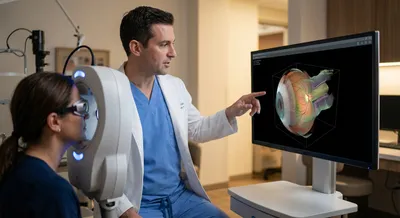
To check the RNFL, doctors commonly use optical coherence tomography (OCT), a non-invasive imaging test that takes cross-sectional “slice” pictures...
Deep research and expert guides on maintaining your visual health.
To check the RNFL, doctors commonly use optical coherence tomography (OCT), a non-invasive imaging test that takes cross-sectional “slice” pictures...
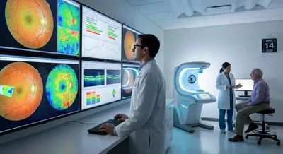
Doctors don’t judge an OCT scan in isolation. Instead, the scan machine compares your eye measurements to a built-in reference database of healthy...
Blind spots often develop gradually without symptoms. Start a free trial and take a quick visual field test to spot changes early.
Find Out NowGlaucoma detection means finding signs of glaucoma early enough to slow or prevent vision loss. Glaucoma is a group of eye conditions that damage the optic nerve, usually linked to pressure inside the eye or poor fluid drainage, and it can cause gradual peripheral vision loss. Detecting glaucoma typically involves a combination of tests: measuring eye pressure, checking peripheral vision, and taking images of the optic nerve and retinal layers. Modern imaging and field tests can reveal nerve fiber thinning or structural changes before a person notices any symptoms, which is crucial because damage is often irreversible. Early detection matters because treatments—such as eye drops, laser procedures, or surgery—can slow progression and preserve sight when started in time. However, diagnosis can be tricky because measurements vary between people and tests can give false alarms or miss early cases. That’s why doctors look at multiple pieces of information over time instead of relying on a single test result. Better tools and larger, well-organized databases of scans and measurements can help clinicians recognize true disease patterns and improve accuracy of detection.