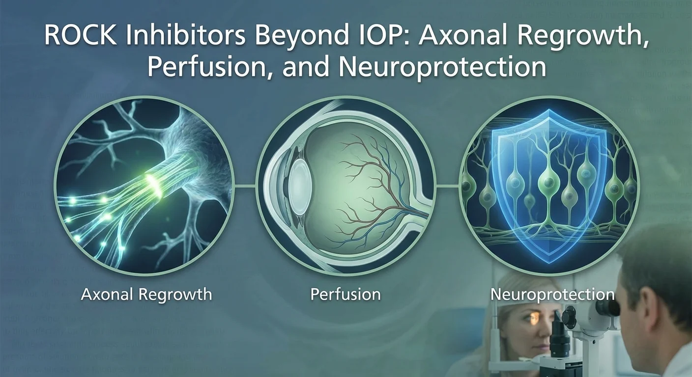
ROCK Inhibitors Beyond IOP: Axonal Regrowth, Perfusion, and Neuroprotection
Glaucoma is an optic nerve disease marked by loss of retinal nerve cells (retinal ganglion cells, or RGCs) and vision loss. Lowering intraocular pressure (IOP) is the only proven way to slow glaucoma, but nerve cells also die from other stresses (poor blood flow, toxins, etc.). Rho kinase (ROCK) inhibitors are a new class of glaucoma drops (e.g. netarsudil, ripasudil) that relax the eye’s drainage channels to lower IOP. Excitingly, laboratory studies suggest these drugs may also protect and help regrow optic nerve fibers (pmc.ncbi.nlm.nih.gov) (pmc.ncbi.nlm.nih.gov). In other words, besides reducing pressure, ROCK inhibitors might boost axon growth, improve optic nerve blood flow, and directly safeguard RGCs. Below we summarize the lab and early clinical findings on these effects, compare netarsudil vs ripasudil, and discuss how clinical trials might test their non-IOP benefits.
Neurite Outgrowth and Axonal Regeneration
In lab models of nerve injury, ROCK inhibitors have repeatedly shown the ability to stimulate nerve regrowth. For example, in rodents with optic nerve crush, daily topical ripasudil greatly increased the number of regenerating RGC axons compared to no treatment (pmc.ncbi.nlm.nih.gov). In fact, three times as many nerve fibers extended past 250 µm in the ripasudil-treated mice (pmc.ncbi.nlm.nih.gov). Another study found that netarsudil (a ROCK/NE–transporter blocker) blocked TNF-induced axon loss in rat optic nerves by activating cellular “cleanup” pathways (autophagy) (pmc.ncbi.nlm.nih.gov). In essence, netarsudil preserved axons under toxic injury.
Likewise, general ROCK inhibition (with other agents like Y-27632) can encourage neurite extension when growth factors are present (pubmed.ncbi.nlm.nih.gov). In an adult rat retinal culture with inhibitory myelin, Y-27632 alone did not grow RGC neurites – but when combined with a growth factor (CNTF) it produced robust nerve sprouting (pubmed.ncbi.nlm.nih.gov). These findings suggest ROCK blockade alone is not magic, but can unleash outgrowth if the environment has support.
More recently, a comprehensive mouse study confirmed that ripasudil eye drops dramatically saved RGCs after injury. Six weeks after glaucoma-model IOP elevation, only ~6.6% of RGCs were lost in ripasudil-treated eyes versus 36% loss with no drug (pmc.ncbi.nlm.nih.gov). After optic nerve crush, ripasudil kept ~68.6% of RGCs alive versus only ~51% in controls (pmc.ncbi.nlm.nih.gov). In short, ROCK inhibition literally doubled or tripled the surviving nerve cells under these insults (pmc.ncbi.nlm.nih.gov). Animal studies like these make a strong case that ROCK inhibitors can support nerve fiber regrowth and RGC survival after injury.
Optic Nerve Head Perfusion
The optic nerve needs steady blood flow. ROCK inhibitors can relax blood vessels and improve circulation. In theory, a drug that improves optic nerve head blood flow could protect RGCs. Indeed, experiments show ROCK blockers do just that. A review notes that applying a ROCK inhibitor may increase vascular tone regulation via endothelin-1 pathways, “improving optic nerve head perfusion and subsequently reducing RGC loss” (pmc.ncbi.nlm.nih.gov).
Animal evidence backs this up. In rabbits, a ROCK inhibitor (called SNJ-1656) significantly increased optic nerve head blood flow after eye drops (pmc.ncbi.nlm.nih.gov). In other tests, toxins that constricted vessels and reduced optic nerve perfusion (like endothelin-1 or phenylephrine) could be counteracted by fasudil or ripasudil eye drops. When ROCK blockers were applied, blood flow recovered and optic disc cupping (a glaucoma damage sign) and RGC loss were reduced (pmc.ncbi.nlm.nih.gov). Notably, one study found the flow improvement from ripasudil did not coincide in time with its IOP drop (pmc.ncbi.nlm.nih.gov), implying the vascular effect can be independent of pressure.
Early clinical data hint at human benefit. In glaucoma patients, one small OCT-angiography trial compared the effects of ripasudil vs an alpha-agonist on peripapillary blood vessels. After treatment, ripasudil eyes showed a significant rise (~12.5%) in superficial retinal capillary density, whereas the control group showed no change (pmc.ncbi.nlm.nih.gov). This suggests low-dose ripasudil can enhance retinal blood perfusion in human eyes (pmc.ncbi.nlm.nih.gov). (Importantly, deep optic nerve circulation measures did not change in that short study (pmc.ncbi.nlm.nih.gov).) Overall, animal and early human data indicate that ROCK inhibition can boost optic nerve head and retinal perfusion, which could help shield RGCs from ischemic damage.
Neuroprotection of RGCs
Laboratory studies consistently show ROCK inhibitors can protect RGCs directly, beyond any blood flow effect. For example, glaucomatous eyes often have high levels of active RhoA signaling. In rats, Rho kinase blockers protected RGCs from both chemical (NMDA) toxicity and from damage caused by an ischemia-reperfusion event (pmc.ncbi.nlm.nih.gov). In other words, RGCs normally stressed by glutamate-like toxins or brief blood loss were spared when ROCK was inhibited.
Further evidence comes from cell and animal models of oxidative stress. A 2025 Japanese study put rat RGCs under oxidative stress in culture and injected NMDA (an excitotoxin) into mice. Ripasudil significantly inhibited RGC death: in cell culture it prevented the loss of living RGCs and suppressed destructive enzyme activity, and in mice it greatly lessened the thinning of the retina and RGC loss caused by NMDA (pmc.ncbi.nlm.nih.gov). The authors concluded ripasudil’s benefit came from antioxidative mechanisms, showing it can protect nerve cells against oxidative injury (pmc.ncbi.nlm.nih.gov).
Altogether, these findings – in rat, mouse, rabbit, and cell models – indicate that ROCK inhibitors can stabilize RGCs and axons in hostile conditions. They seem to counteract toxic signaling and inflammatory glial reactions, keeping RGCs alive longer (pmc.ncbi.nlm.nih.gov) (pmc.ncbi.nlm.nih.gov). If such effects translate to humans, patients might keep more vision for longer even when pressure is controlled.
Comparing Netarsudil and Ripasudil
Netarsudil and ripasudil are both ROCK inhibitors but have some differences. Netarsudil (Rhopressa, 0.02%) was the first approved in the US; it not only blocks ROCK but also inhibits the norepinephrine transporter (pmc.ncbi.nlm.nih.gov). This NE effect helps dilate episcleral veins and lower outflow resistance (pmc.ncbi.nlm.nih.gov). Ripasudil (0.4%) is used in Japan and parts of Asia; it has a very low molecular weight and potently relaxes conventional outflow tissue (pmc.ncbi.nlm.nih.gov). Netarsudil can cause more conjunctival hemorrhages (small bleeds) because of its venous effect, while ripasudil commonly causes redness (hyperemia) (pmc.ncbi.nlm.nih.gov) (pmc.ncbi.nlm.nih.gov).
Dosing also differs: netarsudil is given once daily (often at bedtime to minimize redness) (pmc.ncbi.nlm.nih.gov); ripasudil is typically given twice a day (morning and evening). Whether the dosing schedule affects neuroprotection is unproven. In animal studies, higher concentrations or continuous exposure may be needed for nerve effects (for example, one mouse study used 2% ripasudil drops daily (pmc.ncbi.nlm.nih.gov)). Human trials to date have focused on IOP lowering and used the approved regimens. It remains an open question whether increasing dosing frequency or timing could enhance neuroprotection without unacceptable side effects.
Importantly, not all ROCK inhibitors act the same. In optic nerve injury models, fasudil (a less potent ROCKi) did not promote regeneration, while Y-27632 did (pmc.ncbi.nlm.nih.gov). Likewise, SNJ-1656 and ripasudil each showed axon-protective effects in animals (pmc.ncbi.nlm.nih.gov). Direct comparisons of netarsudil vs ripasudil for nerve effects have not been done in humans. Based on available data, both appear capable of neuroprotection in lab settings, but their efficacies may vary. In practice, netarsudil’s extra NE-blocking action might add vascular benefit, while ripasudil’s stronger ROCK inhibition could be more potent on the cells. More head-to-head studies are needed.
Early Clinical Signals of Functional Recovery
Clinical evidence for non-IOP benefits in patients is still emerging. As noted, the increase in retinal capillary density with ripasudil in glaucoma eyes (pmc.ncbi.nlm.nih.gov) hints at an ocular-perfusion benefit that could translate into function. Beyond imaging, one could look for improved vision or field stability. However, no large trials have yet shown that any ROCK inhibitor reverses visual loss. Visual field tests and optic nerve imaging in the pivotal trials mostly tracked safety and IOP, not neuroprotection. That said, some case reports describe improved perimetry or contrast sensitivity with ROCK inhibitors, but these are anecdotal.
One promising sign is the blood flow effect: since reduced blood flow is a risk factor in normal-tension glaucoma, a drug that boosts ocular perfusion might help these patients especially (pmc.ncbi.nlm.nih.gov) (pmc.ncbi.nlm.nih.gov). The OCT-A finding with ripasudil suggests that a real, measurable eye-bloodflow change is possible. To connect this to “functional recovery,” future studies will need to show that such vascular improvements slow vision loss or restore nerve function (for example, improved pattern ERG or visual acuity). Until then, the lab results offer hope that there are IOP-independent benefits to be tapped in clinical practice.
Designing Trials to Test Neuroprotective Effects
Isolating non-IOP benefits in patients will require careful trial design. One strategy is to minimize differences in IOP, so any change in neuro-function can be attributed to the drug’s other effects. For example, a trial could enroll patients on maximal IOP-lowering therapy (or with normal tension glaucoma) and add netarsudil or placebo. If both arms keep similar pressure, then any slower visual field loss or improved optic nerve blood flow on imaging could be credited to the ROCK inhibitor. Another idea is a crossover design: patients switch from a purely pressure-lowering drop (like a prostaglandin) to one containing ROCK inhibitor, while keeping IOP targets the same.
Endpoints should focus on nerve health, not just pressure. Visual field progression, contrast sensitivity, or low-contrast vision tests could detect subtle functional changes. Imaging biomarkers like OCT angiography (vessel density) or OCT-based nerve fiber layer thickness can be measured over time. Electrophysiologic tests (pattern electroretinogram) directly gauge RGC function and might reveal improvements before vision or field tests do. Trial duration must be long enough to see differences in progression. Finally, combination strategies (ROCK inhibitor plus a standard drop vs standard drop alone) could be used, with all patients matched for mean pressure.
In all cases, the key is to “clamp” the IOP effect. For instance, if one arm has netarsudil on top of a prostaglandin and the other arm has a placebo on the prostaglandin, both should maintain the same IOP (by adjusting other medications as needed). Then investigators compare non-pressure outcomes. As a precedent, a study like the LoGTS trial (which compared two drugs with similar IOP-lowering but different neuro effects) could serve as a model. Ultimately, well-controlled RCTs with neuro-specific endpoints will be needed to prove any vision-preserving benefits of ROCK inhibitors beyond pressure lowering.
Conclusion
In summary, ROCK inhibitors show promise far beyond IOP lowering. In lab studies, they enhance axon regrowth and stabilize RGCs under stress, and they improve optic nerve blood flow. Both netarsudil and ripasudil can trigger these protective effects in animals. Early human data hint at better retinal perfusion with ripasudil and suggest the pathway is worth pursuing. For patients, this means ROCK inhibitors may one day help preserve vision by more than just thinning eye fluid. Ongoing research and cleverly designed clinical trials will tell us if these non-pressure benefits translate into slower vision loss or even some recovery of function. If so, ROCK inhibitors could become a dual-action therapy: lowering pressure and actively protecting the optic nerve.