Introduction
Glaucoma often progresses without symptoms, quietly damaging the optic nerve and shrinking the visual field (the full scope of what you can see). Periodic visual field testing is essential to catch this loss early. These tests map what you see when fixating straight ahead, helping doctors monitor glaucoma and adjust treatment. Visual field tests vary widely in how they work and what they measure. Standard Automated Perimetry (SAP) – the kind done with a Humphrey Field Analyzer – is the most common test in clinics (www.ncbi.nlm.nih.gov) (pmc.ncbi.nlm.nih.gov). Specialized perimeters and new technologies (like virtual reality or tablet apps) are emerging. Each method has strengths and limits in speed, comfort, accuracy, and early detection. This article reviews the main types of glaucoma visual field tests: how they work, what they measure, and how they differ. It will help patients understand the tests they might encounter and guide doctors on which tool best fits different needs.
Conventional Visual Field Testing
Automated Static Perimetry (Humphrey, Octopus)
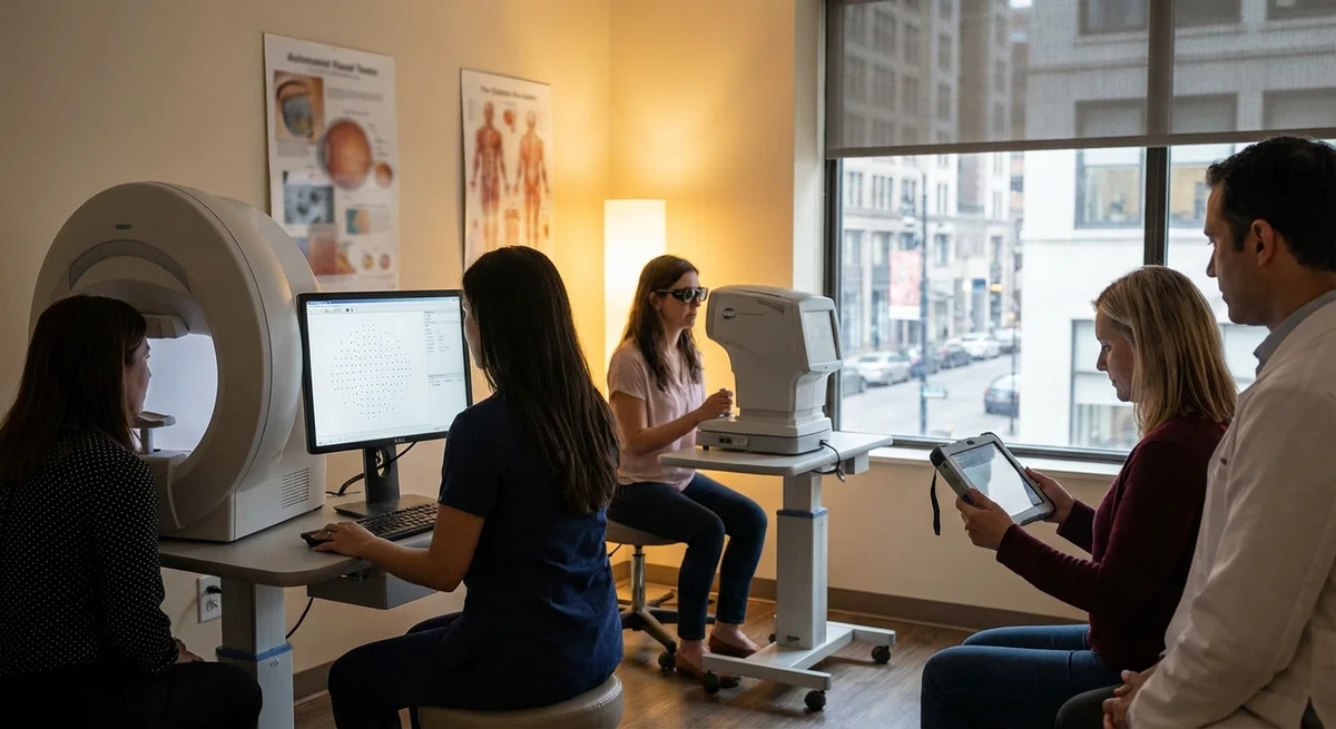
The Humphrey Field Analyzer (HFA) and similar machines (e.g. Octopus) perform static automated perimetry, the current clinical standard (pmc.ncbi.nlm.nih.gov). In these bowl-shaped devices, the patient stares at a fixed central point while small spots of light appear one by one at locations across the field (typically within 24° or 30° of center). For each spot, the patient presses a button if they see the light. The machine automatically adjusts light intensity (“threshold”) to find the dimmest visible spot at each point. Eye-tracking and random “catch” trials (e.g. sometimes no light is shown) check reliability. SAP uses white-on-white stimuli, meaning gray lights on a white background (www.ncbi.nlm.nih.gov). A built-in database compares the patient’s sensitivity map to normal values. The results include measures like Mean Deviation (MD) and a visual field index, which summarize how much vision has been lost overall. In practice SAP detects and tracks the classic glaucomatous defects (such as nasal steps or arcuate scotomas) and shows progression over time (pubmed.ncbi.nlm.nih.gov) (pmc.ncbi.nlm.nih.gov).
Static perimetry is highly quantitative, but it has downsides. The test can take 5–10 minutes per eye, requiring concentration (patients sometimes get tired or distracted) (pmc.ncbi.nlm.nih.gov). Errors from fatigue, tiredness or inattention (“false positives” or “false negatives”) are tracked, but variability remains an issue (pmc.ncbi.nlm.nih.gov). In practice, many patients need multiple tests before a stable baseline is found. On the plus side, SAP results are well-understood: clinicians know how to interpret an HFA printout. Special algorithms like SITA Fast or SITA Faster speed up testing while keeping results accurate (pmc.ncbi.nlm.nih.gov). Newer SAP protocols (e.g. adding extra central test points) may boost early detection and reduce test time (pmc.ncbi.nlm.nih.gov). Overall, automated static perimetry is the workhorse of glaucoma care.
Manual (Kinetic) Perimetry – Goldmann Perimeter
Before computers, Goldmann perimetry was the standard. A trained technician manually moved a bright light of fixed size and intensity across a hemispherical bowl. The patient signaled when they first saw the moving light, tracing out isopters (lines of equal sensitivity) across the field. This kinetic method can map very wide fields easily and tailor the exam on the fly, which helped in earlier eras or in disability evaluations. However, it requires a skilled operator to perform and interpret. In modern practice, Goldmann perimetry is rarely done, especially in glaucoma. Automated tests have largely taken over because they standardize the process and compare easily to normal databases (www.ncbi.nlm.nih.gov). (In some cases where an automated test can’t be done – for example, if a patient must be tested bedside – a semi-automated or even manual perimetry device might still be used (www.ncbi.nlm.nih.gov).) Studies show automated static perimetry usually detects glaucomatous defects faster: one comparison found the Humphrey system found nearly twice as many eyes with defects as a Goldmann test, and it found progression more often (pubmed.ncbi.nlm.nih.gov). In short, the Goldmann test is well-proven but largely supplanted by automated methods that are faster and don’t depend on the examiner’s skill (www.ncbi.nlm.nih.gov).
Specialized Static Perimetry for Early or Specific Detection
Frequency-Doubling Technology (FDT) Perimetry
FDT perimetry uses a unique illusion to test vision. Instead of a small light spot, FDT projects a low-detail (low spatial frequency) striped grating that flickers rapidly. This makes the stripes appear to double in number. The idea is that this stimulus drives the “magnocellular” retinal ganglion cells specially, which may show damage before other cells fail. Early research suggested FDT might catch glaucoma warnings sooner and with high sensitivity (pmc.ncbi.nlm.nih.gov). In fact, some older studies gave it comparable or even greater sensitivity than SAP, with less variability in severely damaged areas (pmc.ncbi.nlm.nih.gov). It became popular as a quick screening tool and is used in some field tests or even on handheld screening machines.
However, FDT is not perfect. It also relies on patient responses and has test-retest variability (some studies found SAP still predicted quality-of-life drops better than FDT did (pmc.ncbi.nlm.nih.gov)). Nowadays most glaucoma specialists rely on SAP, partially because of these reliability concerns and because the pattern (a field located in decibels) is different. Still, clinics may use FDT as an alternative in certain populations (for example, some primary care screening programs use it because of its speed). To patients: an FDT exam feels similar to other perimeters, but the flashing stripe patterns can be an odd sensation.
Short-Wavelength Automated Perimetry (SWAP/Blue-on-Yellow)
Blue-on-yellow or SWAP perimetry was designed to isolate damage to a different retinal cell type. The test flashes a large blue light spot on a bright yellow background. The yellow background temporarily “suppresses” most red and green cones, so detection relies on the short-wavelength (blue-sensitive) cones and their connected retinal ganglion cells. In theory, this tests a subset of retinal cells (the “small bistratified” cells) which glaucoma might affect early.
Research shows SWAP often finds defects sooner than standard perimetry (pmc.ncbi.nlm.nih.gov). One review stated SWAP is “more sensitive than standard… for early glaucoma detection” (pubmed.ncbi.nlm.nih.gov). In practice, a patient doing SWAP sees a bright field and occasionally a blue spot -- it can be more challenging on the eyes because it requires strong yellow illumination. SWAP tests also tend to take longer and can be uncomfortable (patients often find the glare fatiguing). Because of these issues, SWAP is seldom done routinely except at specialty centers or research settings. If used, it is usually alongside SAP in glaucoma suspect cases. For patients, SWAP is a clinical option to catch subtle early loss, but it may not be offered everywhere due to these practical drawbacks.
Central Field and Microperimetry
Microperimetry (or fundus-driven perimetry) is a device that tests the retina point-by-point while simultaneously imaging the retina. It is mainly used for macular disease, but some glaucoma researchers have used it to map the central visual field in detail. In glaucoma, field loss is typically in the mid-periphery first. However, microscopic central defects can exist early. Microperimetry tests many closely spaced points around fixation (often the central 10°) and relates them to the exact retinal location.
Studies suggest microperimetry can detect central sensitivity loss even when a standard 10-2 or 24-2 Humphrey test appears normal (pmc.ncbi.nlm.nih.gov). In one study, glaucoma patients with a single nasal step on standard perimetry showed clear central defects on microperimetry (pmc.ncbi.nlm.nih.gov). The test is highly reproducible with a patient’s own map of vision. In practice, an eye doctor might use microperimetry for a glaucoma patient mainly to study how macular vision is involved – it’s less common as a routine field test. It requires special equipment and expert interpretation. Patients who take a microperimetry test will see lights on a background like any field test, but their eye is being continuously imaged to pinpoint where each spot falls on the retina. Microperimetry reveals detailed central patterns and can correlate field loss with optic nerve anatomy, but it does not replace the standard peripheral field tests for most glaucoma care.
Emerging Technologies
Portable and Head-Mounted Perimetry (Virtual Reality)
New portable perimeters using VR (virtual reality) or head-mounted displays are becoming available. These are compact devices that look like virtual-reality goggles. They present the test patterns inside the headset instead of in a large bowl. With high-resolution screens, the tiny display can emulate the standard field test. Some designs include eye tracking to ensure you keep looking at the central fixation target.
These head-mounted perimeters have notable trade-offs. On the plus side, they do not require a dark room or fixed chinrest, so testing can happen in any quiet room – even at home (www.ncbi.nlm.nih.gov). Many patients find it more comfortable to wear a headset than to lean into a machine’s helmet, especially people with neck/back pain (www.ncbi.nlm.nih.gov). A headset naturally blocks outside light, further eliminating the need for darkness. In one study comparing an “imo” head-mounted device to a Humphrey analyzer, results were closely correlated and the VR test was about 30% faster (pmc.ncbi.nlm.nih.gov). In fact, several VR perimeters (e.g. imo, Vivid Vision, Virtual Field, VIP by Solomon, etc.) have been FDA-cleared or are under development to allow portable glaucoma testing (www.ncbi.nlm.nih.gov) (pmc.ncbi.nlm.nih.gov).
On the other hand, some people dislike the heft of a headset on the face (www.ncbi.nlm.nih.gov). Also, testing outside the eye clinic brings new challenges: ambient noises or distractions in a waiting room might interrupt the test. As one report notes, clinics have already FDA-approved multiple VR perimeters, and more are expected (www.ncbi.nlm.nih.gov). These new devices promise convenient, flexible testing, but they are still being validated. Not every eye doctor has them yet. For patients, VR perimetry may look like wearing a gaming headset and playing a simple video-game-like task for a few minutes each eye.
Tablet/Computer-Based Perimetry
Rather than a bulky machine, ordinary tablets or desktop computers can now run visual field tests. Tablet perimetry apps like Melbourne Rapid Fields (MRF) turn an iPad into a perimeter screen, presenting stimuli through an app. The advantages are obvious: everyone has tablets, they are cheap and portable, and in principle you could test your field at home. The MRF app, for example, is FDA-cleared and runs a full 30° test in about 4–5 minutes per eye (pmc.ncbi.nlm.nih.gov).
Computer-based tests let patients do the exam at home under remote supervision or even unsupervised (there are studies of 3-month home monitoring using MRF online (pubmed.ncbi.nlm.nih.gov)). They can also use creative stimuli (e.g. flickering patterns) that bowl perimeters cannot show (www.ncbi.nlm.nih.gov). Such tests include built-in voice prompts and friendly interfaces, potentially making them more engaging, especially for young or tech-savvy users (pmc.ncbi.nlm.nih.gov) (www.ncbi.nlm.nih.gov).
The trade-offs involve standardization. A clinic’s Humphrey machine carefully controls light level, calibration, and viewing distance. But at home or on a tablet, ambient light can vary and the patient might not fix their eyes the same way (www.ncbi.nlm.nih.gov). Tests may need to pause if the patient moves too much. One advantage of some tablet devices is “blind spot monitors” or frequent fixation checks to ensure the person is looking properly (pmc.ncbi.nlm.nih.gov). Research shows that apps like MRF can give comparable results to a Humphrey on average (pmc.ncbi.nlm.nih.gov). However, individual test variability can be higher than in the sealed clinic environment (pmc.ncbi.nlm.nih.gov). For example, one study found mean deviation scores from an iPad test were a few decibels worse than the Humphrey’s, and a few point locations differed (pmc.ncbi.nlm.nih.gov). That means results between systems shouldn’t be mixed; doctors would track each system’s results separately. Still, for many patients (especially in remote areas or during pandemics), home perimetry via tablets might be a convenient supplement. Work is ongoing to make these apps more robust: one group reported their app remained accurate even when lighting or blur varied, as long as its on-screen instructions were followed (pmc.ncbi.nlm.nih.gov).
Objective Perimetry (Pupillography, Saccadic Tests)
All the above tests rely on you pressing a button when you see a light. But what if someone can’t do that reliably (young children, very disabled patients)? Researchers are exploring objective methods that don’t need a conscious click. One idea is pupil perimetry: shining light stimuli in parts of the visual field and measuring the pupil’s reflex. For instance, a device called RAPDx flashes lights region by region to each eye and tracks the bilateral pupil response. If one hemisphere of vision is weak, the pupil will constrict differently. In studies, automated pupillography has shown some ability to flag glaucoma, especially when one eye is worse than the other (pmc.ncbi.nlm.nih.gov). (It makes sense: the test is particularly good at detecting asymmetry between eyes.) However, the accuracy is still limited: in one study it had a moderate area-under-curve (~0.85) for detecting glaucoma, performing poorly if both eyes were equally damaged (pmc.ncbi.nlm.nih.gov). This method is not standard in routine care yet.
Another concept is tracking-based perimetry: some systems follow eye movements to ensure fixation or use involuntary eye movements as feedback. For example, one experimental test has the patient look naturally at moving spots (like playing an electronic game) while an algorithm infers what they see. This is promising for kids who can’t concentrate on a fixed spot. But these methods are still mostly research tools. Currently, the vast majority of glaucoma clinics use patient-response perimetry (like Humphrey or FDT). If conventional testing isn’t possible, an eye doctor might catch a large defect with simpler confrontational testing or refer for specialized methods.
How the Tests Compare
- Source of Information: SAP/white-on-white testing measures the minimum brightness of a light spot the eye can see at each location (www.ncbi.nlm.nih.gov). FDT measures contrast sensitivity along flickering gratings (targeting certain ganglion cells) (pmc.ncbi.nlm.nih.gov). SWAP measures blue-cone-based sensitivity. Microperimetry maps central retina sensitivity with imaging guidance (pmc.ncbi.nlm.nih.gov).
- Sensitivity and Early Detection: Some tests are designed to catch glaucoma early. SWAP and FDT may find early defects that SAP misses (pmc.ncbi.nlm.nih.gov) (pmc.ncbi.nlm.nih.gov). In practice, SAP is still often “gold standard,” but an early defect on FDT or SWAP can raise suspicion. Regular assessment usually still uses SAP for consistency.
- Reliability and Variability: All subjective tests have variability (how steady your attention is, etc.). Classic Humphrey tests have well-characterized reliability indices. FDT and SWAP have their own norms and can sometimes be more variable if challengingly bright or flickering. Tablet tests have additional sources of inconsistency (lighting, position) (www.ncbi.nlm.nih.gov) (pmc.ncbi.nlm.nih.gov). Generally, clinic-based SAP or VR perimetry yields more repeatable results than ad-hoc home tests, assuming patient cooperation.
- Speed: New algorithms (like SITA Faster) and devices can shorten test time. For example, some tablet tests complete a field in under 5 minutes, compared to ~7–8 minutes per eye on traditional SAP. The IMO head-mounted device cut testing time about 30% compared to an HFA (pmc.ncbi.nlm.nih.gov). Clustering test schedules can also improve efficiency (for clinical trials) (pmc.ncbi.nlm.nih.gov).
- Comfort and Accessibility: Traditional bowl perimeters require leaning forward into a machine with a chinrest. This can be uncomfortable for people with neck/back issues. In head-mounted perimeters, you simply wear goggles with no need for a dark booth (www.ncbi.nlm.nih.gov). Tablets require you to fixate at a closer distance (e.g. 30 cm) but allow sitting comfortably at a desk. VR headsets block outside light and might feel less claustrophobic but some patients report the headset weight as an issue (www.ncbi.nlm.nih.gov). Home tests are convenient (no travel) but require discipline and guidance. Generally, newer devices aim to improve patient comfort and reduce fatigue.
- Objectivity: Currently, SAP/FDT/SWAP all rely on your manual response. This means small kids or very impaired patients may struggle. Objective methods (like pupillography (pmc.ncbi.nlm.nih.gov)) sidestep the need for a button press and can detect afferent defects, but they’re not widely used outside research. If a doctor suspects a patient truly cannot do standard perimetry, they might use bilateral tests or alternative exams (like visual evoked potentials – beyond our scope).
Choosing the Right Test
No single test is best in all situations. The choice depends on patient and clinical needs:
- New glaucoma or suspects: Clinics typically start with standard SAP (Humphrey 24-2 or 30-2). It gives a broad baseline. If central vision is mainly at risk (advanced glaucoma), they might also run a 10-2 test of the central field.
- Early or suspected cases: Some doctors may add an FDT or SWAP field, looking for subtle changes the Humphrey 24-2 might miss. This is especially true if clinical exam (optic nerve appearance) seems worse than the Humphrey VFs suggest.
- Advanced glaucoma: When glaucoma is far progressed, central field becomes crucial. SAP with the 10-2 grid and even microperimetry can map any remaining vision. FDT and SWAP add less information in end-stage eyes.
- Young or uncooperative patients: If a child or very anxious patient cannot do a long fixed fixation test, a doctor might try an easier screening (e.g., FDT screening or optokinetic methods). Some centers use saccadic perimetry or a game-like test with eye tracking for kids. Otherwise, they may focus on structural tests (OCT scans of the nerve) more than visual fields.
- Physical limitations: Patients who can’t sit upright or hold still (wheelchair users, neck/back pain) may benefit from portable head-mounted perimeters. If someone lives far from the clinic, a validated home test (tablet or web-based) might help keep track in between doctor visits.
- Test availability and follow-up: Often the decision is practicality: if the clinic only has a Humphrey, that’s used. If a mobile app test is validated in that practice, it might supplement. The doctor will try to compare like-with-like (meaning if you start monitoring on Humphrey, they will continue on Humphrey for consistency). Switching devices mid-stream can make it hard to tell true change from machine differences. That’s why many clinics adopt new tools slowly and parallel-validate them first.
Practical Barriers and Future Directions
Cost and Equipment: Traditional perimeters (Humphrey, Octopus) are expensive machines and each clinic usually has only one or two. New technologies also cost money: a VR perimeter requires high-resolution displays and tracking, and tablets require calibration tools. Upfront cost can slow adoption, especially in low-resource settings.
Training and Validation: Automated perimetry is operator-friendly, but newer devices need staff training (how to position the patient with a headset, how to calibrate a tablet, etc.). Clinics also need confidence that new tests are valid. Researchers compare results device-by-device (like the study where the iPad test closely matched the Humphrey on average (pmc.ncbi.nlm.nih.gov)). Regulatory approval (like FDA clearance) and published evidence support these devices, but widespread trust takes time.
Standardization: As noted, tablet and home tests lack the controlled environment of a dark room with fixed optics (www.ncbi.nlm.nih.gov). To use these tests reliably, further work on software algorithms and user instructions is needed. For instance, improved eye-tracking during home tests could cancel out fixation errors. Developing robust methods to standardize the distance, brightness, and even the type of input (finger touch vs. spacebar press) is a work in progress (pmc.ncbi.nlm.nih.gov) (www.ncbi.nlm.nih.gov).
Patient Familiarity: Patients new to any perimetry need instruction. Switching from a traditional machine to a tablet might be confusing. Some people might prefer a head-mounted “goggle” as more natural, while others trust the longer-time-tested bowl device. Doctors must guide patients through any test and interpret results in context.
Technology Evolution: The future of visual field testing likely involves a mix of approaches. Virtual reality and AI could make tests faster and smarter. AI could, for example, predict a full field from fewer test points (using patterns learned from large data sets) and so shorten exam time. Already, AI algorithms have shown promise in predicting visual loss from other eye scans (pmc.ncbi.nlm.nih.gov). Deep learning methods combining OCT imaging and visual fields may soon refine perimetry or even provide early warning without an eye-catching test. Wearables and home-testing will probably grow, especially for patient self-monitoring between visits. Still, any new tool must eventually prove it can show real change reliably; otherwise glaucoma management still needs patient responses.
Conclusion
In summary, a variety of visual field tests exist for glaucoma. Standard automated perimetry (Humphrey/Octopus) remains the clinical workhorse for diagnosing and monitoring field loss (www.ncbi.nlm.nih.gov) (pmc.ncbi.nlm.nih.gov). Other methods—FDT, SWAP, microperimetry, etc.—target specific cell types or regions and can reveal certain defects earlier (pmc.ncbi.nlm.nih.gov) (pmc.ncbi.nlm.nih.gov). Emerging technologies like virtual reality perimeters and tablet-based tests promise more comfort and accessibility (www.ncbi.nlm.nih.gov) (pmc.ncbi.nlm.nih.gov), though they bring logistical challenges (environmental control, standardization) (www.ncbi.nlm.nih.gov) (pmc.ncbi.nlm.nih.gov). Each approach measures visual sensitivity in slightly different ways, so results are not always directly interchangeable.
For patients, the take-home point is: multiple testing options may be offered depending on your situation. Don’t be surprised if one visit you sit at a Humphrey machine, and another time you put on special goggles or even do a test on a tablet. The doctor may choose the method based on your age, the stage of glaucoma, or practical factors. All tests aim to do the same thing—map your field so that even subtle vision loss becomes apparent. As technology advances, visual field testing may become quicker and more patient-friendly, but the goal remains clear: detect any vision loss as early as possible and follow it carefully to protect your sight (pmc.ncbi.nlm.nih.gov) (www.ncbi.nlm.nih.gov).