Introduction
Glaucoma is a leading cause of irreversible blindness, marked by progressive death of retinal ganglion cells (RGCs) and damage to the optic nerve. It often involves elevated intraocular pressure (IOP) due to dysfunction in the trabecular meshwork (TM) outflow system, as well as age‐related neurodegeneration of RGC axons. Age is the strongest risk factor: aging causes oxidative stress, mitochondrial decline, accumulation of damaged proteins and cells, and chronic inflammation – all of which contribute to glaucoma pathophysiology (pmc.ncbi.nlm.nih.gov) (pmc.ncbi.nlm.nih.gov).
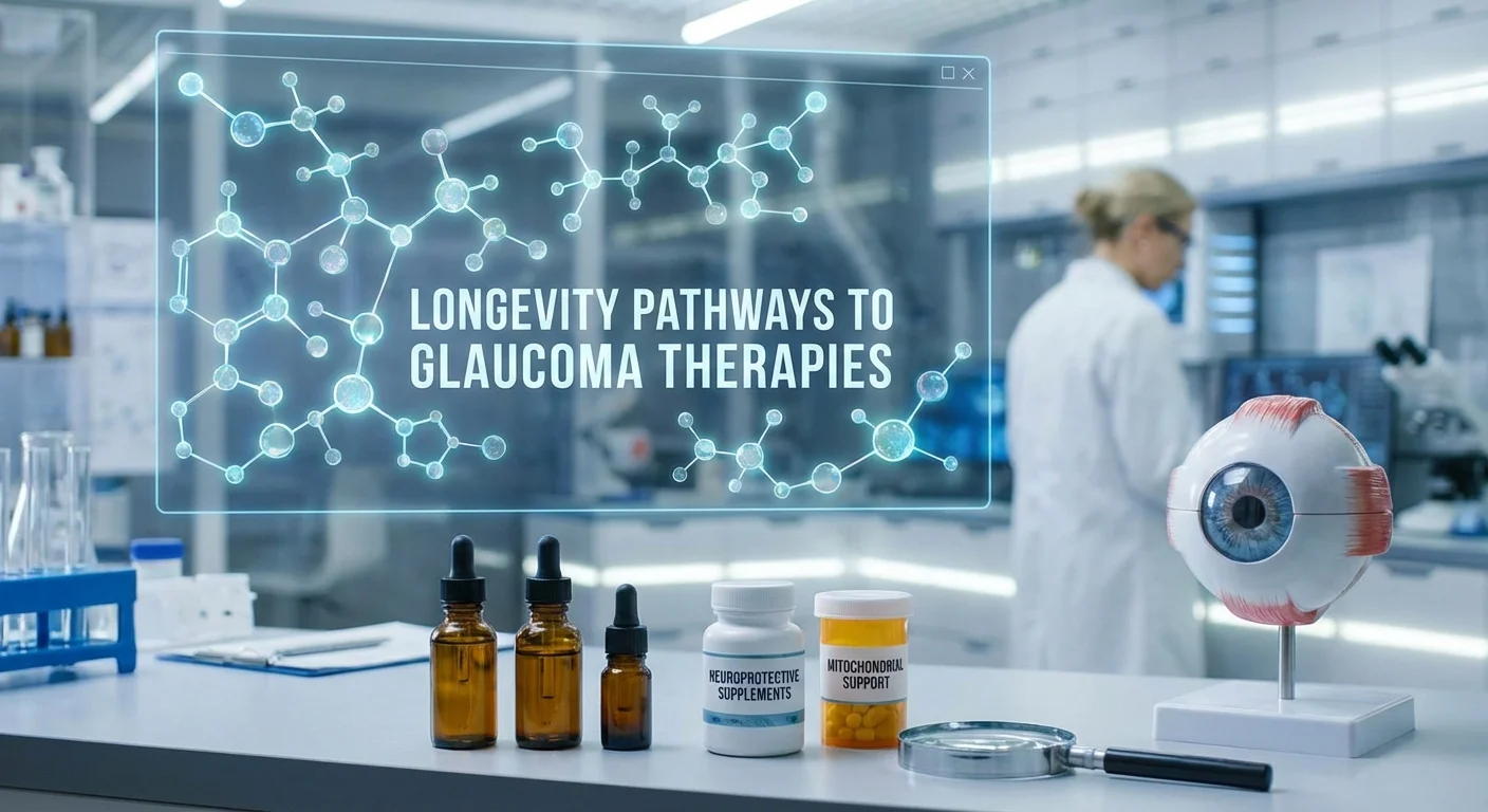
Biologists studying aging (“longevity pathways”) have identified key regulators – AMPK, mTOR, sirtuins, autophagy, and cellular senescence – that govern metabolic health and tissue maintenance. These pathways overlap with mechanisms in glaucoma: for example, autophagy dysfunction and inflammation are linked to both neuronal loss and TM failure (pmc.ncbi.nlm.nih.gov) (pmc.ncbi.nlm.nih.gov). Translational research now asks whether nutrition or supplements that modulate these pathways can protect the aging optic nerve and TM. This article maps each core pathway to glaucoma biology, highlights supplements that influence them, and suggests biomarkers (like NAD⁺ levels, cytokines, and OCT imaging) to measure effects. We also discuss critical gaps – notably, the lack of controlled trials comparing these supplements to standard IOP‐lowering care – that must be addressed to move from bench to bedside.
Longevity Pathways in Glaucoma Pathophysiology
Energy Sensing: AMPK and mTOR
AMPK (adenosine monophosphate–activated protein kinase) and mTOR (mechanistic Target of Rapamycin) are nutrient/energy sensors that regulate cell survival and growth. AMPK is activated by low energy (high AMP/ADP) and promotes catabolism and autophagy, whereas mTOR is active with abundant nutrients and encourages growth and protein synthesis. In aging tissues, AMPK signaling tends to decline while mTOR signaling is relatively enhanced (pmc.ncbi.nlm.nih.gov), suppressing autophagy and stress resistance. In glaucoma, dysregulated AMPK/mTOR contributes to disease: for example, increased mTOR activity can drive fibrotic scarring in the optic nerve head and TM extracellular matrix, worsening IOP elevation and axonal injury (pmc.ncbi.nlm.nih.gov) (pmc.ncbi.nlm.nih.gov). Conversely, activating AMPK (e.g. with drugs like metformin) has anti–fibrotic and neuroprotective effects. Notably, large observational studies have found that diabetic patients on metformin had a significantly lower risk of developing glaucoma than those on other medications (pmc.ncbi.nlm.nih.gov), implicating AMPK-mediated metabolism in optic nerve vulnerability. Reported mechanisms include AMPK’s promotion of autophagy and antioxidant defenses in stressed RGCs and TM cells. Nutraceutical modulators of this pathway include berberine and alpha-lipoic acid, which activate AMPK in metabolic tissues, though direct glaucoma data are limited. (Rapamycin inhibits mTOR and can induce autophagy in neurons, but as a potent immunosuppressant drug it is not a dietary supplement.) In summary, rebalancing energy sensing toward AMPK activation and mTOR inhibition may protect the aging TM and optic nerve by enhancing autophagy and reducing fibrosis (pmc.ncbi.nlm.nih.gov).
Sirtuins and NAD⁺ Metabolism
Sirtuins are NAD⁺-dependent deacetylases that regulate stress resistance and mitochondrial function. For example, SIRT1 deacetylates transcription factors to boost antioxidant genes, and SIRT6 in RGCs maintains chromatin stability and metabolism. Glaucoma studies show that sirtuins decline with age: deletion of Sirt6 in mice led to accelerated RGC loss and optic nerve degeneration even without high IOP (pmc.ncbi.nlm.nih.gov). Conversely, enhancing Sirt6 (genetically or by small‐molecule activators) markedly protected RGCs in both normal‐tension and high‐IOP glaucoma models (pmc.ncbi.nlm.nih.gov).
Because sirtuins require NAD⁺, cellular NAD⁺ levels are crucial. Aging and glaucoma are associated with systemic NAD⁺ decline (pmc.ncbi.nlm.nih.gov). In a mouse glaucoma model, nicotinamide (vitamin B3), a precursor in NAD⁺ biosynthesis, dramatically protected RGC soma, axons, and dendrites across multiple injury paradigms (pmc.ncbi.nlm.nih.gov). Nicotinamide prevented metabolic failure and mitochondrial dysfunction in glaucomatous RGCs, effectively “reversing” the disease‐related metabolic disturbances (pmc.ncbi.nlm.nih.gov). These findings suggest that NAD⁺ metabolism/SIRT pathways are critical in glaucoma: loss of NAD⁺ makes RGCs vulnerable, whereas boosting NAD⁺ (via nicotinamide or related compounds) enhances cellular repair and survival (pmc.ncbi.nlm.nih.gov) (pmc.ncbi.nlm.nih.gov).
Supplements targeting this pathway include nicotinamide (vitamin B3) itself and next-generation NAD⁺ precursors like nicotinamide riboside or mononucleotide. A landmark mouse study even showed that dietary niacinamide prevented glaucoma in aged mice by bolstering retinal NAD⁺ and mitochondrial health (pmc.ncbi.nlm.nih.gov). Human research is emerging: clinical trials are underway to test nicotinamide riboside for glaucoma neuroprotection. Other sirtuin activators like resveratrol (a polyphenol in grapes) mimic some aging benefits by enhancing SIRT1 activity. In multiple rodent models of optic nerve injury, resveratrol increased SIRT1 expression, suppressed RGC apoptosis, and reduced oxidative stress (pmc.ncbi.nlm.nih.gov). A recent systematic review and meta-analysis of preclinical studies confirms that resveratrol treatment retards retinal thinning and improves RGC survival in experimental glaucoma (pmc.ncbi.nlm.nih.gov). However, human trials of resveratrol in glaucoma are lacking. Still, these data support the concept that supporting NAD⁺/sirtuin function (with B3 vitamins or SIRT-activating phytochemicals) could mitigate age-linked neurodegeneration in glaucoma.
Autophagy and Proteostasis
Autophagy is the cellular “recycling” system that clears damaged proteins and organelles. It is closely linked to both AMPK/mTOR and sirtuin pathways: AMPK activation and sirtuin activity can induce autophagy, while mTOR suppresses it. Autophagy efficiency typically declines with age, leading to accumulation of toxic waste. In glaucoma, autophagy is indeed dysregulated in both TM cells and the optic nerve (pmc.ncbi.nlm.nih.gov) (pmc.ncbi.nlm.nih.gov). For example, aged or stressed TM cells show impaired autophagic flux and accumulation of oxidized proteins, which contributes to outflow resistance (pmc.ncbi.nlm.nih.gov). Similarly, RGCs under high pressure exhibit defective autophagy that precedes apoptosis (pmc.ncbi.nlm.nih.gov).
Animal studies indicate that enhancing autophagy can protect the eye. For instance, systemic treatment with rapamycin or fasting (both autophagy stimulators) sustained autophagy after retinal injury and promoted RGC survival (pmc.ncbi.nlm.nih.gov). Another study showed that daily spermidine intake (a dietary polyamine that induces autophagy) significantly reduced RGC death after optic nerve crush in mice (pmc.ncbi.nlm.nih.gov). Spermidine-treated eyes had less oxidative stress, reduced inflammatory signaling, and even improved axon regeneration (pmc.ncbi.nlm.nih.gov). These findings suggest that autophagy enhancers could help clear cellular damage in glaucoma.
Potential supplements to induce autophagy include spermidine (found in soy, mushrooms, aged cheese) and plant polyphenols like resveratrol (already noted) and curcumin. Many of these compounds show overlapping effects: for example, resveratrol as a SIRT1 activator can also trigger autophagy, and curcumin reduces protein aggregation and boosts cellular cleanup pathways. A recent review emphasizes that established autophagy inducers (including caloric restriction mimetics) are promising for ocular diseases (pmc.ncbi.nlm.nih.gov). Thus, targeting autophagy may simultaneously alleviate TM cell damage and RGC stress by clearing misfolded proteins and dysfunctional mitochondria.
Cellular Senescence and Inflammation
Cellular senescence is an irreversible cell cycle arrest that occurs in response to stress or damage. Senescent cells accumulate with age and secrete a pro‐inflammatory mix of cytokines and proteases known as the senescence-associated secretory phenotype (SASP). This can drive chronic low-grade inflammation and tissue dysfunction. In glaucoma, evidence points to senescence in both TM and neural cells (pmc.ncbi.nlm.nih.gov) (pmc.ncbi.nlm.nih.gov). Senescent TM cells have been observed in eyes with elevated IOP; they stiffen the outflow pathways and secrete inflammatory factors that may worsen trabecular failure (pmc.ncbi.nlm.nih.gov). Likewise, stressed RGCs exhibit markers of senescence, and aged optic nerves accumulate senescent glial cells forming a toxic environment (pmc.ncbi.nlm.nih.gov).
Importantly, eliminating senescent cells has shown benefit in experimental glaucoma. In a key senescence review, therapies that remove or suppress senescent cells ameliorated RGC loss and improved vision in glaucoma models (pmc.ncbi.nlm.nih.gov). This underscores that senescence likely plays a causal role. Supplements targeting senescence or inflammation may thus help. Known senolytic compounds include quercetin and fisetin (plant flavonols) which selectively kill senescent cells in aging tissues. Although direct glaucoma trials are lacking, these senolytics (often combined with the drug dasatinib in research) have shown promise in other age-related models and could theoretically reduce SASP-driven damage in the eye.
In practice, anti-inflammatory nutraceuticals also intersect here. Curcumin (turmeric) is a classic anti-inflammatory antioxidant. In cultured TM cells under oxidative stress, curcumin sharply suppressed SASP factors (such as IL-6, IL-8, and ELAM-1) and prevented senescence marker activation (iovs.arvojournals.org). These TM cells treated with curcumin had lower reactive oxygen species and fewer apoptotic cells (Fig. 1). Green tea polyphenol EGCG is another anti-inflammatory: animal glaucoma models show that oral EGCG significantly improved RGC survival, reducing pro-apoptotic proteins (Bax) and inflammatory signals (iNOS) in the optic nerve (pmc.ncbi.nlm.nih.gov). Thus, antioxidant‐anti-inflammatory supplements (curcumin, EGCG, etc.) can mitigate the chronic inflammation associated with aging TM and neurons, complementing direct senescence-targeting.
Supplements and Their Evidence
Some dietary supplements have been proposed to modulate these longevity pathways in glaucoma. The evidence varies widely by compound and ranges from cell/animal experiments to small human studies. Here we summarize examples, noting the evidence hierarchy (preclinical vs clinical):
-
Nicotinamide (Vitamin B3): As discussed, high-dose nicotinamide dramatically protected RGCs in mouse glaucoma models (pmc.ncbi.nlm.nih.gov). This is strong preclinical evidence (peer-reviewed in Redox Biology). Epidemiological evidence (in diabetes patients) suggests a link to lower glaucoma risk (pmc.ncbi.nlm.nih.gov). Human trials are emerging now: a randomized trial of nicotinamide riboside (another NAD⁺ precursor) in glaucoma patients is underway. Currently, no large RCT data exist for nicotinamide in human glaucoma, so clinical efficacy is unproven.
-
Resveratrol/Pterostilbene: These sirtuin‐activating polyphenols show consistent benefit in animal models. A Frontiers meta-analysis found that resveratrol treatment in rodents increased SIRT1 levels, suppressed inflammatory cytokines, and protected RGCs from death (pmc.ncbi.nlm.nih.gov). Thus preclinical evidence is clear. However, human trials have not been done (and resveratrol’s oral bioavailability is low), so it remains a compelling hypothesis with only basic science support.
-
Coenzyme Q10: A mitochondrial antioxidant often classified as a supplement. Animal models of ocular hypertension have shown that CoQ10 can preserve mitochondrial function and reduce RGC loss (pmc.ncbi.nlm.nih.gov). Some small clinical studies (for example, topical CoQ10 drops with vitamin E in pseudoexfoliation glaucoma) report improved electrophysiological markers, but firm trial evidence is limited. CoQ10 illustrates an antioxidant approach aligned with longevity, but more trials are needed.
-
Citicoline (CDP-choline): A precursor for membrane phospholipids, citicoline is thought to stabilize neuronal membranes and neurotransmitters. In a prospective clinical trial (n≈22), oral citicoline given alongside standard IOP therapy improved visual-evoked potentials and showed trends toward thicker nerve fiber layers over 6 months (pmc.ncbi.nlm.nih.gov). This suggests possible neuroprotection in patients. However, that study lacked a placebo control, and the results were modest. We count citicoline as having some human data (class II evidence) but no large randomized trial.
-
Curcumin: Numerous lab studies show protective effects on the TM and retina. In culture, curcumin prevented TM cell death and senescence under oxidative stress (iovs.arvojournals.org). In animal glaucoma or retinal injury models, curcumin reduced ROS, caspase activity, and maintained retinal structure (pmc.ncbi.nlm.nih.gov). These translational anecdotes are encouraging, but clinical testing in glaucoma is virtually absent. Curcumin’s poor absorption in normal form is also a limitation (researchers are studying nano-formulations to address this (pmc.ncbi.nlm.nih.gov)).
-
EGCG (Green Tea Extract): In rodent glaucoma models, oral EGCG promoted RGC survival and increased neurofilament proteins in the optic nerve (pmc.ncbi.nlm.nih.gov). It acted as an ROS scavenger and anti‐apoptotic agent. One small human study (not large enough to be definitive) has tested GTE supplements for normal‐tension glaucoma with mixed results. The preclinical data are solid, but clinical endorsement awaits controlled trials.
-
Berberine: An alkaloid (from plants like goldenseal) that activates AMPK and has anti-inflammatory properties. Preclinical retina studies indicate berberine protects RGCs in diabetic and excitotoxic models by modulating oxidative stress and inflammation (pmc.ncbi.nlm.nih.gov). No direct human data in glaucoma are available. Berberine is often taken by metabolic syndrome patients, which might indirectly benefit ocular perfusion, but again no trials exist.
-
Spermidine: A naturally occurring polyamine (high in certain cheeses, soy, etc.) that induces autophagy. A striking mouse study gave daily spermidine in drinking water and found reduced RGC apoptosis after optic nerve injury (pmc.ncbi.nlm.nih.gov). Spermidine also dampened inflammation in the retina and even enhanced axon regeneration (pmc.ncbi.nlm.nih.gov). To our knowledge, no human glaucoma studies exist, but the animal evidence is a proof-of-concept for autophagy-oriented supplementation.
-
Senolytics (e.g. Quercetin, Fisetin): These flavonoids can selectively kill senescent cells in aging tissues. While senolytics have shown promise in age-related disorders (and the senescence hypothesis is strong in glaucoma (pmc.ncbi.nlm.nih.gov)), specific glaucoma data are lacking. Nonetheless, these compounds are included in some longevity supplement regimens and might theoretically reduce SASP in the aging eye. It’s an area needing research.
In summary, the hierarchy of evidence is largely preclinical. Most supplements have animal or in vitro support (as cited above), while clinical evidence in human glaucoma is extremely limited or only pilot-level. No high-level randomized trials have yet compared these agents against placebo or standard therapy in glaucoma patients (pmc.ncbi.nlm.nih.gov) (pmc.ncbi.nlm.nih.gov). This is a major gap in translating longevity science to clinical practice.
Biomarkers for Translational Studies
To test these ideas in humans, appropriate biomarkers and endpoints are essential. Three general strategies emerge:
-
NAD⁺ and Metabolic Markers. Since the NAD⁺/sirtuin axis is central, measuring NAD⁺ levels (or the NAD⁺/NADH ratio) in blood or tissues could indicate whether an intervention “hits” the target. Glaucoma experts propose that systemic NAD⁺ redox state might correlate with optic nerve susceptibility (pmc.ncbi.nlm.nih.gov). In practice, clinical studies could measure plasma NAD⁺ (or its vitamers nicotinamide, nicotinic acid) before and after supplementation to gauge metabolic impact. Other assays could track cellular bioenergetics (e.g. PBMC mitochondrial function).
-
Inflammatory/SASP Panels. Since aging glaucoma involves inflammation and senescence, profiling cytokines in blood or ocular fluids could serve as a readout. For example, levels of IL-6, TNF-α, IL-1β, CCL2 (MCP-1), or β-galactosidase (a senescence marker) might reflect the tissue environment. Some studies have measured TGF-β, TNF-α, and chemokines in aqueous humor or vitreous of glaucoma eyes (pmc.ncbi.nlm.nih.gov), but even peripheral (serum) panels can give hints of systemic inflammation. A translational trial could include a multiplex cytokine assay to see if a supplement reduces pro-inflammatory markers or SASP factors compared to baseline.
-
OCT Structural Metrics. Optical coherence tomography (OCT) is a noninvasive imaging biomarker already used clinically. Circumpapillary RNFL thickness (the retinal nerve fiber layer around the optic disc) is a quantitative measure of axons. RNFL loss occurs early in glaucoma, often years before visual field loss (pmc.ncbi.nlm.nih.gov). Thus, in a clinical trial, tracking RNFL thickness (or macular ganglion cell layer thickness) by OCT is a powerful structural endpoint. If a supplement truly protects neurons, it should slow the rate of RNFL thinning over time. Additional OCT‐based measures (like optic nerve head morphology or OCT-A vascular flow) might also be explored.
Together, these biomarkers (metabolic, inflammatory, and imaging) could be incorporated into translational trials. For example, a study might randomize glaucoma patients to high-dose nicotinamide versus placebo (on top of IOP-lowering drops) and measure serum NAD⁺, a panel of inflammatory cytokines, and OCT RNFL at baseline and at 6-12 months. Consistent changes could then link the longevity pathway modulation to clinical outcomes. At present, such integrated studies are largely hypothetical, but the framework exists.
Gaps and Future Directions
Translating longevity science to glaucoma care faces several gaps. First and foremost, high-quality clinical trials are lacking. To date there are no randomized, double-blind studies comparing longevity-targeted supplements head-to-head with standard glaucoma treatment (i.e. IOP-lowering drops or surgery) or placebo. Most available human data are case reports, small open-label series, or epidemiological associations (pmc.ncbi.nlm.nih.gov) (pmc.ncbi.nlm.nih.gov). Without RCTs, we cannot assess true efficacy or optimal dosing.
Second, the dosage, formulation, and safety of these supplements for glaucoma patients need clarification. For instance, nicotinamide at neuroprotective levels (1.5–3 g/day) is much higher than typical dietary intakes and may have side effects. Resveratrol and curcumin have poor bioavailability. Long-term safety in the elderly (who often take multiple medications) must be proven.
Third, how to integrate with standard care is open. Any supplement trial would likely be adjunctive to IOP control; designing these head-to-head (supplement + IOP therapy vs IOP therapy alone) is essential. Endpoints must be chosen carefully: slowing visual field loss and RNFL thinning over 1–2 years, along with patient-reported outcomes.
Finally, biomarkers themselves need validation. For example, it remains to be proven that raising blood NAD⁺ translates to retinal NAD⁺ or to neuroprotection. Similarly, which cytokines best reflect glaucomatous stress is not firmly established.
In short, there is encouraging bench research suggesting that targeting AMPK/mTOR, sirtuins, autophagy, and senescence could benefit glaucoma (Figure 1). Supplements like nicotinamide, resveratrol, curcumin, EGCG, and citicoline have plausible mechanisms and some supportive evidence (pmc.ncbi.nlm.nih.gov) (iovs.arvojournals.org). But rigorous bench-to-bedside translation remains to be done. Well-designed clinical trials using the biomarkers discussed here are essential to determine whether these longevity‐based interventions truly add value beyond conventional IOP lowering.
By illuminating the connections between aging pathways and glaucoma damage, we can chart a research path. Ideally, future studies will test targeted supplement regimens (alone or in combination) against placebo in patients, stratify by risk biomarkers (e.g. low NAD⁺, high inflammation), and use OCT/RGC function as outcomes. Such work could finally validate – or refute – the hope that modulating lifespan pathways may slow the “silent thief of sight”.