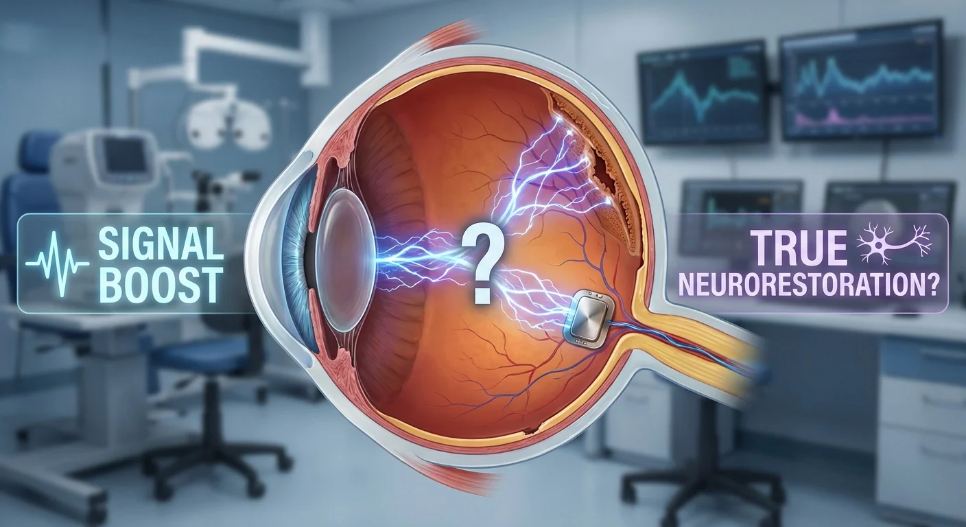
Electrical Stimulation for Glaucoma: Signal Boost or True Neurorestoration?
Glaucoma is a leading cause of irreversible vision loss (affecting >70 million people worldwide) characterized by loss of retinal ganglion cells and optic nerve damage (pmc.ncbi.nlm.nih.gov). Presently, the only proven treatment slows damage by lowering intraocular pressure (IOP) (pmc.ncbi.nlm.nih.gov); no therapy can actually restore lost vision. This has spurred interest in neurostimulation therapies to protect or even revive retinal neurons. Two main approaches are under study: transcorneal electrical stimulation (TES, via corneal electrodes) and transorbital or transcranial alternating-current stimulation (ACS, via electrodes near the eyes). We review sham-controlled studies of these methods in glaucoma, their proposed mechanisms, typical stimulation parameters, and the observed effects on vision (visual field and contrast sensitivity), plus practical issues of safety and availability.
How Might Electrical Stimulation Help?
Experimental work suggests several ways brief currents can boost neural survival and plasticity. One class of effects is neurotrophic upregulation: stimulation prompts the retina and optic nerve to produce growth factors that nourish neurons. For example, in animal models of optic injury, TES or ACS increases levels of neurotrophins such as brain-derived neurotrophic factor (BDNF), ciliary neurotrophic factor (CNTF) and insulin-like growth factor (IGF-1) (pmc.ncbi.nlm.nih.gov). BDNF in particular is critical for retinal ganglion cell (RGC) survival and synaptic plasticity, so its upregulation may help “revive” dysfunctional but living cells. In one study, alternating currents applied to injured rats raised BDNF and CNTF in the eye (pmc.ncbi.nlm.nih.gov).
Electrical stimulation also appears to trigger anti-apoptotic (anti-cell-death) signaling. Gene analyses in rodent retina after TES have shown downregulation of apoptotic factors and upregulation of cell-survival proteins (pmc.ncbi.nlm.nih.gov) (pmc.ncbi.nlm.nih.gov). For instance, TES can increase Bcl-2 (an anti-apoptotic protein) and decrease Bax (a pro-apoptotic protein) in retinal cells (pmc.ncbi.nlm.nih.gov). In practical terms, these molecular shifts correlate with greater neuronal survival: in a glaucomatous injury model, TES-treated eyes had significantly more surviving RGCs at one month post-injury than untreated eyes, along with higher levels of anti-inflammatory IL-10 and less NF-κB activity (pmc.ncbi.nlm.nih.gov). In other words, electrical pulses suppress damaging inflammation and cell-death pathways, helping to preserve RGCs (pmc.ncbi.nlm.nih.gov) (pmc.ncbi.nlm.nih.gov).
Finally, electrical stimulation may engage cortical plasticity. Glaucoma deprives the brain of input from the damaged optic nerve, but some visual pathways remain intact (“residual vision”). By sending rhythmic currents to the eyes, rtACS may entrain brain waves (especially alpha-band oscillations) in visual cortex, potentially reactivating underused circuits. In one controlled trial, study authors noted that claimed vision gains from 10 Hz ACS were attributed to “increased neuronal synchronization and coherent oscillatory activity via entrainment of alpha frequencies” in occipital cortex (pmc.ncbi.nlm.nih.gov). This kind of neuromodulation-inspired idea – boosting brain connectivity with surviving inputs – is actively studied, although evidence in glaucoma patients remains indirect (pmc.ncbi.nlm.nih.gov).
In summary, laboratory data suggest electrical stimulation could promote neuroprotection by (1) raising growth factors like BDNF, (2) blocking cell-death signals (e.g. by upregulating Bcl-2), (3) reducing inflammation, and (4) harnessing brain plasticity. These effects are hypothetical in humans, but provide a rationale for clinical trials.
Clinical Studies
Transcorneal Electrical Stimulation (TES)
In TES, a conductive contact (such as a corneal lens electrode) delivers brief pulses or sinusoidal currents through the cornea to the retina. In glaucoma, most TES studies have been small and preliminary. One Japanese pilot case series treated five eyes (four men) with open-angle glaucoma by quarterly 30-minute TES sessions over several years (pmc.ncbi.nlm.nih.gov). In that uncontrolled study, the amount of cumulative stimulation strongly correlated with better visual fields: eyes receiving more sessions showed greater improvement in mean defect (MD) (pmc.ncbi.nlm.nih.gov). However, without a control group this could reflect slow inherent changes or learning effects. By contrast, a sham-controlled RCT of TES in 14 glaucoma patients found no significant visual field benefit (pubmed.ncbi.nlm.nih.gov). In that trial the TES “dose” was weekly 30-minute sessions for 6 weeks at either 66% or 150% of phosphene threshold, and outcomes (acuity and Humphrey field) did not differ from sham (pubmed.ncbi.nlm.nih.gov). No serious adverse events occurred, and apart from one spontaneous optic disc hemorrhage (in a control eye) no safety signals appeared (pubmed.ncbi.nlm.nih.gov).
Another small series (K. Ota 2018) followed five eyes with quarterly suprathreshold TES over ~4 years; these showed a gradual MD improvement proportional to the number of treatments (pmc.ncbi.nlm.nih.gov). Thus far, the evidence for TES in glaucoma is mixed: some small case studies hint at a stabilization or modest gain in field with repeated sessions (pmc.ncbi.nlm.nih.gov), but the single published RCT did not confirm an effect (pubmed.ncbi.nlm.nih.gov). Importantly, no TEC study has compared beyond a few months or tested long-term retention of benefit.
Typical TES parameters in glaucoma trials have been on the order of 20–30 minutes per session, often delivered weekly or monthly, with currents adjusted to induce phosphenes. (For example, one protocol used 20 Hz biphasic pulses at each subject’s phosphene threshold level for 30 min once weekly (pubmed.ncbi.nlm.nih.gov).) No dose–response standard has been set, and devices vary. As of 2025, TES for glaucoma remains experimental and is offered only within trials or specialty clinics.
Transorbital/Transcranial Alternating-Current Stimulation (rtACS)
An alternate approach is noninvasive transorbital ACS: electrodes are placed on the skin around the eye (often in a goggle-like frame) to send weak alternating currents into the visual pathway. Over the last decade, several sham-controlled trials have studied rtACS in optic neuropathies (usually mixed diagnoses), including a few focused on glaucoma.
A landmark randomized trial (Gall et al., 2016) enrolled 82 patients with various partially blind optic neuropathies and applied rtACS daily for 10 consecutive weekdays. The treated group showed an average 24% improvement in visual field sensitivity (mean defect) compared to baseline, lasting at least two months (pmc.ncbi.nlm.nih.gov). This was significantly better than sham. (This study included some glaucoma patients, but also other causes of field loss.) A follow-up long-term retrospective analysis of many patients also found that nearly two-thirds of treated eyes “halted” progression for ~1 year after a similar rtACS course (pmc.ncbi.nlm.nih.gov): median MD improved from 14.0 to 13.4 dB (p<0.01) over one year, with about 63% of eyes showing stable or better MD (pmc.ncbi.nlm.nih.gov). By comparison, typical glaucoma patients decline by ~0.5 dB per year on average, so this stability is noteworthy.
However, other studies have tempered enthusiasm. A smaller RCT (Ramos-Cadena et al., 2024) in 16 advanced glaucoma patients applied 10 rtACS sessions over 2 weeks (10 Hz sine wave at 0.45–1.5 mA through forehead/cheek electrodes) and followed up to 1 month (pmc.ncbi.nlm.nih.gov) (pmc.ncbi.nlm.nih.gov). That trial found no significant changes in objective vision tests – neither visual acuity, contrast sensitivity, nor Humphrey field MD improved beyond placebo (pmc.ncbi.nlm.nih.gov). (The sham group actually showed a slight early field gain that later receded, suggesting a practice effect (pmc.ncbi.nlm.nih.gov).) The treated group did report higher patient-reported quality of life related to vision (near activities, dependency, mental health) (pmc.ncbi.nlm.nih.gov), but without accompanying functional gains. Notably, no serious side effects occurred in these patients, and only mild tingling or phosphene sensations were reported (pmc.ncbi.nlm.nih.gov).
In summary, the magnitude of benefit in rtACS trials has been modest and inconsistent. The Gall study’s 24% VF gain sounds large, but it represents an average relative improvement that lasted only a couple months (pmc.ncbi.nlm.nih.gov). By contrast, Ramos-Cadena’s double-masked trial saw no significant visual field or contrast benefit over 1–4 weeks (pmc.ncbi.nlm.nih.gov). Likewise, a 2021 German “real life” cohort suggested stabilization (no mean decline) over 1 year (pmc.ncbi.nlm.nih.gov), but without a control arm this could partly reflect expected variability. In practice, any field gains reported with rtACS are small (a few decibels) and short-lived, often disappearing after weeks if therapy is not repeated. Contrast sensitivity changes have been even less evident: in the 2024 RCT neither group showed measurable contrast threshold improvements (pmc.ncbi.nlm.nih.gov).
A key issue is the placebo/practice effect. Performing repeated perimetric tests can itself produce small “learning” improvements. In the Ramos-Cadena study, the sham group had a temporary field gain that then declined, illustrating this phenomenon (pmc.ncbi.nlm.nih.gov). Thus, any modest field gain with real stimulation must be judged against what happens in controls. So far, only a few trials are large enough to judge this – and their results are mixed. On balance, therapies claim statistical improvements over sham in some studies (e.g. Gall 2016 (pmc.ncbi.nlm.nih.gov)) but not others (e.g. Ramos 2024 (pmc.ncbi.nlm.nih.gov)). The clinical significance (how much better a patient sees) of the modest gains reported is still uncertain.
Typical rtACS parameters in glaucoma studies have been roughly: 10 sessions, each ~25–40 minutes long, of low-intensity (under 2 mA) alternating currents at ~5–20 Hz. For example, Ramos-Cadena used a 10 Hz sine wave with gradually increasing amplitude (0.45–1.5 mA) over 5 consecutive days (30 min each), then 5 more days at 40 min each (pmc.ncbi.nlm.nih.gov). Other protocols have varied frequency (often ~10 Hz, sometimes alternating bands up to 37 Hz) and electrode placement. In practice, investigators choose currents just strong enough to elicit phosphenes (brief flashes) in the patients.
Safety
Across trials, electrical stimulation has been well-tolerated. In the TES RCT, no treatment-related serious adverse events occurred (pubmed.ncbi.nlm.nih.gov). The main side effects are mild: eyelid tingling or twitch, a few patients can feel the current or slight headache during stimulation. The 2024 rtACS trial reported no serious adverse events at all (pmc.ncbi.nlm.nih.gov). Indeed, in Europe more than 1,000 patients have already received 10-day rtACS courses (10×60 min) under medical supervision, with zero reports of serious harm (www.ophthalmologytimes.com). Overall, the risk to patients appears negligible apart from temporary discomfort – one reason these methods are appealing to patients eager for novel therapies.
Next-Generation Therapies
Devices and availability: Currently, electrical stimulation for glaucoma is mostly a research or niche clinical service. One commercial system, Eyetronic Nextwave, delivers transorbital ACS via goggles and is CE-marked in Europe for all optic neuropathies (including glaucoma) (ichgcp.net). It is used in Germany and some other countries, though not covered by insurance, so patients typically pay out-of-pocket. In the US, Eyetronic therapy is only available in clinical trials. Notably, Dr. Sunita Radhakrishnan (Glaucoma Center of SF) recently treated the first US patient in such a trial (www.ophthalmologytimes.com). The registered Eyetronic trial plans 10 sessions of 1-hour stim (daily) and will track Humphrey fields for a year (ichgcp.net).
Other “next-gen” research approaches include implantable stimulators. For example, a recent preclinical study tested a suprachoroidal retinal implant (an electrode array placed between the retina and choroid) delivering continual pulses (pmc.ncbi.nlm.nih.gov). In cats, chronic suprathreshold stimulation via this implant produced no retinal damage or safety issues (pmc.ncbi.nlm.nih.gov). Thus an implantable device might one day provide ongoing neuroprotective currents without requiring daily clinic visits (pmc.ncbi.nlm.nih.gov). Meanwhile, trials like the Hong Kong GREAT study are exploring head-worn transcranial stimulators paired with vision training (perceptual learning) to enhance any residual vision. In short, efforts are underway to make neurostimulation more personalized (e.g. MRI-tailored electrode placement (pmc.ncbi.nlm.nih.gov)) and user-friendly.
Conclusion
Electrical stimulation therapies offer an intriguing signal-boosting strategy for glaucoma, but it is still uncertain whether they achieve true neurorestoration. Early studies show occasional small gains in visual fields and patient-reported vision, but results have been inconsistent, and gains (if any) are typically short-lived. The scientific rationale (BDNF upregulation, anti-apoptosis, cortical plasticity) is solid in animals (pmc.ncbi.nlm.nih.gov) (pmc.ncbi.nlm.nih.gov), but proof in patients is modest so far. More large, sham-controlled trials are needed to determine how much beyond placebo these therapies truly benefit. For now, electrical stimulation remains experimental – safe but unproven – and should not replace standard IOP-lowering treatment. Clinicians and patients should watch ongoing trials (such as the VIRON study) for stronger evidence. If confirmed, noninvasive neuromodulation could become a valuable adjunct for preserving vision beyond IOP control, finally offering glaucoma patients a chance at actual vision improvement.