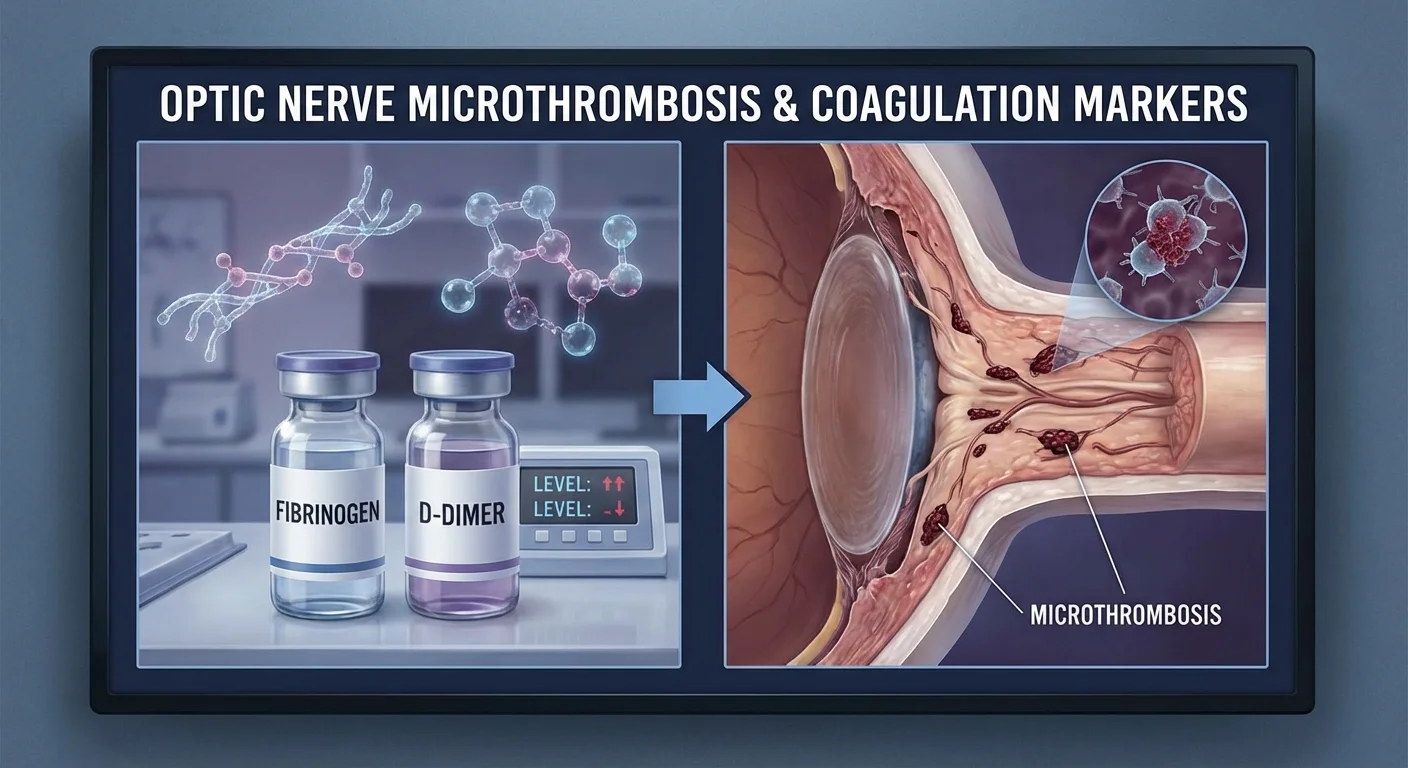
Coagulation markers (fibrinogen and D-dimer) and optic nerve microthrombosis
The optic nerve is the cable connecting your eye to your brain. If it does not get enough blood, sudden vision loss can occur – this is called optic nerve ischemia (often seen in NAION, non-arteritic anterior ischemic optic neuropathy (pmc.ncbi.nlm.nih.gov)). Many common risk factors (high blood pressure, diabetes, high cholesterol, smoking) reduce eye blood flow (pmc.ncbi.nlm.nih.gov). Doctors are now also checking if a tendency for extra blood clotting – a hypercoagulable state (sometimes called thrombophilia) – might contribute to tiny clots (microthrombosis) around the optic nerve. In simple terms, if your blood clots too easily, it might block small blood vessels feeding your optic nerve, leading to damage. For example, several case reports note that abnormal clotting factors were found in patients with acute optic nerve events (pmc.ncbi.nlm.nih.gov).
As a result, researchers have suggested measuring clotting markers like fibrinogen and D-dimer to see if they predict optic nerve problems. This article explains these tests in everyday language, how they relate to optic nerve health, and how you or your doctor might use them safely.
What is hypercoagulability (thrombophilia)?
Blood clotting is a normal repair process, but when “too much” clotting factor is present, it is called thrombophilia or a hypercoagulable state (www.reviewofoptometry.com). In a hypercoagulable state, your blood has extra clotting factors or fewer clot-dissolving factors, so it can clot more easily. People with thrombophilia often never have problems until something triggers a clot. For example, inherited conditions like Factor V Leiden or high homocysteine can be present from birth, but clots may only form if another risk (like smoking or hormonal birth control) is present (www.reviewofoptometry.com). Acquired factors (surgery, cancer, pregnancy, severe infections) can also temporarily tilt the balance toward clotting (www.reviewofoptometry.com).
In the eye, clotting can cause blockages in retinal or optic nerve blood vessels. Conditions like central retinal vein occlusion (CRVO) or NAION are sometimes linked to clotting problems (www.reviewofoptometry.com). An eye care review notes that when we see unexplained optic nerve or retinal occlusions (sudden vision loss), we should consider a blood clotting tendency (www.reviewofoptometry.com). In fact, one clinical case report concluded: “In young patients without other health issues, unexplained optic nerve ischemia should prompt careful testing for clotting disorders” (pmc.ncbi.nlm.nih.gov). In plain words, if you are young and suddenly lose vision due to optic nerve swelling, your doctor should check if your blood is clotting too much.
Because clot markers can change with illnesses or treatment, doctors suggest controlling for things like recent infections, trauma or surgery when interpreting results (pmc.ncbi.nlm.nih.gov) (www.medicalnewstoday.com). For example, after a bone fracture or surgery, studies show that fibrinogen and D-dimer can rise dramatically (pmc.ncbi.nlm.nih.gov) (www.medicalnewstoday.com). Similarly, being on blood thinners (anticoagulants) will lower clotting markers. In practice, any lab test for clot risk is interpreted in context.
Blood tests for clotting: fibrinogen and D-dimer
To check clot risk, doctors use specific blood tests:
-
Fibrinogen test – Fibrinogen is a protein made by the liver that helps blood clots form (my.clevelandclinic.org) (emedicine.medscape.com). When you bleed, fibrinogen is converted to fibrin strands that make a clot. Normal fibrinogen in adults is roughly 200–400 mg/dL (emedicine.medscape.com). If your fibrinogen is very low (e.g. <100 mg/dL), you may bruise or bleed too easily (not a clot problem). But an elevated fibrinogen can mean your blood is “stickier” and more prone to clot. In fact, levels above ~700 mg/dL have been linked to higher risk of dangerous clots (e.g. stroke, heart attack) (www.webmd.com). Fibrinogen is also an “acute phase reactant”, meaning it naturally rises if you have inflammation, infection, or even cancer (emedicine.medscape.com). In other words, high fibrinogen could come from injury or illness as well as from clot risk. WebMD explains that very high fibrinogen often occurs with conditions like infection or heart disease (www.webmd.com).
-
D-dimer test – D-dimer is a small protein fragment made when a blood clot dissolves. A very useful test, it tells us if clotting and clot breakdown has recently been happening in the body. The Cleveland Clinic describes D-dimer as a “protein fragment your body makes when a blood clot dissolves” (my.clevelandclinic.org). Normally, D-dimer is almost undetectable (close to 0) because your body only makes a little of it after minor clot breakdown. A high D-dimer level means your body recently formed and dissolved significant clots (my.clevelandclinic.org) (my.clevelandclinic.org).
Laboratory ranges for D-dimer are usually given in mg/L (fibrinogen equivalent units). Values below ~0.50 mg/L are generally normal (www.medicalnewstoday.com) (www.medicalnewstoday.com). Readings above 0.50 mg/L are called positive and suggest that clots may be present somewhere in the body (www.medicalnewstoday.com) (www.medicalnewstoday.com). Medical sources note that a positive D-dimer prompts further testing (like ultrasound or scans) to find a clot (www.medicalnewstoday.com). It's important to know that many things besides a dangerous clot can raise D-dimer. For example, D-dimer levels are known to be higher in cases of recent surgery, infection, cancer, or even simply with age (pmc.ncbi.nlm.nih.gov) (www.medicalnewstoday.com). Because of this, doctors always interpret a high D-dimer in context: if you’ve just been ill or had surgery, a high D-dimer by itself might not mean an ongoing clot. Conversely, a very low D-dimer makes a significant clot very unlikely.
Interpreting fibrinogen and D-dimer results
- If fibrinogen is high, consider whether you have any inflammatory or clot-related conditions. Are you recovering from an infection, injury or surgery? Those can raise it (pmc.ncbi.nlm.nih.gov) (www.medicalnewstoday.com). If you have none of those and fibrinogen is high (and other clot tests are abnormal), your doctor may suspect an underlying clotting tendency.
- If D-dimer is high, it may mean clots are forming or dissolving in your body. Your doctor will likely check you for things like deep vein thrombosis, pulmonary embolism, or other clot causes (my.clevelandclinic.org) (www.medicalnewstoday.com). At the same time, they will rule out other reasons (like recent operation, cancer, etc) that could explain the high value (pmc.ncbi.nlm.nih.gov) (www.medicalnewstoday.com).
- A normal or low D-dimer is reassuring – it usually rules out significant clots at that moment (especially if you are not bleeding heavily) (www.medicalnewstoday.com).
Both tests require only a standard blood draw. For patients, a D-dimer test is very accessible; it is routinely drawn in hospitals when clots are suspected (my.clevelandclinic.org). A fibrinogen blood test is also available via most hospital labs or specialized clinics (my.clevelandclinic.org). In many areas, you can have these tests done through your doctor. There are even online lab services (for example, UltaLabTests and similar) where patients can order a D-dimer test without a doctor’s prescription — you would simply get a blood draw at a lab and receive the results.
Normal ranges: As a quick guide, D-dimer in healthy people is usually well under 0.50 mg/L (or under 500 ng/mL in older units) (www.medicalnewstoday.com) (www.medicalnewstoday.com). Fibrinogen normal range is about 200–400 mg/dL (emedicine.medscape.com). Results reports should list the lab’s reference range. Discuss any abnormal result with your doctor, who will consider your full history and any recent illnesses.
Platelet and white cell markers (MPV, PLR, NLR)
Besides fibrinogen and D-dimer, doctors often look at simple blood count numbers for clues about clotting. A complete blood count (CBC) is a routine test that includes platelets and white blood cells. Two ratios in a CBC are growing in interest:
-
Mean Platelet Volume (MPV): This measures the average size of your platelets (cells that clump in clots). Larger platelets are more active and likely to form clots. Elevated MPV means more of the “big and sticky” platelets. Studies have found that MPV is often higher in people with clot-related eye disease. For example, patients with NAION (optic nerve ischemia) had significantly higher MPV than healthy people (pmc.ncbi.nlm.nih.gov). Another study found that both NAION and arteritic AION groups had elevated MPV compared to controls (pmc.ncbi.nlm.nih.gov). In plain terms, unusually large average platelet size can signal a higher clot risk (pmc.ncbi.nlm.nih.gov) (pmc.ncbi.nlm.nih.gov).
-
Platelet-to-Lymphocyte Ratio (PLR): This is calculated by dividing the number of platelets by the number of lymphocytes (a type of white blood cell). It is a marker of inflammation/clotting balance. A higher PLR means more platelets relative to immune cells. Some researchers have suggested PLR could add information about clot risk in stroke and vascular disease. However, in general hospital populations, high PLR by itself was not consistently linked to more clots (pubmed.ncbi.nlm.nih.gov) (pubmed.ncbi.nlm.nih.gov). For instance, a large study of venous blood clots found that patients with high PLR did not have a significantly increased clot risk overall (pubmed.ncbi.nlm.nih.gov) (pubmed.ncbi.nlm.nih.gov). In the eye context, PLR is still an experimental marker.
Doctors may look at these alongside other inflammatory markers like the neutrophil-to-lymphocyte ratio (NLR) when considering clot risk (pmc.ncbi.nlm.nih.gov). These counts are all part of the CBC, so they are very easy to obtain. The MPV is reported on many CBCs and can give a hint of platelet activation. If MPV is high in an optic nerve case, it might point to clot-related damage as a factor.
Tracking the eye: OCTA and visual field tests
If clotting is suspected as a cause of optic nerve ischemia, specialists use advanced imaging and vision tests to see how the eye is affected:
-
Optical Coherence Tomography Angiography (OCTA): This is a non-invasive scan that maps blood flow in the retina and optic nerve head. In OCTA, moving blood cells are detected by a specialized camera, creating images of tiny blood vessels in different layers of the retina (www.ncbi.nlm.nih.gov). Without any dye injection, OCTA can highlight areas where blood flow is reduced. Studies of chronic NAION patients have shown that OCTA metrics are significantly lower than normal. For example, one 2023 study found that vessel density (how much blood vessel area is visible) and blood flow (flux) around the optic nerve were much lower in affected eyes. These OCTA measures strongly correlated with how much vision was lost (pmc.ncbi.nlm.nih.gov) (pmc.ncbi.nlm.nih.gov). In other words, OCTA can objectively show microvasculature damage in NAION. Researchers believe parameters like vessel density might even predict disease severity (pmc.ncbi.nlm.nih.gov).
-
Visual field testing: This is how patients go to an eye doctor and press a button each time they see a flash of light in their peripheral vision. It maps out the “field” or shape of the person’s vision. Optic nerve damage causes blind spots or areas of vision loss. In NAION, visual field tests typically show significant deficits. For example, the “mean deviation” score (a summary of field loss) was about –13.5 dB in NAION eyes versus –0.5 dB in normal eyes in one study (pmc.ncbi.nlm.nih.gov). (In these tests, more negative means more loss; 0 is basically normal.) Regular visual field exams track if any new vision loss is happening over time. In a research setting, doctors use both OCTA and visual field results over time to see if patients are getting better or worse.
By combining eye tests (OCTA images and visual fields) with blood markers (D-dimer, fibrinogen, MPV, etc.), researchers hope to capture transient risk periods. For instance, D-dimer might spike around an illness or surgery, and if an OCTA done at that time shows reduced flow, it could suggest a cause-effect link. Emerging studies use “time-updated” markers – taking repeated blood tests at different visits – to catch these temporary surges in clotting risk. This approach is akin to checking blood pressure or blood sugar multiple times rather than only once.
Managing other factors
Several factors can confuse clot-marker results, so good studies and medical practice adjust for them:
-
Recent illness or injury: As noted above, even minor injuries or infections can raise fibrinogen and D-dimer (pmc.ncbi.nlm.nih.gov) (www.medicalnewstoday.com). For example, fracture patients awaiting surgery had average D-dimer ~1283 ng/mL (very high) and fibrinogen ~321 mg/dL, compared to ~98 ng/mL and 277 mg/dL in healthy controls (pmc.ncbi.nlm.nih.gov). This means that if you’ve had any recent trauma or illness, your doctor will consider that a potential cause of high clot markers before blaming the optic nerve event.
-
Recent surgery or immobilization: After an operation or if you’ve been bedridden, clot risk goes up and markers rise. The medical literature notes that D-dimer is often elevated by surgery, cancer, or intensive illness (pmc.ncbi.nlm.nih.gov) (www.medicalnewstoday.com). For example, hip or knee surgery patients routinely get D-dimer testing because of clot risk.
-
Genetic clotting predisposition: If you have a known thrombophilia (like Factor V Leiden, Protein S/C deficiency, or antiphospholipid antibodies), this background matters. A study mentioned that very high PLR was problematic mainly when combined with known thrombophilia (pubmed.ncbi.nlm.nih.gov). In practice, if you have a thrombophilic gene mutation, your doctor will interpret your clot tests with that in mind. Sometimes they will test for those inherited factors directly.
-
Anticoagulant (blood thinner) use: Drugs like warfarin, heparin, or newer anticoagulants affect clot test results. While they primarily prolong clotting times (INR/PTT, etc.), they can also lower D-dimer and fibrinogen indirectly (since the drug is preventing clot formation). If you are on a blood thinner, a low D-dimer doesn’t completely rule out clots, because the medication is doing its job. Always inform any doctor doing clot testing if you take such medication.
Because of these factors, serious studies and careful doctors “control for” them. In plain terms, that means they make sure patients are not in the middle of an infection, cancer flare, or right after surgery when taking these tests. If they are, they note it and may exclude the test result or interpret it differently.
Getting and understanding these tests
Patients often wonder: How do I get these tests and what do I do with the results? Here’s a practical guide:
-
Who can order these tests? Typically, a physician (your family doctor, an eye doctor, or a hematologist) orders fibrinogen and D-dimer. You might have seen a D-dimer ordered in the emergency room if clots were suspected. Fibrinogen is ordered when unusual clot or bleeding symptoms occur. Some patients use online lab companies (which allow self-requested tests) to get a D-dimer or a broader “clotting panel.” But even if you order your own lab, you should review the results with a healthcare provider who understands clotting.
-
How are the tests done? Both are simple blood tests. A nurse will draw a small vial of blood from your arm and send it to the lab. A D-dimer test is available almost everywhere: hospital labs, outpatient labs, even some pharmacies have lab draw stations. The fibrinogen test is less common but still widely available. Because they aren’t routine, you may have to ask for them specifically if you feel you need them.
-
What do the results look like? Test reports will list your number and the lab’s normal range. For example, a D-dimer report might say: Result: 0.30 mg/L, Reference: <0.50 mg/L. A fibrinogen report might say 300 mg/dL (normal 200–400). It’s vital to compare to the “normal range” on the report. If your D-dimer is above the range (often noted as “positive”), discuss with your doctor. If your fibrinogen is near the top or above normal, or if it’s very low, that is also noteworthy.
-
Interpreting results:
- A normal D-dimer (within range) is usually reassuring – it means active clotting is unlikely at that time (www.medicalnewstoday.com).
- A high D-dimer (above normal) warrants further investigation. It could mean clots somewhere, but might also be from another cause. Doctors would not diagnose a blood clot just from one D-dimer; they would likely order imaging (e.g. an ultrasound or CT scan) or look for sources of inflammation or recent surgery.
- Normal fibrinogen is expected (200–400 range). A high fibrinogen suggests increased clot potential or inflammation. A doctor might then check other clotting factors to see if multiple pieces point to a clotting tendency. Sometimes very high fibrinogen is found with heavy smoking, obesity, or metabolic syndrome, reflecting chronic inflammation.
- Low fibrinogen (well below normal) is rare but would raise concern for bleeding problems or a consumptive clotting process (like DIC).
If you get these lab results yourself (e.g., through an online lab service), do not panic. Review them with your doctor. Clotting markers are complex: they rarely stand alone for a diagnosis. They are part of the puzzle.
Accessible tests for patients: In the US and other countries, many regions now allow individuals to order lab tests directly online and pay out of pocket. Services like Ulta Lab Tests, Walk-In Lab, or local private labs often list D-dimer and fibrinogen by name. The prices vary (for example, D-dimer might cost on the order of ~$50–100 non-insurance). You would still need to get a blood draw at a partner lab. For patients outside the US, availability depends on local medical practice. Regardless, the process is the same: blood draw → lab analysis → result report.
Conclusion
In summary, there is a plausible link between hypercoagulability (a tendency to form clots) and optic nerve ischemia. Some patients with unexplained optic nerve strokes have been found to have clotting abnormalities (pmc.ncbi.nlm.nih.gov). Measuring clotting markers like fibrinogen and D-dimer may help identify people at higher risk. However, these markers must be interpreted with care, controlling for recent illness, surgery, or medications. Modern eye imaging (like OCT angiography) and vision tests can capture how these clot-related changes affect the optic nerve.
For patients, the take-home message is: talk to your doctor if you have had an optic nerve issue (like NAION) and wonder about clot risk. Ask if checking fibrinogen or D-dimer makes sense in your case. Remember to mention any factors like recent surgeries or chronic conditions. If you have risk factors for clots (personal or family history, or thrombophilia), monitoring these blood tests over time might offer early clues. Ultimately, studies are ongoing, but these tests are accessible and could add valuable information alongside standard eye exams.
Finally, living a “heart-healthy” lifestyle also protects your eyes. Control your blood pressure, cholesterol, and blood sugar, and don’t smoke. These steps reduce blood-vessel stress and clot risk. And keep up with regular eye check-ups, including visual field tests. That way, if any changes occur, you and your doctor can catch them early – possibly using the very blood tests and imaging tools discussed here.