Introduction
Glaucoma is an eye disease that damages the optic nerve, causing peripheral vision loss. Once damage occurs, conventional treatments (like lowering eye pressure) cannot restore the lost vision. Researchers have therefore explored whether non-invasive brain stimulation might help improve remaining vision. Two common methods are transcranial direct current stimulation (tDCS) and transcranial magnetic stimulation (TMS), which apply weak electrical or magnetic pulses to the scalp to modulate the brain’s activity. Small studies have tested such techniques on glaucoma patients to see if visual processing (contrast sensitivity, field defects, etc.) can be enhanced. We review these pilot and controlled trials, noting where electrodes or coils were placed, the stimulation settings, the measured vision gains, and how long those gains lasted. We also discuss possible mechanisms (like boosting brain plasticity or reducing neural “noise”) and the importance of good sham-controlled study designs (since practice or placebo effects can mimic improvement).
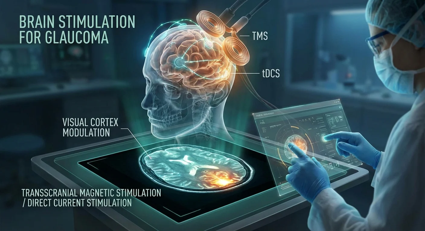
Brain Stimulation Techniques
tDCS uses a mild constant electrical current applied through electrodes on the scalp. Depending on polarity, it can increase (anodal) or decrease (cathodal) cortical excitability. Typically, one electrode is placed over the target brain region (often the occipital visual cortex), and the other electrode (reference) is placed elsewhere (e.g. the cheek or forehead). Treatment sessions often last 10–20 minutes at 1–2 mA. TMS uses brief magnetic pulses through a coil to induce electrical currents in the underlying cortex. Both methods have been used for many brain disorders; for vision, they aim to “boost” residual visual function by recruiting plasticity in visual pathways.
tDCS in Glaucoma
In glaucoma studies, researchers have generally targeted the visual cortex (occipital lobe). A recent randomized trial had patients receive one session of anodal tDCS (a-tDCS) at 2 mA for 20 minutes. The anode was placed at Oz (midline occiput) and the cathode on the cheek. This single session modestly improved visual field detection accuracy (about 3–5% gain in high-resolution perimetry) compared to sham (pmc.ncbi.nlm.nih.gov). The multifocal visual-evoked potentials (mfVEP) also showed slightly higher signal-to-noise and faster responses after a-tDCS. These gains were statistically significant versus sham, but very small in magnitude, roughly on the order of test-retest variability (pmc.ncbi.nlm.nih.gov). In other words, vision improved on some tests, but only by a few percent, which may not be noticeable in daily life.
Session parameters: Typical pilot studies used a single 20-minute session of 1–2 mA a-tDCS to the occiput (Oz). One study also tried alternative waveforms (alternating current tACS at 10 Hz, and random-noise tRNS) versus sham, but only a-tDCS showed any clear effect (pmc.ncbi.nlm.nih.gov). No study has used very high intensity or very long duration beyond 20–30 minutes.
Vision outcomes: Measured outcomes have included visual field indices (e.g. detection accuracy or mean defect in perimetry) and sometimes contrast sensitivity or visual acuity. In the above trial, a-tDCS produced a small increase in detection accuracy on a high-res perimetry test (pmc.ncbi.nlm.nih.gov). No large change in standard automated perimetry (mean defect) was shown, nor in visual acuity. Contrast sensitivity was not always measured in glaucoma trials, though in other eye disorders tDCS can transiently boost contrast thresholds. Crucially, the Glaucoma RCT noted that the tiny improvements “may not be clinically meaningful” (pmc.ncbi.nlm.nih.gov).
Duration of effects: In these studies effects were tested immediately before and after the stimulation session. No sustained follow-up beyond hours was reported in this trial, so it’s unclear how long the benefit lasts from one session. Other research (in optic nerve damage generally) suggests any improvement often fades over days or weeks once stimulation ends (pmc.ncbi.nlm.nih.gov).
TMS and Other Modalities
TMS: To date, there are few published trials of repetitive TMS (rTMS) for glaucoma specifically. TMS can excite visual cortex neurons and has been used experimentally to induce phosphenes (flashes of light) even in blind individuals. In theory, rTMS could be applied in multiple sessions to the occipital lobe to enhance cortical excitability and possibly unmask residual vision. However, no well-controlled studies in glaucoma have yet shown clear vision gains from TMS. (Most visual-field research with TMS has been in stroke-related vision loss rather than glaucoma.)
Alternate electrical stimulation: Some trials have used transorbital alternating current stimulation (rtACS), where electrodes are placed on the closed eyelids to stimulate the retina/optic nerve. Although this mainly targets the eye rather than the brain, it has been combined with brain monitoring. In one large randomized trial of rtACS in optic nerve damage (including many glaucoma patients), subjects received 10 daily sessions of 50 minutes each. Both the real-stimulation and sham groups improved their visual field on routine testing, with a slightly larger mean gain in the rtACS group (median ~41.3% vs 29.3% detection increase (pmc.ncbi.nlm.nih.gov)). The difference did not reach statistical significance for the main outcome (pmc.ncbi.nlm.nih.gov). Interestingly, at 2-month follow-up there was a modest between-group advantage in one measure (static perimetry sensitivity) favoring rtACS (pmc.ncbi.nlm.nih.gov). In words, this suggests some lingering benefit, but most gains were also seen in the sham group, indicating learning or placebo effects. The authors concluded that rtACS appears “to partially restore vision” by promoting brain plasticity (pmc.ncbi.nlm.nih.gov), but overall the clinical impact was mild.
Study Results – Gains and Limits
Across studies, any improvements in vision have generally been modest and short-lived. For instance, in the transcranial trials above, contrast sensitivity was not majorly changed, and field improvements were only a few percentage points higher than baseline. Patients rarely notice such small changes. Most reports describe immediate post-stimulation gains, with little evidence on long-term durability. In the rtACS trial, a small field improvement persisted at 2 months in one measure (pmc.ncbi.nlm.nih.gov), but many other measures regressed. Single-session tDCS effects also are expected to fade without repeated sessions.
Moreover, placebo effects are important. Some studies found that vision tests improved even with sham (inactive) stimulation (pmc.ncbi.nlm.nih.gov) (pmc.ncbi.nlm.nih.gov). That is why the larger trial saw 29% gain in sham responders. A recent review of non-invasive stimulation across eye diseases concluded that the small average benefits (for acuity, field detection, etc.) may partly reflect placebo or practice effects (pmc.ncbi.nlm.nih.gov). In other words, “active” stimulation often outperformed sham by only a tiny margin, and sometimes sham improvements were as large. This uncertainty means we must interpret early pilot results cautiously.
Possible Mechanisms
If brain stimulation truly boosts vision, how might it work? One idea is cortical plasticity: the visual cortex may strengthen weak pathways and unmask “backup” circuits after the eye injury. Stimulation could increase levels of growth factors or change neurotransmitters, making it easier for the brain to adapt (pmc.ncbi.nlm.nih.gov). For example, anodal tDCS is thought to slightly depolarize neurons, potentially enhancing synaptic plasticity in visual areas. Another idea is noise reduction: in degenerating vision, the remaining signals from the eye may be buried in “neural noise.” Some studies (in other retinal diseases) suggest that reducing noise can quickly improve perception. For instance, one trial in proliferative diabetic retinopathy found that applying cathodal tDCS (which can inhibit hyperactive neurons) improved visual tasks. The authors proposed that tDCS likely lowered the level of random neural activity, thereby clarifying the actual visual signal (pmc.ncbi.nlm.nih.gov). By analogy, if surviving retinal ganglion cells in glaucoma are noisy, tDCS might help “silence” that noise and enhance contrast or field sensitivity.
On the other hand, some effects may not be physiological at all. Stimulation can increase alertness or the placebo sensation of “something happening,” which can improve test performance. Indeed, the optic nerve stimulation trial noted that much of the current actually travels through the retina and optic nerve, not deep cortex (pmc.ncbi.nlm.nih.gov). Those authors still claim changes in brain synchrony (EEG rhythms in visual areas) after treatment, but it’s hard to rule out non-specific effects. To disentangle these possibilities, future studies must combine brain measures (like EEG or fMRI) with vision tests.
Future Trials – Improving Rigor
Given the modest and mixed results so far, future trials must be carefully designed. Key elements include:
- Randomized sham-controlled design: Every real stimulation group must have a sham treatment that mimics the sensation (e.g. brief current ramp-up but no ongoing stimulation). Both patients and examiners should be masked. This is crucial to account for learning and placebo.
- Multiple sessions: Single sessions give only short-lived effects. Trials should test repeated sessions (for example, daily for 1–2 weeks) since neuroplastic changes often require repetition. The VIRON trial is doing 10 sessions of 25 minutes each for glaucoma (pubmed.ncbi.nlm.nih.gov).
- Objective outcomes: Use standardized vision tests like automated perimetry (mean defect, total deviation), contrast sensitivity charts, and even electrophysiology (VEP or EEG) as secondary measures. High-resolution perimetry can detect small changes, but results must exceed normal test variability. Including patient-reported vision questionnaires can gauge real-world impact.
- Follow-up measurements: To assess durability, vision should be re-tested weeks after the last stimulation. If benefits last, then visual field (or acuity) should be better than baseline at follow-up.
- Neuroimaging / physiology: Combining with functional MRI or EEG can show whether the brain’s visual networks change after stimulation. For example, one could do fMRI while presenting visual stimuli pre- and post-treatment, or measure resting-state connectivity of visual areas. This helps verify that any perceptual changes have a neural correlate, and can distinguish plastic changes from mere test practice.
Such rigorous trials will clarify whether brain stimulation truly helps glaucoma or is simply a placebo-like effect. Until then, tDCS and TMS remain promising research tools but unproven therapies for patients.
Conclusion
In summary, pilot studies of brain stimulation in glaucoma report small improvements in visual field tests or contrast tasks, but these are often similar to improvements seen with sham stimulation (pmc.ncbi.nlm.nih.gov) (pmc.ncbi.nlm.nih.gov). A recent randomized trial found that a single session of occipital a-tDCS yielded only a few percent better detection accuracy than sham (pmc.ncbi.nlm.nih.gov). A larger optic-nerve study showed some visual field gains after multiple days of transorbital current, but the difference vs sham was not significant immediately after treatment (pmc.ncbi.nlm.nih.gov). Reported “durability” of these gains varies; one trial found a small advantage for real stimulation at 2 months on one measure (pmc.ncbi.nlm.nih.gov), but most effects did not last.
Mechanistically, improvements could reflect real neuroplastic changes – the brain rewiring to make better use of the remaining retinal signals (pmc.ncbi.nlm.nih.gov) – or simply the reduction of aberrant neural noise (pmc.ncbi.nlm.nih.gov). Alternatively, motivational or placebo factors may account for some gains. The existing evidence is still preliminary. Future research needs well-controlled, repeat-session trials, with objective measures and brain imaging, to prove definitively if tDCS or TMS can help glaucoma patients.