
Macronutrient Patterns and Intraocular Pressure: A Systematic Evaluation
This article reviews the latest evidence on macronutrient patterns and glaucoma. We will survey epidemiologic studies of diet patterns...
Deep research and expert guides on maintaining your visual health.
This article reviews the latest evidence on macronutrient patterns and glaucoma. We will survey epidemiologic studies of diet patterns...
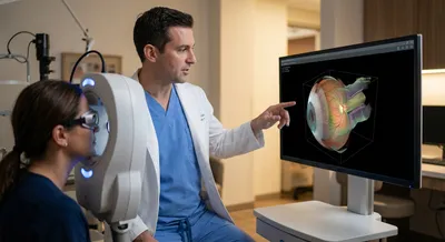
To check the RNFL, doctors commonly use optical coherence tomography (OCT), a non-invasive imaging test that takes cross-sectional “slice” pictures...
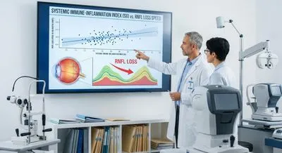
SII = (Platelet count × Neutrophil count) / Lymphocyte count ().
Screen your peripheral vision from home — no downloads, no waiting rooms. Sign up for a free trial and test in under 5 minutes.
The retinal nerve fiber layer is a thin layer in the eye made up of the long fibers from the nerve cells that carry visual information to the brain. Those fibers run along the surface of the retina and converge at the optic disc to form the optic nerve, which transmits the signals your brain uses to form images. Measuring the thickness of this layer is important because thinning usually indicates loss of these nerve fibers, which can mean damage to the optic nerve. Conditions such as glaucoma, optic neuritis, and some neurodegenerative diseases can cause the layer to thin, often before people notice vision loss. Eye doctors use imaging tools like optical coherence tomography to measure the layer noninvasively and track changes over time. Detecting thinning early helps clinicians intervene sooner to try to slow or prevent further vision loss. Thickness can vary with age, eye size, and other factors, so measurements are interpreted against normal ranges and other clinical findings. While useful, the measurement is only one piece of the evaluation, and doctors combine it with vision tests, pressure checks, and other exams to understand eye health. Overall, the retinal nerve fiber layer is a key structural indicator of optic nerve health and a valuable tool in preventing permanent vision damage.