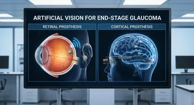
Artificial Vision for End-Stage Glaucoma: Retinal vs. Cortical Prostheses
In conditions like retinitis pigmentosa or macular degeneration, the photoreceptors die but RGCs and optic nerve remain intact () (). Retinal...
Deep research and expert guides on maintaining your visual health.
In conditions like retinitis pigmentosa or macular degeneration, the photoreceptors die but RGCs and optic nerve remain intact () (). Retinal...
Screen your peripheral vision from home — no downloads, no waiting rooms. Sign up for a free trial and test in under 5 minutes.
Visual prosthetics are medical devices designed to give people who have lost sight some ability to perceive light, shapes, or movement by replacing or bypassing damaged parts of the eye or visual system. They typically combine a camera or sensor, a processor that converts images into electrical signals, and an implanted electrode array that stimulates remaining neural tissue. Different approaches aim at different levels of the visual pathway, such as the retina, optic nerve, or visual cortex, depending on where the damage is. The output is not the same as natural vision; it is often low-resolution and requires training for the brain to interpret the new signals. Researchers and clinicians work closely with recipients to adjust settings and develop rehabilitation strategies so that the user can learn to make everyday use of the new sensations. Visual prosthetics matter because they offer a chance for people with severe blindness to regain useful visual information for tasks like detecting obstacles, locating light sources, or recognizing basic shapes. They also drive advances in neuroscience, engineering, and rehabilitation methods that can benefit other sensory disorders. Limitations include surgical risks, variable outcomes between individuals, and current technological constraints on resolution and battery life. Ongoing research seeks to improve image quality, miniaturize components, and expand who can benefit from these devices.