How Useful Is OCT at Each Stage of Glaucoma?
Each OCT result comes with color-coded maps and numbers. Green usually means “within normal limits,” yellow means “borderline,” and red indicates...
Deep research and expert guides on maintaining your visual health.
Each OCT result comes with color-coded maps and numbers. Green usually means “within normal limits,” yellow means “borderline,” and red indicates...
Track peripheral vision changes between eye doctor visits. Start your free trial and get results in under 5 minutes.
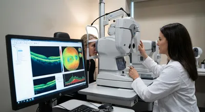
Optical coherence tomography is a medical imaging method that uses light waves to create detailed cross‑section pictures of the eye. It works a bit like ultrasound, but it uses light instead of sound, so it can show very thin layers inside the retina and optic nerve area. The test is noninvasive, quick, and painless: you rest your chin and look at a target while the machine collects the images. The result is a precise map of the eye’s internal structure that doctors can review on a computer. This technology matters because it lets clinicians see changes that are too small to detect with a regular exam. It is commonly used to diagnose and monitor conditions that affect the retina and the optic nerve, and it helps doctors decide whether treatments are working. By comparing images taken over time, providers can spot progression early and adjust care before significant vision is lost. For patients, that means earlier detection, more tailored treatment plans, and better chances of preserving sight.