Deep research and expert guides on maintaining your visual health.
retinal imaging
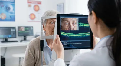
Retinal imaging refers to a set of techniques that take pictures or maps of the back of the eye, where the retina and optic nerve are located. Common methods include color fundus photography, optical coherence tomography (OCT), fluorescein angiography, and newer scans that show blood flow without dye. These images let doctors see the layers of the retina, detect swelling, bleeding, abnormal blood vessels, and other structural changes that are invisible through a simple eye exam. Most retinal imaging is quick, painless, and can be performed in a clinic; some scans are noncontact while others may require a dye injection for more detailed blood vessel views.
Retinal imaging matters because many sight-threatening conditions—such as diabetic retinopathy, age-related macular degeneration, retinal vein occlusions, and early glaucoma—produce changes that these technologies can detect early. Early detection often allows treatment that prevents or slows vision loss, and imaging provides objective records to track disease progression over time. In addition, retinal scans can sometimes reveal signs of systemic diseases like high blood pressure or stroke risk, making them useful beyond eye care. With advances in portable cameras and telemedicine, retinal imaging is becoming more accessible, helping reach patients who might otherwise delay eye care.