How Useful Is OCT at Each Stage of Glaucoma?
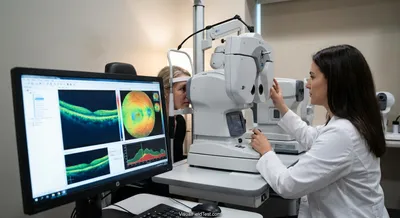
Each OCT result comes with color-coded maps and numbers. Green usually means “within normal limits,” yellow means “borderline,” and red indicates...
Deep research and expert guides on maintaining your visual health.
Each OCT result comes with color-coded maps and numbers. Green usually means “within normal limits,” yellow means “borderline,” and red indicates...
Our visual field test is inspired by the perimetry methods eye care professionals use. Check for blind spots and track changes over time.
Test Your VisionThe retinal nerve fiber layer is the innermost layer of the retina made up of long, thin fibers that carry visual information from the eye’s light‑sensing cells to the brain. Those fibers are the axons of retinal ganglion cells, and they bundle together to form the optic nerve at the back of the eye. In a healthy eye this layer has a predictable thickness and structure, so changes in its appearance can signal trouble. Measuring the thickness of this layer is important because thinning is a common early sign of diseases that damage the optic nerve, such as glaucoma. Eye care professionals often use imaging tests to measure the layer and compare results over time to detect progression. Knowing whether the layer is thinning can help guide treatment decisions and prevent further vision loss. While measurements are a valuable tool, they are interpreted alongside eye exams and visual field tests because individual differences and imaging limitations can affect results.