A New Way to Read Eye Scans in Glaucoma: Can 3D Nerve Fiber Shape Improve Detection?
To check the RNFL, doctors commonly use optical coherence tomography (OCT), a non-invasive imaging test that takes cross-sectional “slice” pictures...
Deep research and expert guides on maintaining your visual health.
To check the RNFL, doctors commonly use optical coherence tomography (OCT), a non-invasive imaging test that takes cross-sectional “slice” pictures...
Track peripheral vision changes between eye doctor visits. Start your free trial and get results in under 5 minutes.
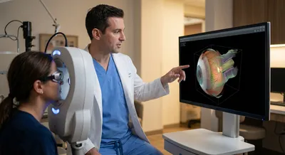
3D imaging creates a three-dimensional picture of an object or body part by combining many slices or angles into a single view. In medicine, this technique builds a depth-aware model that shows shape, thickness, and spatial relationships more clearly than a flat picture. For eyes and other organs, 3D views can reveal subtle features like curved surfaces or irregularities that are missed in two-dimensional images. That extra depth helps doctors measure structures more precisely, see where tissue is thinning or bulging, and plan treatments or surgeries with greater accuracy. Building accurate 3D models requires good equipment, careful image processing, and sometimes computer algorithms to highlight the most relevant details. While powerful, 3D imaging can also produce large amounts of data that need storage and expert interpretation. Overall, 3D imaging matters because it often improves diagnosis and decision-making by giving a fuller, more realistic picture of anatomy and disease.