Can Eye Blood Vessel Changes Warn of Future Glaucoma Damage Before Vision Tests Turn Abnormal?
Glaucoma is often called a “silent thief of sight.” It quietly damages the optic nerve before any vision loss shows up. Structural damage means...
Deep research and expert guides on maintaining your visual health.
Glaucoma is often called a “silent thief of sight.” It quietly damages the optic nerve before any vision loss shows up. Structural damage means...

Below we break down each common glaucoma-related test, roughly how long it takes, and why it matters. We then explain how these pieces add up to...
Screen your peripheral vision from home — no downloads, no waiting rooms. Sign up for a free trial and test in under 5 minutes.
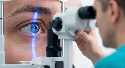
Optic nerve imaging refers to noninvasive scans and photographs that show the structure of the optic nerve and the surrounding nerve fiber layer at the back of the eye. Common technologies include optical coherence tomography (OCT), fundus photography, and scanning laser devices, which create detailed pictures or maps of tissue thickness and shape. These images help doctors see changes such as thinning of nerve fibers, increased cupping of the nerve head, swelling, or other abnormalities that are not always obvious on a routine exam. The scans are quick, painless, and provide objective measurements that can be stored and compared over time. This kind of imaging matters because many eye and neurological conditions cause subtle changes in the optic nerve before symptoms appear. Regular imaging gives a baseline and allows clinicians to detect progression early, monitor treatment effectiveness, and decide when to change a plan of care. High-quality images and proper interpretation are essential, since factors like cataracts or poor fixation can affect the results. For patients, optic nerve imaging offers a clear, repeatable way to track eye health and support timely treatment when problems arise.