Glaukomos regos lauko tyrimo apribojimai: dažnumas, subjektyvumas ir tai, kas lieka nepastebėta
Glaukoma yra lėtinė regos nervo liga, dažnai vadinama „tyliuoju regos vagimi“. Ji sukelia laipsnišką, negrįžtamą regos praradimą. Pagrindinis būdas, kuriuo gydytojai stebi glaukomos progresavimą, yra regos lauko (RL) tyrimai: automatiniai perimetrijos tyrimai, kurie nustato paciento periferinio regėjimo žemėlapį. Teoriškai šie tyrimai leidžia klinikams anksti pastebėti regėjimo praradimą ir koreguoti gydymą. Tačiau praktikoje standartiniai regos lauko tyrimai turi svarbių trūkumų. Šiame straipsnyje aptariama, kodėl RL tyrimai dažnai atliekami per retai, kaip jų subjektyvumas ir paciento veiksniai padidina „triukšmą“, ir kokius regos praradimo tipus šie tyrimai gali praleisti. Taip pat apžvelgsime tyrimų apie tyrimo patikimumą rezultatus ir tai, ką mokslininkai bei gydytojai daro, kad atskirtų tikrąjį progresavimą nuo atsitiktinių svyravimų. Galiausiai, atkreipsime dėmesį į naujas technologijas, kurios šiuo metu tiriamos, ir pateiksime praktinių patarimų pacientams bei specialistams, kaip kuo geriau išnaudoti regos lauko tyrimus.
Regos lauko tyrimo dažnumas
Gairės ir realioji praktika
Most glaucoma guidelines stress frequent monitoring, especially soon after diagnosis. For example, expert recommendations suggest newly diagnosed patients get about three VF tests per year in the first two years to establish a reliable baseline and detect “fast progressors” early (www.ncbi.nlm.nih.gov). In fact, one modeling study concluded that six tests in two years (i.e. three per year) are needed to reliably measure a typical glaucoma progression rate of ~1 dB/year (www.ncbi.nlm.nih.gov). The European Glaucoma Society (EGS) adopted this schedule into its guidelines.
However, surveys and audits show that in practice glaucoma patients are tested far less often. In one large UK audit (n≈90,000 patients), VF testing was done on average only once per year (www.ncbi.nlm.nih.gov). In the United States, a national insurance-data study found a median frequency of only 0.63 VF tests per year among open-angle glaucoma patients (pmc.ncbi.nlm.nih.gov). Over 75% of patients had less than one test per year, falling short of recommended annual monitoring (pmc.ncbi.nlm.nih.gov) (www.ncbi.nlm.nih.gov). In other words, most patients go more than a year between fields, even though an earlier analysis suggests annual testing would already delay detection by years (see below). Clinicians often cite constraints on time and resources for the low testing cadence (www.ncbi.nlm.nih.gov).
Reto tyrimų atlikimo poveikis
Why does frequency matter? Because glaucoma usually progresses slowly, doctors rely on multiple VF tests over time to detect a meaningful trend. Sparse testing greatly delays awareness of vision loss. For example, Che Hamzah et al. estimate that detecting a loss of 1 dB/year would take about 6 years if fields are done once a year, but only around 2 years if tests are done 3 times/year (pmc.ncbi.nlm.nih.gov). In other words, infrequent fields can leave patients at risk of unnoticed loss. Delays in spotting progression can mean delayed treatment changes — and once nerve fibers die, vision cannot be recovered. In economic modeling, more frequent early testing (3x/year) in high-risk patients was actually cost-effective by catching “fast progressors” sooner (www.ncbi.nlm.nih.gov).
Nevertheless, many ophthalmologists and clinics do not follow these intensive protocols. UK and US survey data found that providers view thrice-yearly testing as impractical with current resources (www.ncbi.nlm.nih.gov). Patients themselves often dread the test (it is time-consuming and tedious), even though they recognize its importance (www.ncbi.nlm.nih.gov). In short, there is a gap between what modeling and guidelines recommend and the reality of busy clinics: tests are done too rarely to catch small changes before significant vision loss occurs.
Tyrimo kintamumas ir subjektyvumas
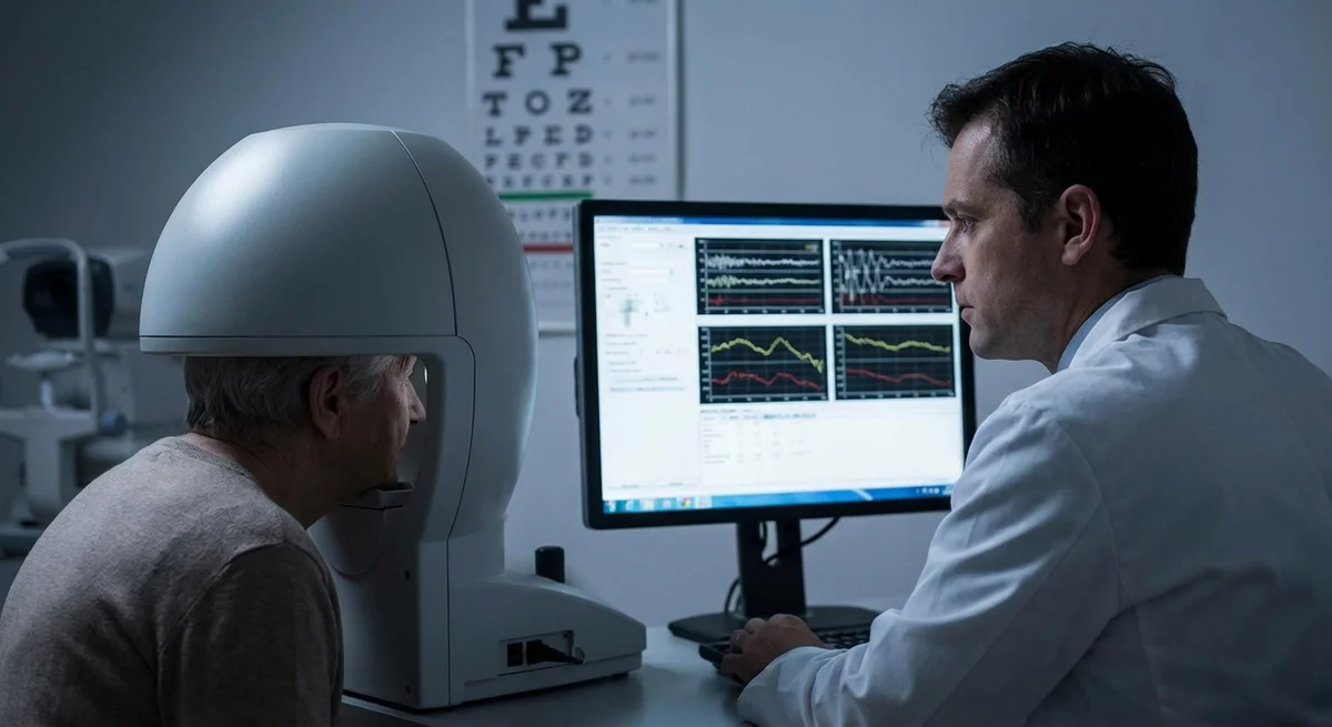
Automated perimetry is powerful but inherently noisy. Each visual field result is a threshold sensitivity map assembled from the patient’s responses to hundreds of light stimuli. Many factors – both physiological and situational – cause significant variability from test to test. In fact, test-retest differences can be large enough to mask true vision changes or, conversely, create false progression signals.
Pakartotinių tyrimų kintamumas
Studies have repeatedly shown that standard automated perimetry (SAP) suffers from considerable test-retest variability. Guimarães et al. reported that worse visual field status itself increases variability: eyes with more severe VF defects (lower MD or VFI) showed greater fluctuation between tests (pmc.ncbi.nlm.nih.gov). In general, even a stable patient can exhibit 2–3 dB swings in sensitivity at any given point of the field from one visit to the next. This “noise” means small true changes are hard to distinguish from chance fluctuation. As one review explained, “detection of progression depends on separating true change (signal) from test-retest variability (noise)” (pmc.ncbi.nlm.nih.gov). If variability is large, real deterioration can be missed, delaying treatment escalation (pmc.ncbi.nlm.nih.gov). Conversely, spurious jumps in the data can falsely trigger concern.
Paciento ir aplinkos veiksniai
Because the test relies on the patient’s responses, many human factors affect reliability. Older patients and those with general health problems tend to have less reliable fields (pmc.ncbi.nlm.nih.gov). In one study, worse sleep quality and older age were each linked to more errors: poorer sleep was associated with fixation lapses and increased false responses, and older patients showed more false negatives, likely from fatigue (pmc.ncbi.nlm.nih.gov). Patient anxiety and mood also play a role: many patients find perimetry stressful. Kaliaperumal et al. found that automated perimetry induced slightly higher anxiety in glaucoma patients compared to an OCT scan, and that anxiety was higher in patients with fewer prior visual field tests (pmc.ncbi.nlm.nih.gov). Notably, patients with fewer than two prior fields had the highest anxiety scores, which fell off after five or more tests (pmc.ncbi.nlm.nih.gov). This suggests unfamiliarity and nervousness degrade early test performance.
Other factors can also skew results. Vision testers note that poor instruction or an uncomfortable testing environment (bright lights, noise, long waits) can distract patients. An audit found that patients complained about inconsistent instructions, distracting setups, and unclear explanations of results (www.ncbi.nlm.nih.gov). Standard reliability indices (fixation losses, false positives, false negatives) try to catch some issues, but those too are influenced by patient factors like blink rate, depression, or inattentiveness. In summary, each VF test is a subjective effort that depends on patient cooperation, understanding, and focus.
Mokymosi efektai
Another important source of variability is the learning effect. Many patients perform poorly on their very first field and improve over the next few tests as they learn what to do. Rana et al. (2023) demonstrated a clear learning curve: patients (both glaucoma patients and normal controls) showed significantly better reliability indices (fewer fixation losses, false positives) and more stable global indices by the third test compared to the first (pmc.ncbi.nlm.nih.gov). They concluded that at least three baseline fields are needed before the results stabilize, especially in glaucoma patients (pmc.ncbi.nlm.nih.gov). In practice, this means the very first VF exam on a patient – or after a long break – may underestimate true sensitivity. Clinicians often discount the first field or ensure the patient practices, because these learning improvements are well-documented (pmc.ncbi.nlm.nih.gov) (pmc.ncbi.nlm.nih.gov).
Progresavimo atskyrimas nuo triukšmo
Given all this variability, how do clinicians decide when vision loss is real and when it is just noise? In routine care, doctors look for consistent patterns over time. Modern perimeters include statistical tools (such as the Guided Progression Analysis, GPA) that flag progression if certain points decline repeatedly. However, these algorithms assume a level of measurement consistency and can still generate false alarms. For example, GPA’s “possible progression” alert mode may produce a false-positive in roughly 15–20% of cases purely from fluctuation (pubmed.ncbi.nlm.nih.gov). (In other words, some patients will trigger an alarm even if they are stable.) Reliance on event-based rules alone can therefore mislead.
In response, clinicians often take growth curves across serial visual fields (trend analysis) into account. They also double-check suspect results. If a patient’s field shows a sudden drop, a common practice is to repeat the test relatively soon to see if it persists. Consistency across multiple tests raises confidence that a change is real. Eye doctors also examine the standard reliability indices on each report: a field with high fixation losses or false positives is interpreted cautiously. If indices exceed rough thresholds (often fixations >20%, false positives >15–33%), many clinicians will either discount isolated changes or ask for retesting immediately (pmc.ncbi.nlm.nih.gov). In practice, this means no single VF result is acted on without considering its context and confirming it.
Combining functional and structural data also helps. If VF data are ambiguous but optical coherence tomography (OCT) shows clear retinal nerve fiber loss, a clinician may lean toward true progression. Ultimately, judgment about change often requires pattern recognition over time. Many glaucoma specialists use two or three consecutive fields with similar decline before escalating treatment. This cautious approach helps avoid unnecessary interventions from a one-off “bad” field, but it also highlights the risk: significant damage could accumulate while clinicians wait for confirmation. In summary, no tester’s magic marker can completely separate signal from noise – it remains partly an art honed by experience and aided by statistical rules.
Ar dabartiniai protokolai yra pakankami?
Taken together, the limited frequency of testing and the inherent variability mean standard perimetry may miss early glaucoma progression until it becomes moderate or severe. European experts argue that the official recommendation of three tests per year (in early disease) is evidence-based (www.ncbi.nlm.nih.gov), but in reality this often doesn’t happen (www.ncbi.nlm.nih.gov) (pmc.ncbi.nlm.nih.gov). Even annual perimetry (the US guideline minimum) may be too slow for some patients. In low-risk, stable cases, infrequent testing might be acceptable, but in high-risk scenarios (e.g. very high pressure or advanced field loss) more vigilance is needed.
In fact, some researchers point out that visual field changes lag behind structural damage. By the time a VF defect appears, many retinal ganglion cells may already have been lost. OCT can detect thinning of nerve fiber layers earlier than a field defect showing up. Thus, VF testing has limitations in early detection. Also, the standard 24-2 testing grid does not densely assess central vision; a patient could lose small central island of vision or macular fibers without clear 24-2 changes. (Detecting those often requires a 10-2 Humphrey test or other methods.) In practice, clinicians realize that VF is only part of the story, and they monitor intraocular pressure and imaging closely too.
Ultimately, evidence suggests that current testing protocols are only marginally adequate. Many eyes with progressing glaucoma are discovered late enough that significant vision is already lost. There is continuing debate about optimal intervals and whether we should stratify patients by risk – faster progressors get more frequent tests. Organizations like NICE in the UK have noted a lack of solid clinical trials on monitoring intervals, and call for more research. Meanwhile, reviews consistently emphasize that too-infrequent or inconsistent testing can overlook vision loss until it is serious (pmc.ncbi.nlm.nih.gov) (pmc.ncbi.nlm.nih.gov).
Atsirandančios alternatyvos ir papildymai
To overcome the limits of standard perimetry, new approaches are being explored. These include both alternative testing methods and leveraging technology for more frequent monitoring.
-
Struktūrinis vaizdavimas (OCT): Optical coherence tomography provides high-resolution images of the retinal nerve fiber layer and optic nerve head. Unlike perimetry, OCT is objective and requires minimal patient input (pmc.ncbi.nlm.nih.gov). It has become a routine part of glaucoma care. While structural measures do not perfectly predict functional vision, they often show progression earlier. For example, a thinning in the nerve fiber layer on OCT can signal damage even if the VF is still “normal.” In practice, comparing VF trends to OCT trends gives a more complete view of the disease. (Patients should note: if a doctor points out OCT thinning despite “normal” fields, it may mean early glaucoma damage.)
-
Namų stebėjimas ir naujoviška perimetrija: Recognizing that clinic visits are infrequent, researchers have developed devices and apps for patients to test vision at home. One review highlights tablet- and computer-based perimetry tools, such as the Moorfields Motion Displacement Test (MMDT) and the Melbourne Rapid Fields (MRF) app (pmc.ncbi.nlm.nih.gov). MMDT runs on a laptop with specialized stimuli, and MRF runs on an iPad; both mimic aspects of Humphrey fields but can be done at home. Head-mounted virtual reality perimeters are also under development. Early studies show these approaches are promising: they are portable, user-friendly, and can generate real VF data. The idea is that patients could test themselves (for example, weekly or monthly) and send results to their doctor, catching changes earlier. These tools are still in validation, but they represent a way to increase VF data points without overburdening clinics (pmc.ncbi.nlm.nih.gov) (pmc.ncbi.nlm.nih.gov).
-
Šluojančiosios technikos ir grupinis tyrimas: Some clinical research suggests doing multiple short field exams (clustered over a few weeks) can improve sensitivity to change. By concentrating several tests in close succession, variability can be averaged out, making progression stand out. This approach is still experimental but has shown that more frequent, clustered data points can detect change sooner without increasing total test time.
-
Pažangi analizė: Artificial intelligence and statistical methods are also being applied. For instance, combining OCT and VF data through machine learning might predict progression earlier. Enhanced progression algorithms (beyond standard GPA) are also in development, aiming to define significant change considering each patient’s variability profile. These are mostly in research stages.
In summary, new technologies are on the horizon. OCT amplifies what VF can miss; at-home perimetry could supply more frequent VF data; and smarter software might untangle noise from real change. But none of these has yet replaced standard VF testing in routine practice.
Praktiniai patarimai pacientams ir klinikams
Given these challenges, both patients and doctors can take steps to improve visual field testing outcomes.
-
Pacientams:
- Poilsis ir mityba: Arrive well-rested and fed. Good sleep and a relaxed state help concentration. One study found that poor sleep quality increased VF errors (pmc.ncbi.nlm.nih.gov). If possible, avoid scheduling the test at the end of a long day.
- Valdykite nerimą: It’s normal to feel anxious about the test. Remember that multiple people worry about it. Knowing that anxiety tends to drop after the first few tests can help (pmc.ncbi.nlm.nih.gov). Some clinics offer practice runs or videos – taking advantage of those can reduce stress. (For example, Sherafat et al. showed that a brief instructional video before testing significantly improved reliability (pmc.ncbi.nlm.nih.gov).)
- Atidžiai laikykitės instrukcijų: Sit properly, use any prescription glasses provided by the clinic, and keep your head steady on the chinrest. Focus on the central fixation light. If you see a light or stimulus, press the button without second-guessing. Don’t press when you’re unsure; false presses can create misleading results. If the patient has an established test strategy, try to replicate it each time.
- Klauskite: If you don’t understand something, speak up. Better to clarify than guess. Many reliability problems come from misunderstanding instructions (pmc.ncbi.nlm.nih.gov). Nowadays, educational materials (videos or demos) are often available. It doesn’t hurt to request one.
- Keli tyrimai: Know that your doctor may order two or three “baseline” fields in a short span. This may seem redundant, but it’s to overcome the learning effect (pmc.ncbi.nlm.nih.gov). Treat these as practice to help later exams be more reliable.
-
Klinikams:
- Patikrinkite patikimumo indeksus: Always check fixation losses, false positives, and false negatives on the printout. High error rates (e.g. FP >15–20% or FN >33%) should prompt caution. If indices are poor, consider repeating the test immediately (pmc.ncbi.nlm.nih.gov).
- Pakartokite įtartinus laukus: If a field shows a new focal defect or a big change (e.g. MD drop >2 dB) but the reliability is marginal, retesting or even short-term repeat testing can confirm whether it’s real. Do not make major treatment changes based on a single abnormal field.
- Naudokite progresavimo programinę įrangą, bet su išmanymu: While tools like the Guided Progression Analysis provide quick alerts, recognize their limits. An “upgrade” to likely progression often requires confirmation on follow-up fields.
- Integruokite visus duomenis: Look at OCT and optic nerve appearance alongside VF results. Concordance between structural and functional damage strengthens confidence. If fields and OCT disagree, plan to investigate further (perhaps with specialist testing like microperimetry or a second opinion).
- Pritaikykite tyrimų intervalus: Consider risk factors. Rapid progressors, advanced disease, or highly asymmetric fields may warrant more frequent testing (toward the 3/year recommendation) (www.ncbi.nlm.nih.gov). In contrast, stable early glaucoma at target pressure might be seen yearly or stopped if truly stable over 5+ years.
- Pagerinkite paciento patirtį: A bit of encouragement goes a long way. Explain that the test is important for their care and praise their effort afterward. Comfortable lighting and a friendly testing environment can reduce fatigue. Remember that we are asking patients to perform a challenging task. Even a 5-minute rest halfway through a long test can improve results.
- Išmintingai naudokite naujus įrankius: Stay informed about advances like home perimetry. Discuss these options in complex cases or clinical trials. Coordinate with clinic workflows to potentially integrate validated home tests or tablet apps that allow extra data points between visits.
Išvada
Standartinis automatinis regos lauko tyrimas išlieka auksiniu standartu funkciniam glaukomos įvertinimui, tačiau jis turi realių apribojimų. Tyrimai dažnai atliekami retai, o kiekvienas tyrimas turi didelį paciento sukeltos kintamumo potencialą. Dėl to subtilus progresavimas per ilgai gali likti nepastebėtas. Klinikai turi atsargiai interpretuoti RL rezultatus, patvirtinti įtartinus pokyčius ir dažnai pasikliauti papildoma informacija (vaizdavimas, slėgio tendencijos) kad priimtų sprendimus. Pacientai gali padėti būdami pasiruošę, laikydamiesi instrukcijų ir suprasdami tyrimo tikslą. Žvelgiant į priekį, atsirandančios technologijos – namų perimetrija, geresnė analizė ir patobulintas struktūrinis tyrimas – žada užpildyti spragas. Iki tol šių apribojimų suvokimas yra esminis: „triukšmo“ atpažinimas regos lauko tyrimuose padeda apsaugoti pacientų regėjimą, skatinant savalaikius pakartotinius tyrimus ir gydymo koregavimus.
.