뇌 가소성과 지각 학습: 뇌는 시신경 손상을 보상할 수 있는가?
흥미롭게도 많은 녹내장 환자는 맹점을 거의 인지하지 못합니다. 뇌가 누락된 주변 정보를 "채워 넣는" 이러한 지각적 채워넣기(perceptual filling-in)는 신경 보상을 반영하는 것으로 여겨집니다. 예를 들어, 한 뇌 영상 연구에서는 녹내장 환자(심각한...
시각 건강을 유지하기 위한 심층 연구 및 전문가 가이드.
흥미롭게도 많은 녹내장 환자는 맹점을 거의 인지하지 못합니다. 뇌가 누락된 주변 정보를 "채워 넣는" 이러한 지각적 채워넣기(perceptual filling-in)는 신경 보상을 반영하는 것으로 여겨집니다. 예를 들어, 한 뇌 영상 연구에서는 녹내장 환자(심각한...
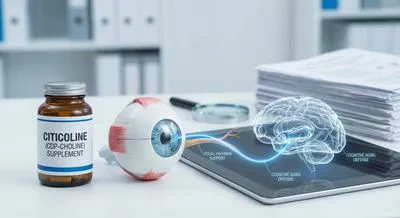
시티콜린(사이티딘-5′-디포스포콜린)은 체내에서 사이티딘과 콜린으로 대사됩니다. 콜린은 주요 막 지질인 포스파티딜콜린과 신경전달물질인 아세틸콜린의 합성에 사용됩니다 (). 이러한 구성 요소를 공급함으로써 시티콜린은 세포막을 복구하고 유지하는 데 도움을 줍니다....
집에서 주변 시야를 검사하세요 — 다운로드도, 대기실도 필요 없습니다. 무료 평가판에 가입하고 5분 이내에 검사하세요.
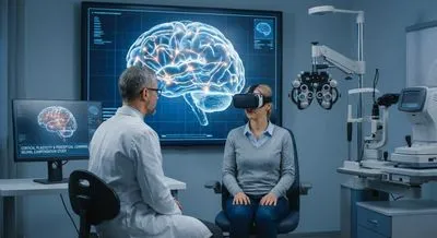
VEP는 눈으로 들어온 시각 정보가 뇌에서 어떻게 처리되는지를 전기 신호로 기록하는 검사입니다. 두피에 전극을 붙여 시각 자극에 대한 뇌의 반응을 측정하므로 시신경에서 뇌까지의 전달 상태를 평가할 수 있습니다. 시각 자극은 깜빡이는 빛이나 패턴 무늬로 제공되며 검사 중 편안히 있으면 됩니다. 이 검사는 시신경의 전도 속도나 효율에 문제가 있는지 보여주어 여러 신경계 질환을 구별하는 데 도움이 됩니다. 시력 저하의 원인이 눈 자체인지 아니면 신경 전달 문제인지 판단할 때 유용합니다. 예를 들어 눈에는 이상이 없지만 뇌로의 신호 전달이 느리거나 약해진 경우 VEP에서 이상 소견이 보일 수 있습니다. 검사 결과는 신경과나 안과 전문의가 다른 검사와 함께 종합해 해석합니다. 어린아이부터 성인까지 적용 가능하고, 비언어적인 방법으로 시각 기능을 평가할 수 있다는 장점이 있습니다. VEP를 통해 신경계의 문제를 조기에 발견하면 적절한 치료나 재활 계획을 세우는 데 큰 도움이 됩니다.