การวิจัยเชิงลึกและคู่มือผู้เชี่ยวชาญเกี่ยวกับการรักษาสุขภาพสายตาของคุณ
OCT-Angiography
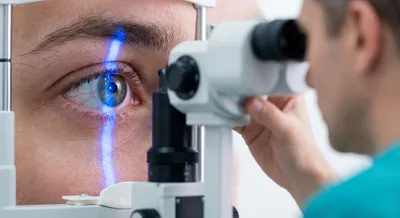
OCT-Angiography เป็นเทคโนโลยีการถ่ายภาพที่ใช้แสงเพื่อเห็นโครงข่ายหลอดเลือดภายในดวงตาโดยไม่ต้องฉีดสารสีเข้าเส้นเลือดตรงๆ ทำให้แพทย์สามารถมองเห็นการไหลเวียนของเลือดในชั้นจอประสาทตาและชั้นอื่นๆ ได้อย่างละเอียดและรวดเร็ว การตรวจนี้มักทำได้ในคลินิกโดยไม่เจ็บและไม่มีความเสี่ยงจากการแพ้สารสีเหมือนการฉีดสารสีแบบเดิม
ความสำคัญของการตรวจนี้คือช่วยให้เห็นสัญญาณของโรคที่ทำให้หลอดเลือดผิดปกติ เช่น เบาหวานขึ้นตา ต้อหินบางชนิด หรือโรคจอประสาทตาเสื่อมในระยะแรก แพทย์จึงสามารถวางแผนการรักษา ติดตามผลการรักษา และประเมินความเสี่ยงการสูญเสียการมองเห็นได้ดีขึ้น อย่างไรก็ตาม ภาพที่ได้อาจมีข้อจำกัด เช่น เกิดภาพผิดพลาดจากการเคลื่อนไหวของตาหรือไม่สามารถแสดงการรั่วของหลอดเลือดได้ชัดเจนเสมอไป ทำให้ต้องใช้ข้อมูลร่วมกับการตรวจอื่นๆ เพื่อการวินิจฉัยที่ถูกต้องและการดูแลที่เหมาะสม